. 1986 May 30;232(4754):1138-40.
doi: 10.1126/science.3518061.

Brain glutamate decarboxylase cloned in lambda gt-11: fusion protein produces gamma-aminobutyric acid

Brain glutamate decarboxylase cloned in lambda gt-11: fusion protein produces gamma-aminobutyric acid

D L Kaufman et al. Science. .

Abstract

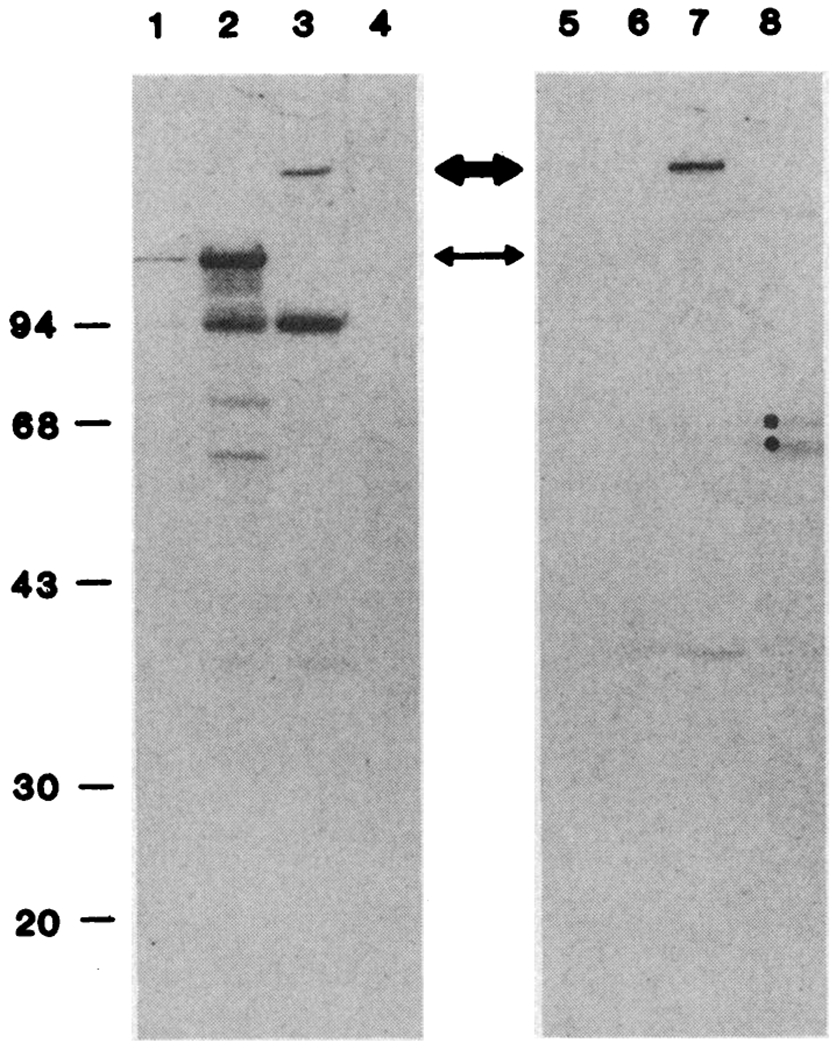
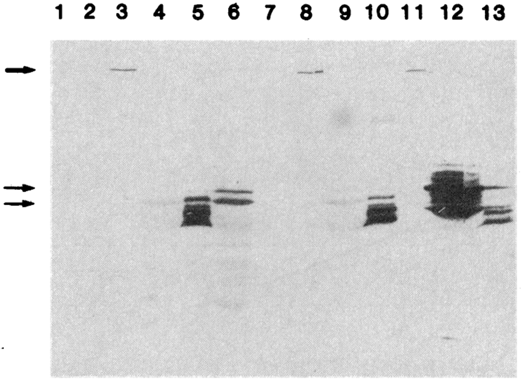
Glutamate decarboxylase (GAD; E.C. 4.1.1.15) converts glutamate to gamma-aminobutyric acid (GABA), the major inhibitory neurotransmitter in the vertebrate central nervous system. This report describes the isolation of a GAD complementary DNA clone by immunological screening of a lambda gt-11 brain complementary DNA expression library. The fusion protein produced by this clone catalyzes the conversion of glutamate to GABA and carbon dioxide, confirming its identity as GAD. Antibodies to beta-galactosidase remove GAD enzymatic activity from solution, showing that this activity is associated with the fusion protein. In immunoblotting experiments all three available antisera to GAD reacted with the fusion polypeptide and with two major polypeptides (molecular size, 60,000 and 66,000 daltons) in brain extracts.


Figures

Fig. 1.
Fig. 1.
Immunological verification of GAD and β-galactosidase epitopes in the same polypeptide. Protein was extracted from λ-lysogens as described in the legend to Table 1, except that 50 mM tris-HCl, pH 7.2, replaced potassium phosphate. Brain was homogenized in a solution containing 0.2 mM PMSF, 150 mM NaCl, and 50 mM tris-HCl, pH 8.0, and the protein was extracted. Cell debris was removed by centrifugation. The proteins were separated by electrophoresis in a 10% polyacrylamide gel in SDS (23) and electrophoretically transferred to nitrocellulose (13, 24). The unreacted sites were blocked with bovine serum albumin and gelatin, and the nitrocellulose sheet was cut in half. One half was incubated overnight with a 1:1000 dilution of affinity-purified rabbit antibody to β-galactosidase and the other half with a 1:500 dilution of sheep antibody to GAD; the sheets were then incubated for 1 hour with a 1:1000 dilution of rabbit antiserum to sheep IgG. The sheet was extensively washed, and bound antibodies were detected with 125I-labeled protein A and autoradiography. (Lanes 1 to 4) Detection of β-galactosidase epitopes. (Lanes 5 to 8) Detection of GAD epitopes. Each lane contained the following: lanes 1 ana 5, purified β-galactosidase (200 ng); lanes 2 and 6, λgt-11 lysogen extracts (20 μg); lanes 3 and 7, λGAD lysogen extracts (20 μg); and lanes 4 and 8, extracts of cat brain (20 μg). A ninth lane (not shown) contained 125I-labeled molecular weight standards. The thin arrow marks the position of wild-type β-gaiactosidase, and the thick arrow marks die fusion protein produced by λGAD.
Fig. 2.
Fig. 2.
Immunoblotting of brain extracts with three antibodies to GAD. Extracts of lysogens and brain were analyzed by electrophoresis in 10% polyacrylamide gel in SDS followed by immunoblotting as described in the legend to Fig. 1. The nitrocellulose sheet, was cut into three parts, and each was incubated with a 1:500 dilution of a different antiserum to GAD: lanes 1 to 7 with sheep antiserum to rat GAD (4), lanes 8 to 10 with sheep antiserum to pig GAD (25), and lanes 11 to 13 with rabbit antiserum to mouse GAD (3). These antisera specifically recognize the fusion polypeptide (thick arrow) and at least two major polypeptides (60 and 66 kD) in cat brain (thin arrows). Although these two polypeptides are weak in lane 7, they can be seen on longer exposures. Similar results were obtained when electrophoresis was performed in 7.5% polyacrylamide. The following extracts were examined: Lane 1, λgt-11 lysogen (20 μg); lane 2, λGAD lysogen without IPTG induction (20 μg); lanes 3, 8, and 11, λGAD lysogen after IPTG induction (20 μg); lanes 4, 9, and 12, cat brain (20 μg); lanes 5, 10, and 13, partially purified rat GAD (25) (4 μg); lane 6, mouse brain (20 μg); and lane 7, human brain (20 μg).

References

    1. Roberts E, Chase TN, Tower DB, Eds., GABA in Nervous System Function (Raven, New York, 1976).
    1. Hertz L, Kvamme E, McGeer EG, Schousboe A, Eds., Glutamine, Glutamate and GABA in the Central Nervous System (Liss, New York, 1983).
    1. Saito K et al. , Proc. Natl. Acad. Sci. U.S.A 71, 269 (1974).
    1. Oertel WH, Schmechel DE, Tappaz ML, Kopin IJ, Neuroscience 6, 2689 (1981). - PubMed
    1. Oertel WH, Schmechel DE, Mugnaini E, Tappaz ML, Kopin IJ, ibid., p. 2715. - PubMed

Publication types

LinkOut - more resources