. 2022 Feb 18;14(1):17.
doi: 10.1186/s13073-022-01022-0.

PLCG2 is associated with the inflammatory response and is induced by amyloid plaques in Alzheimer's disease

Affiliations

PLCG2 is associated with the inflammatory response and is induced by amyloid plaques in Alzheimer's disease

Andy P Tsai et al. Genome Med. .

Abstract

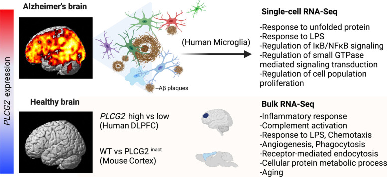
Background: Alzheimer's disease (AD) is characterized by robust microgliosis and phenotypic changes that accompany disease pathogenesis. Accumulating evidence from genetic studies suggests the importance of phospholipase C γ 2 (PLCG2) in late-onset AD (LOAD) pathophysiology. However, the role of PLCG2 in AD is still poorly understood.

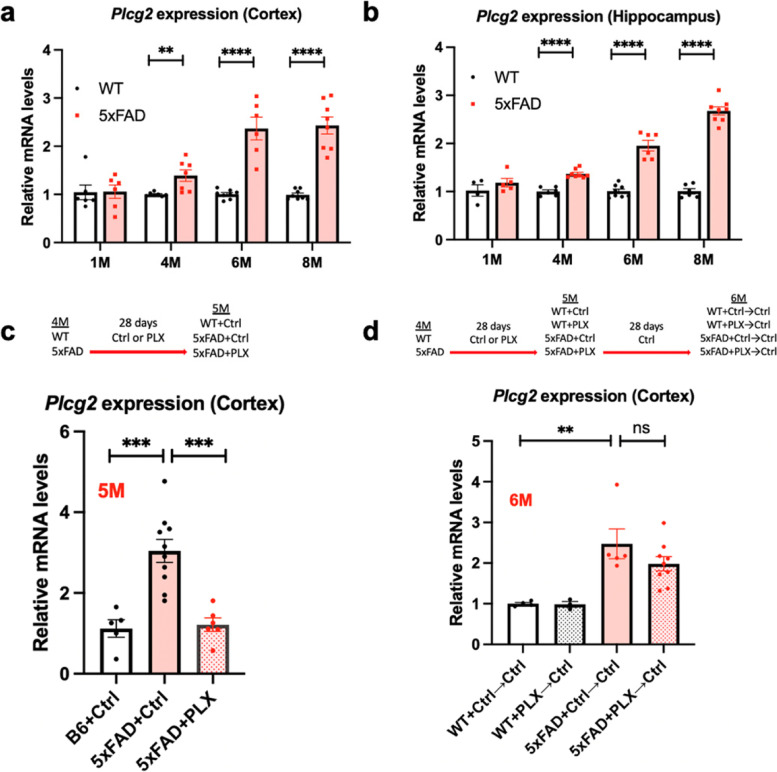
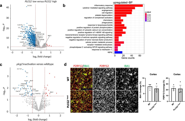
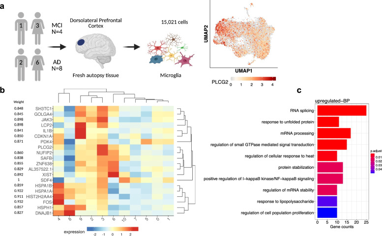
Methods: Using bulk RNA-Seq (N=1249) data from the Accelerating Medicines Partnership-Alzheimer's Disease Consortium (AMP-AD), we investigated whether PLCG2 expression increased in the brains of LOAD patients. We also evaluated the relationship between PLCG2 expression levels, amyloid plaque density, and expression levels of microglia specific markers (AIF1 and TMEM119). Finally, we investigated the longitudinal changes of PLCG2 expression in the 5xFAD mouse model of AD. To further understand the role of PLCG2 in different signaling pathways, differential gene expression and co-expression network analyses were performed using bulk RNA-Seq and microglial single-cell RNA-Seq data. To substantiate the human analyses, we performed differential gene expression analysis on wild-type (WT) and inactivated Plcg2 mice and used immunostaining to determine if the differentially expressed genes/pathways were altered by microglial cell coverage or morphology.

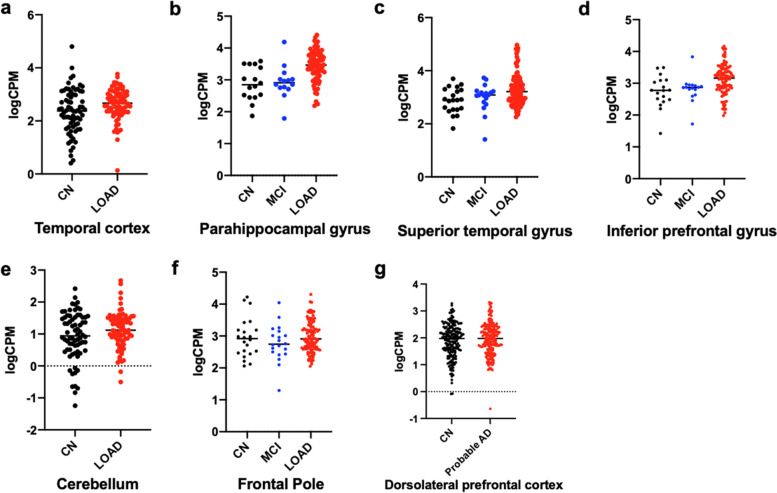
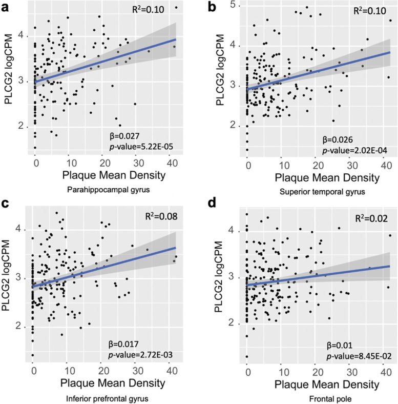
Results: We observed significant upregulation of PLCG2 expression in three brain regions of LOAD patients and significant positive correlation of PLCG2 expression with amyloid plaque density. These findings in the human brain were validated in the 5xFAD amyloid mouse model, which showed disease progression-dependent increases in Plcg2 expression associated with amyloid pathology. Of note, increased Plcg2 expression levels in 5xFAD mice were abolished by reducing microglia. Furthermore, using bulk RNA-Seq data, we performed differential expression analysis by comparing cognitively normal older adults (CN) with 75th percentile (high) and 25th percentile (low) PLCG2 gene expression levels to identify pathways related to inflammation and the inflammatory response. The findings in the human brain were validated by differential expression analyses between WT and plcg2 inactivated mice. PLCG2 co-expression network analysis of microglial single-cell RNA-Seq data identified pathways related to the inflammatory response including regulation of I-kappaB/NF-kappa B signaling and response to lipopolysaccharide.

Conclusions: Our results provide further evidence that PLCG2 plays an important role in AD pathophysiology and may be a potential target for microglia-targeted AD therapies.

Keywords: Alzheimer’s disease; Co-expression network analysis; Inflammatory response; Microglia; PLCG2; Single-cell RNA-Seq analysis.


Conflict of interest statement

The authors declare that they have no competing interests. The stipend for A.P.T. was provided in part through a fellowship funded by Eli Lilly and Company; however, the project was not sponsored by Eli Lilly and Company and they had no influence on the design, conduct, or reporting of the research.

Figures

Fig. 1
Fig. 1
Difference of PLCG2 expression levels across the diagnostic groups. Gene expression levels are shown as logCPM values. a Temporal cortex - Mayo, b Parahippocampal gyrus - MSBB, c Superior temporal gyrus - MSBB, d Inferior prefrontal gyrus - MSBB, e Cerebellum - Mayo, f Frontal pole - MSBB, and g Dorsolateral prefrontal cortex - ROS/MAP. CN cognitively normal older adults, AD Alzheimer’s disease, MCI mild cognitive impairment
Fig. 2
Fig. 2
Associations of PLCG2 expression levels with mean amyloid plaque density. The scatter plots show positive associations between PLCG2 expression levels and mean plaque density in the a parahippocampal gyrus, b superior temporal gyrus, and c inferior prefrontal gyrus and d frontal pole from the MSBB cohort
Fig 3.
Fig 3.
Plcg2 expression is increased exclusively in microglia in the brains of 5xFAD mice. Plcg2 levels were assessed in cortical and hippocampal lysates from 5xFAD mice. There were significant changes in Plcg2 gene expression in both the a cortex and b hippocampus at 4, 6, and 8 months of age (n=4–8 mice). However, increased Plcg2 expression levels were abolished after the PLX5622 treatment (c) and restored after switching the PLX diet to a normal diet (d) (n=3–10). **p<0.01; ***p<0.001; ****p<0.0001, ns not significant
Fig. 4
Fig. 4
Differential expression analysis in cognitively normal older adults and Plcg2- inactivated mouse model identified inflammation-related pathways. The volcano plot shows significant DEGs (fold change>1.5, FDR-corrected p<0.05) in cognitively normal older adults (CN) with lower (bottom 25%; n=50; low) versus higher (top 25%; n=50; high) expression of PLCG2 (a). Gene-set enrichment analysis was performed on the DEGs (fold change>1.5, FDR-corrected p<0.05) to identify GO terms, including inflammatory response (b). The volcano plot shows significant DEGs derived from the NanoString analysis of the brains of Plcg2 inactivation (PLCG2 inact, n=3) and WT (n=3) mice (1-month male) (c). Immunostaining of microglial markers with IBA1 (green) and P2RY12 (red) and quantification of percent area in brain sections of 1-month-old male WT and PLCG2 inact demonstrate unchanged microglial morphology and area coverage (d). GO gene ontology, BP biological process, DEGs differentially expressed genes, FDR false discovery rate
Fig. 5
Fig. 5
PLCG2 co-expression network analysis of microglial single-cell RNA-Seq data identified inflammatory response-related pathways. In the microglia single-cell RNA-Seq data, cells in the dorsolateral prefrontal cortex were isolated from 12 donors of both sexes. After quality control procedures, 15,021 cells were used for analysis. The UMAP plot shows the expression of PLCG2, and each dot represents a cell (a). The heatmap shows gene expression levels of the top 20 genes identified as co-expressed with PLCG2 using GENIE3 (b). The top ten GO terms from gene-set enrichment analysis of the top 20% of upregulated genes co-expressed with PLCG2 in human microglia in the disease condition (c). UMAP Uniform Manifold Approximation and Projection, MCI mild cognitive impairment, AD Alzheimer's disease
Fig. 6
Fig. 6
Overview of our findings for PLCG2 in the brain and plaque-associated microglia. PLCG2 expression was increased in LOAD patients and was associated with plaque density. PLCG2 was increased exclusively in microglia in the AD mouse model. The inflammation-related pathways were identified in human and Plcg2-inactivated mouse brains and PLCG2 co-expression network analysis of human microglia single-cell RNA-Seq

References

    1. Isik AT. Late onset Alzheimer’s disease in older people. Clin Interv Aging. 2010;5:307–311. doi: 10.2147/CIA.S11718. - DOI - PMC - PubMed
    1. Riedel BC, Thompson PM, Brinton RD. Age, APOE and sex: triad of risk of Alzheimer’s disease. J Steroid Biochem Mol Biol. 2016;160:134–147. doi: 10.1016/j.jsbmb.2016.03.012. - DOI - PMC - PubMed
    1. Reiman EM, Arboleda-Velasquez JF, Quiroz YT, Huentelman MJ, Beach TG, Caselli RJ, Chen Y, Su Y, Myers AJ, Hardy J, et al. Exceptionally low likelihood of Alzheimer’s dementia in APOE2 homozygotes from a 5,000-person neuropathological study. Nat Commun. 2020;11:667. doi: 10.1038/s41467-019-14279-8. - DOI - PMC - PubMed
    1. Kim J, Basak JM, Holtzman DM. The role of apolipoprotein E in Alzheimer’s disease. Neuron. 2009;63:287–303. doi: 10.1016/j.neuron.2009.06.026. - DOI - PMC - PubMed
    1. Corrada MM, Brookmeyer R, Paganini-Hill A, Berlau D, Kawas CH. Dementia incidence continues to increase with age in the oldest old: the 90+ study. Ann Neurol. 2010;67:114–121. doi: 10.1002/ana.21915. - DOI - PMC - PubMed

Publication types