. 2022 Apr;34(16):e2109252.
doi: 10.1002/adma.202109252. Epub 2022 Mar 7.

In Situ Super-Resolution Imaging of Organoids and Extracellular Matrix Interactions via Phototransfer by Allyl Sulfide Exchange-Expansion Microscopy (PhASE-ExM)

Affiliations

In Situ Super-Resolution Imaging of Organoids and Extracellular Matrix Interactions via Phototransfer by Allyl Sulfide Exchange-Expansion Microscopy (PhASE-ExM)

Michael R Blatchley et al. Adv Mater. 2022 Apr.

Abstract

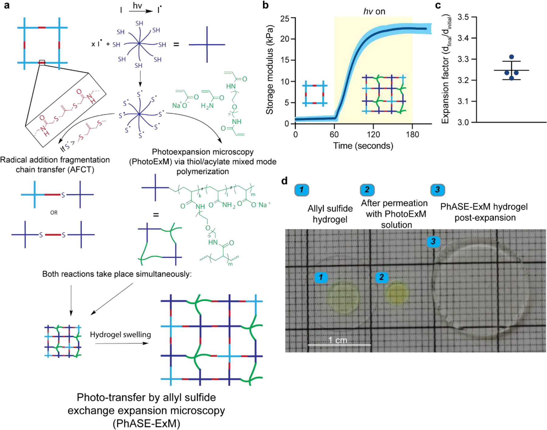
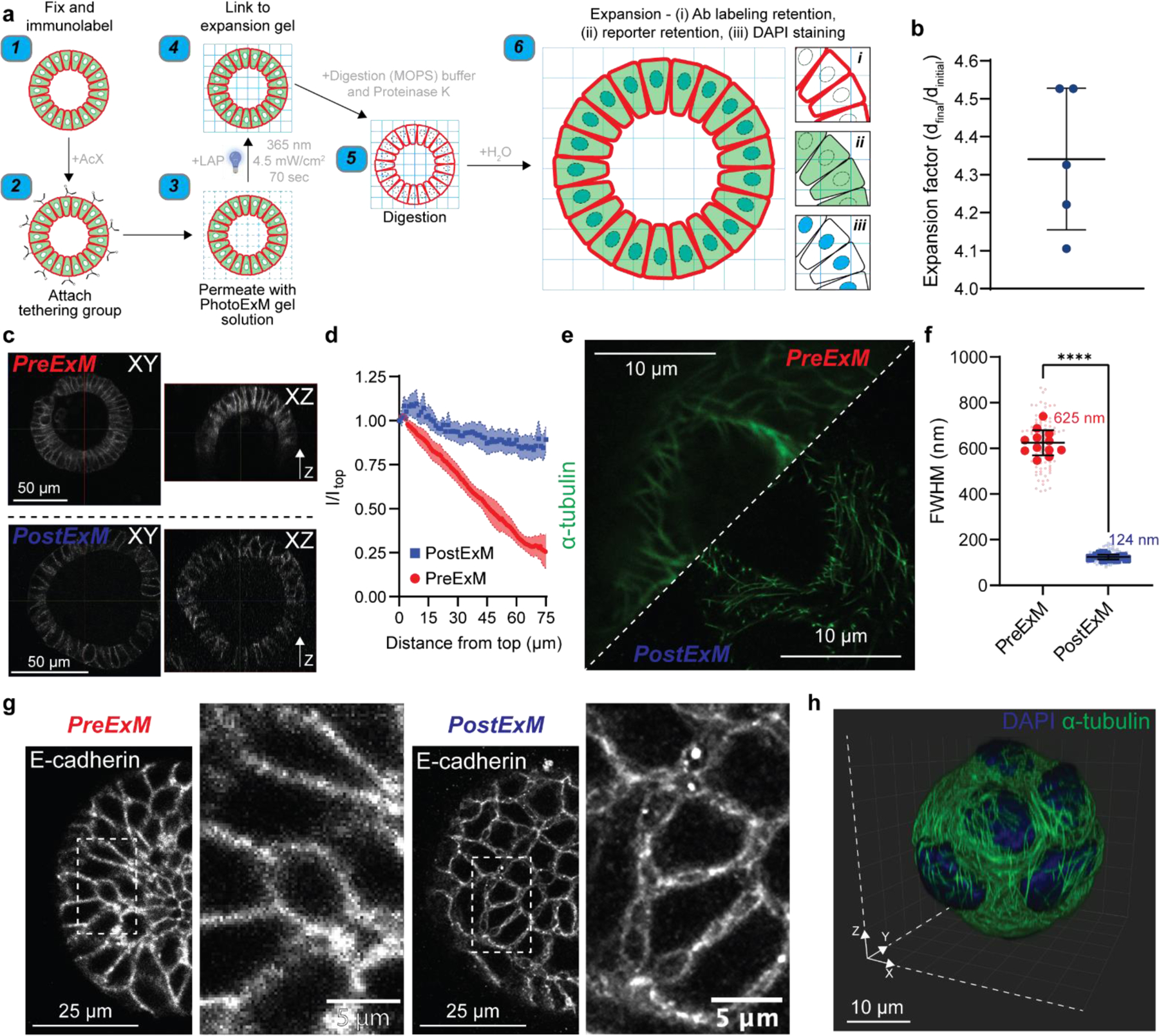
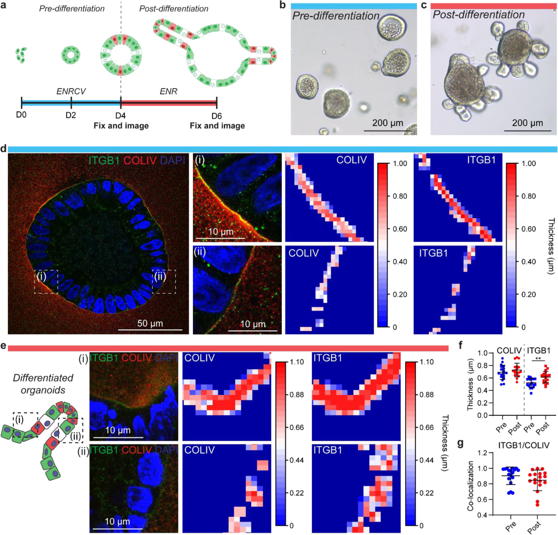
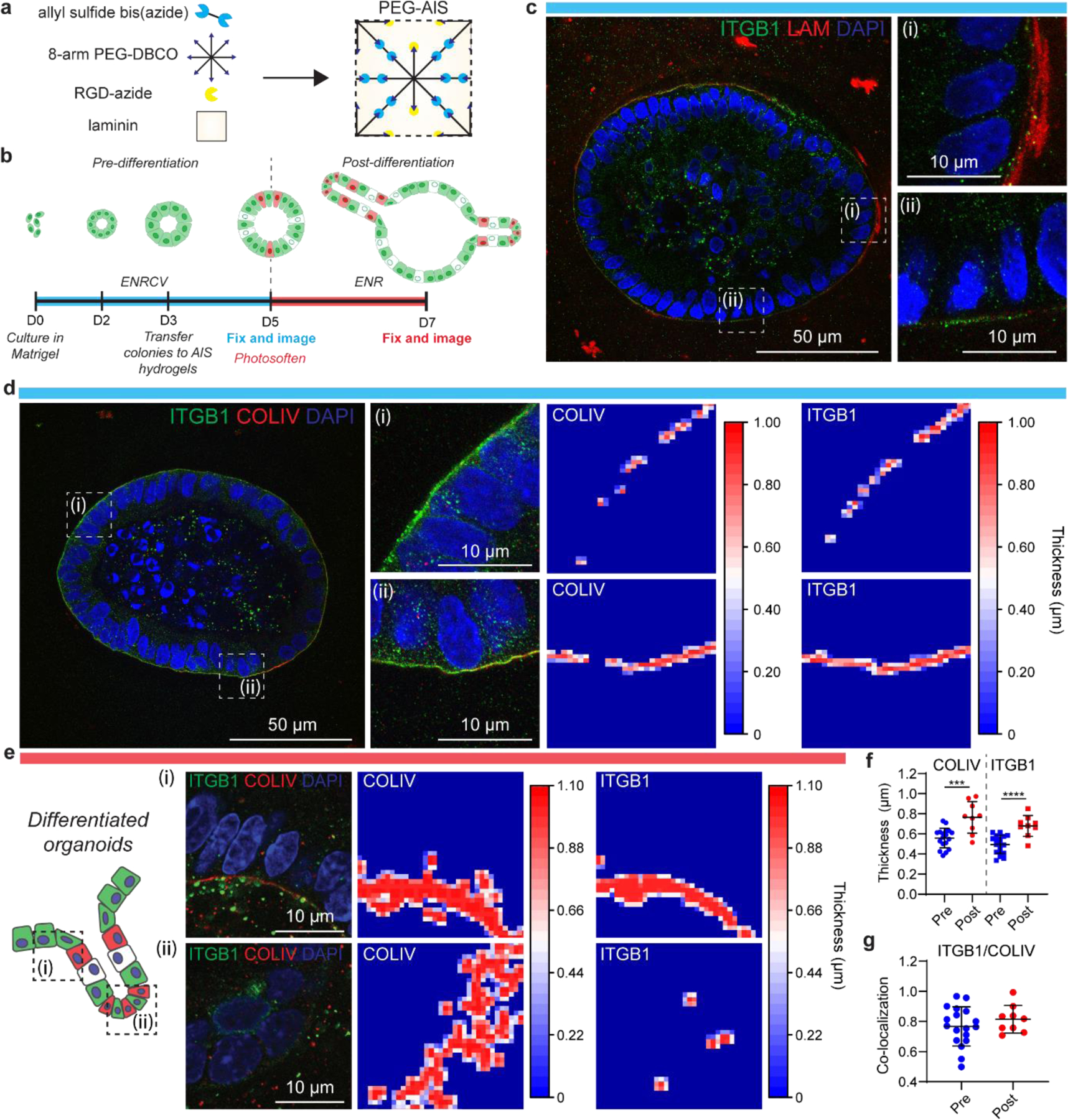
3D organoid models have recently seen a boom in popularity, as they can better recapitulate the complexity of multicellular organs compared to other in vitro culture systems. However, organoids are difficult to image because of the limited penetration depth of high-resolution microscopes and depth-dependent light attenuation, which can limit the understanding of signal transduction pathways and characterization of intimate cell-extracellular matrix (ECM) interactions. To overcome these challenges, phototransfer by allyl sulfide exchange-expansion microscopy (PhASE-ExM) is developed, enabling optical clearance and super-resolution imaging of organoids and their ECM in 3D. PhASE-ExM uses hydrogels prepared via photoinitiated polymerization, which is advantageous as it decouples monomer diffusion into thick organoid cultures from the hydrogel fabrication. Apart from compatibility with organoids cultured in Matrigel, PhASE-ExM enables 3.25× expansion and super-resolution imaging of organoids cultured in synthetic poly(ethylene glycol) (PEG) hydrogels crosslinked via allyl-sulfide groups (PEG-AlS) through simultaneous photopolymerization and radical-mediated chain-transfer reactions that complete in <70 s. Further, PEG-AlS hydrogels can be in situ softened to promote organoid crypt formation, providing a super-resolution imaging platform both for pre- and post-differentiated organoids. Overall, PhASE-ExM is a useful tool to decipher organoid behavior by enabling sub-micrometer scale, 3D visualization of proteins and signal transduction pathways.

Keywords: expansion microscopy; extracellular matrix; intestinal organoids; photochemistry; synthetic hydrogels.


Figures

Figure 1.
Figure 1.. Photo-transfer by allyl sulfide exchange expansion microscopy (PhASE-ExM).
(a) Schematic illustration of photo-transfer by allyl sulfide exchange expansion microscopy (PhASE-ExM): A cytocompatible hydrogel prepared using allyl-sulfide crosslinking groups (top left) is permeated with the photoexpansion microscopy (PhotoExM) formulation, which contains an 8-arm, 10 kDa PEG-SH as the chain transfer agent, sodium acrylate as the polyelectrolyte monomer, PEG-diacrylamide as the crosslinker, acrylamide, and LAP as the photoinitiator. Upon light irradiation, two processes simultaneously occur. While photopolymerization of PhotoExM formulation yields a swellable polyelectrolyte network (middle right), radical addition fragmentation chain transfer (AFCT) reaction between the allyl-sulfide and thiyl radicals incorporates the chains of the cytocompatible hydrogel to the polymerizing PhotoExM network (middle left), which can be homogeneously expanded with repetitive H2O washes upon termination of light irradiation (bottom), enabling expansion microscopy of organoids cultured in synthetic hydrogels without requiring another degradation step for the hydrogel. (b) Gel evolution of PhASE-ExM hydrogels as measured by in situ photo-rheology: The first 60 s shows the storage modulus (G’) of the allyl-sulfide crosslinked hydrogels permeated with the PhotoExM formulation, and upon light irradiation (Io = 4.5 mW/cm2, λ = 365 nm), PhASE-ExM hydrogels reach > 95% conversion within 70 s. n = 3 independent measurements, dark blue line = mean, blue shaded area = s.d. Yellow shaded area shows the duration of light irradiation. (c) Overall size expansion factor of the PhASE-ExM hydrogels. n = 4 hydrogels, graph presented as mean +/− s.d. (d) Representative images of allyl sulfide crosslinked hydrogels on a glass coverslip (left), which shrinks ~ 30% by size following permeation with the PhotoExM solution (middle) and post-expansion (right). The allyl sulfide crosslinked hydrogels were functionalized with AlexaFluor 488 to determine its retention post-expansion.
Figure 2.
Figure 2.. PhotoExM of intestinal organoids cultured in Matrigel.
(a) Schematic for PhotoExM of intestinal organoids. Organoids are first fixed and immunolabeled using standard protocols, then the tethering group, Acryloyl X (AcX), is added. Next, the PhotoExM solution is permeated into the sample. Upon 70 s light irradiation (Io = 4.5 mW/cm2, λ = 365 nm) and the addition of a photoinitiator, LAP, the sample is linked to the expansion hydrogel. The addition of Proteinase K (16 U/mL) in the digestion buffer degrades and digests, as well as optically clears the organoids. Finally, immersion in H2O promotes dissociation of the electrolyte (sodium acrylate) monomer, resulting in physical expansion. Spatial information is retained, and immunolabeled whole organoids can be imaged in 3D. Additionally, retention of DNA allows for PostExM staining with DAPI, and retention of eGFP reporters allows for imaging of reporter organoid lines. (b) Expansion factor of organoids expanded in Matrigel. n = 5 hydrogels, graph presented as mean +/− s.d. (c) PreExM and PostExM images of E-cadherin labeled organoids. XY (single plane cross section) and XZ (orthogonal projection). (d) Fluorescent intensity of E-cadherin immunolabeling for PreExM and PostExM images across Z-stacks. Intensity normalized to the intensity at depth z=0 (Itop). The line represents the mean, with shaded regions representing 95% CI. (e) α-tubulin imaging PreExM and PostExM of organoids cultured for 2 days, starting from single cell suspensions. (f) Resolution of PreExM and PostExM samples quantified by the diameter of microtubules. n = 90–100 line scans per condition (smaller data points) across 12 individual organoids (larger data points). Graph presented as mean +/− s.d. **** p < 0.0001. (g) Single z-stack PreExM and PostExM imaging of E-cadherin. (h) 3D reconstruction in IMARIS of a whole PostExM organoid immunolabeled with α-tubulin and stained with DAPI. Organoids were cultured for 2 days, starting from a single cell encapsulation.
Figure 3
Figure 3. PhotoExM of cell-ECM interactions of organoids cultured in Matrigel.
(a) Timeline of organoid growth. Organoids are generated starting from a population of single ISCs. For the first 4 days, cells were cultured in ENRCV media, favoring maintenance of stemness (“pre-differentiation”). On day 4, the media composition was changed to ENR to promote cell differentiation and crypt formation (“post-differentiation”). (b) Pre-differentiation, spherical organoids at day 4 in culture. (c) Post-differentiation organoids, containing crypts at day 6 in culture. (d) PhotoExM imaging of pre-differentiation organoids. Insets (i) and (ii) shows regions of high and low COLIV/ITGB1 thickness, respectively. (e) PhotoExM of post-differentiation organoids. The hinge region adjacent to an intestinal crypt (i) shows thicker COLIV/ITGB1 compared to the crypt region (ii). (f) Global quantification of the mean thickness of COLIV and ITGB1 in pre- and post-differentiated organoids. n = 18 organoids per condition. Graph presented as mean +/− s.d. ** p < 0.01. (g) Co-localization of ITGB1 and COLIV. Reported values are Mander’s coeffecient for the overlap of ITGB1 and COLIV. n = 18 organoids per condition. Graph presented as mean +/− s.d.
Figure 4.
Figure 4.. PhASE-ExM of organoids in PEG-AlS hydrogels.
(a) PEG-AlS are synthesized by reaction of an 8-arm, 20 kDa PEG-dibenzocyclooctyne (PEG-DBCO), a bis-azide functionalized allyl sulfide, an azide functionalized RGD, and laminin. (b) Timeline of organoid growth. Organoids were generated starting from a population of single ISCs, and cultured in Matrigel for 3 days in ENRCV media, favoring stemness. At day 3, organoids were transferred to PEG-AlS hydrogels and cultured for 2 days. These organoids were the “pre-differentiation” population. On day 5, hydrogels were photosoftened and the media composition was changed to ENR to promote cell differentiation and crypt formation (“post-differentiation”). (c) Pre-differentiation PhASE-ExM imaging of LAM and ITGB1. (d) Pre-differentiation PhASE-ExM imaging and quantification of COLIV and ITGB1. (e) Post-differentiation PhASE-ExM imaging and quantification of COLIV and ITGB1. The hinge region adjacent to an intestinal crypt (i) shows thicker COLIV/ITGB1 compared to the crypt region (ii), where the COLIV is discontinuous and there is minimal ITGB1. (f) Global quantification of mean thickness of COLIV and ITGB1 in pre- and post-differentiated organoids. n = 9–18 organoids per condition. Graph presented as mean +/− s.d. *** p<0.001, **** p<0.0001. (g) Co-localization of ITGB1 and COLIV. Reported values are Mander’s coeffecient for the overlap of ITGB1 and COLIV. n = 9–18 organoids per condition. Graph presented as mean +/− s.d.

References

    1. Hofer M and Lutolf MP, Engineering organoids. Nature Reviews Materials, 2021. 6(5): p. 402–420. - PMC - PubMed
    1. Clevers H, Modeling Development and Disease with Organoids. Cell, 2016. 165(7): p. 1586–1597. - PubMed
    1. Gjorevski N, et al., Designer matrices for intestinal stem cell and organoid culture. Nature, 2016. 539(7630): p. 560–564. - PubMed
    1. Cruz-Acuña R, et al., Synthetic hydrogels for human intestinal organoid generation and colonic wound repair. Nature Cell Biology, 2017. 19(11): p. 1326–1335. - PMC - PubMed
    1. Hushka EA, et al., Relaxation of Extracellular Matrix Forces Directs Crypt Formation and Architecture in Intestinal Organoids. Advanced Healthcare Materials, 2020. 9(8): p. 1901214. - PMC - PubMed

LinkOut - more resources