. 2022 Jun 27;43(6):557-570.
doi: 10.1093/carcin/bgac023.

Chemopreventive efficacy of silibinin against basal cell carcinoma growth and progression in UVB-irradiated Ptch+/- mice

Affiliations

Chemopreventive efficacy of silibinin against basal cell carcinoma growth and progression in UVB-irradiated Ptch+/- mice

Sandeep Paudel et al. Carcinogenesis. .

Abstract

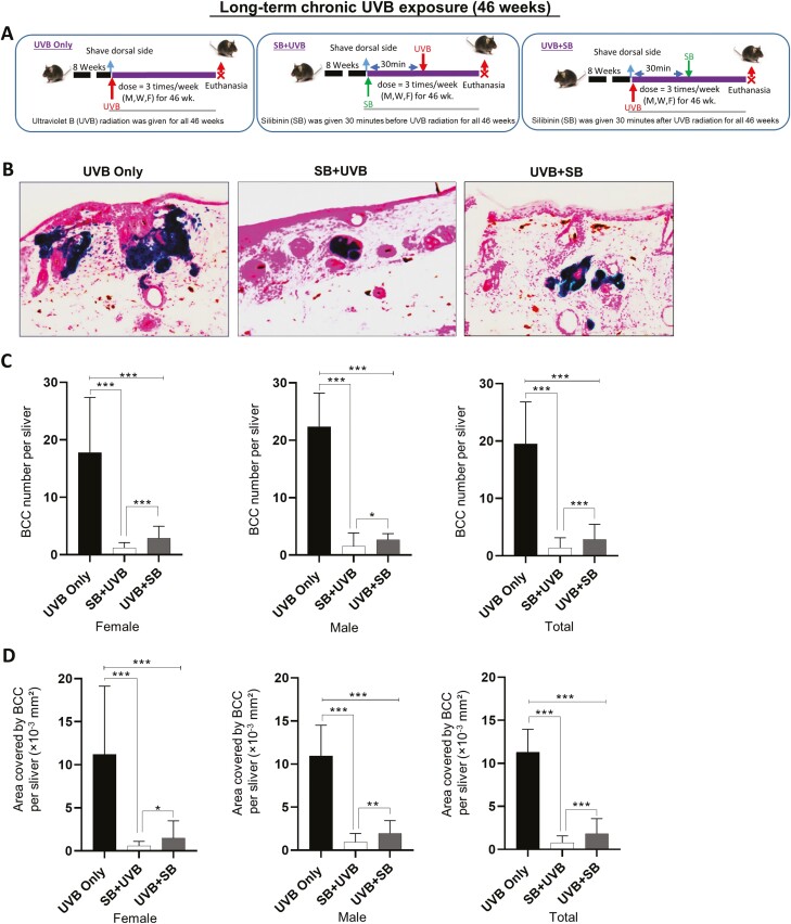
The factors (environmental and genetic) contributing to basal cell carcinoma (BCC) pathogenesis are well-established; however, effective agents for BCC prevention are marred by toxic side-effects. Herein, we assessed the efficacy of flavonolignan silibinin against ultraviolet B (UVB)-induced BCC in Ptch+/- (heterozygous patched homolog 1 gene) mouse model. Both male and female Ptch+/- mice were irradiated with a 240 mJ/cm2 UVB dose 3 times/week for 26 or 46 weeks, with or without topical application of silibinin (9 mg/200 µl in acetone, applied 30 min before or after UVB exposure). Results indicated that silibinin application either pre- or post-UVB exposure for 26 weeks significantly decreased the number of BCC lesions by 65% and 39% (P < 0.001 for both) and the area covered by BCCs (72% and 45%, P < 0.001 for both), respectively, compared to UVB alone. Furthermore, continuous UVB exposure for 46 weeks increased the BCC lesion number and the BCC area covered by ~6 and ~3.4 folds (P < 0.001), respectively. Notably, even in this 46 week prolonged UVB exposure, silibinin (irrespective of pre- or post-UVB treatment) significantly halted the growth of BCCs by 81-94% (P < 0.001) as well as other epidermal lesions; specifically, silibinin treated tissues had less epidermal dysplasia, fibrosarcoma, and squamous cell carcinoma. Immunohistochemistry and immunofluorescence studies revealed that silibinin significantly decreased basal cell proliferation (Ki-67) and the expression of cytokeratins (14 and 15), and Hedgehog signaling mediators Smo and Gli1 in the BCC lesions. Together, our findings demonstrate strong potential of silibinin to be efficacious in preventing the growth and progression of UVB-induced BCC.


Figures

Graphical Abstract
Graphical Abstract
Figure 1.
Figure 1.
Effect of topical application of silibinin on short-term chronic UVB exposure -induced microscopic basal cell carcinoma formation: (A) Experimental protocol for the study. Study groups (both male and female mice) are: [UVB only, Ptch+/– mice were irradiated with 240 mJ/cm2 UVB dose, 3 times per week (M, W, F) for 26 weeks]; [SB + UVB, Ptch+/– mice were irradiated with 240 mJ/cm2 UVB dose, 3 times per week (M, W, F) for 26 weeks. 9 mg silibinin in 200 µl acetone/mouse was applied topically 30 min prior to the UVB irradiation]; [UVB + SB group, Ptch+/– mice were irradiated with 24 0mJ/cm2 UVB dose, 3 times per week (M, W, F) for 26 weeks. 9 mg silibinin in 200 µl acetone/mouse was applied topically 30 min after the UVB irradiation]. (B) Representative images of dark blue BCCs following β-galactosidase staining. All images were captured at x100 magnification, (C) quantification of average BCC number in female (F), male (M) and total (F + M) mice, (D) quantification of area covered by BCC in female (F), male (M) and total (F + M) mice. n =55 (33 male/22 female, UVB only group); n = 43 (26 male/17 female, SB + UVB group); n = 39 (25 male/14 female, UVB + SB group). Columns indicate median [95% CI]; nsP > 0.05; *P ≤ 0.05; **P ≤ 0.01; ***P ≤ 0.001. SB, silibinin (9 mg/200µl acetone).
Figure 2.
Figure 2.
Effect of topical application of silibinin on long-term chronic UVB exposure -induced basal cell carcinoma lesions. (A) Experimental protocol for the study. Study groups (both male and female mice) are: [UVB only, Ptch+/– mice were irradiated with 240 mJ/cm2 UVB dose, 3 times per week (M, W, F) for 46 weeks]; [SB + UVB, Ptch+/– mice were irradiated with 240 mJ/cm2 UVB dose, 3 times per week (M, W, F) for 46 weeks. 9 mg silibinin in 200 µl acetone/mouse was applied topically 30 min prior to the UVB irradiation]; [UVB + SB group, Ptch+/– mice were irradiated with 240 mJ/cm2 UVB dose, 3 times per week (M, W, F) for 46 weeks. 9 mg silibinin in 200 µl acetone/mouse was applied topically 30 min after the UVB irradiation]. (B) Representative images of dark blue BCCs processed for β-galactosidase staining. All images were captured at ×100 magnification, (C) Quantification of BCC numbers per sliver in female (F), male (M) and total (F + M) mice, (D) quantification of area covered by BCCs per sliver in female (F), male (M) and total (F + M) mice. n = 53 (32 male/21 female, UVB only group); n = 56 (30 male/26 female, SB + UVB group); n = 60 (33 male/27 female, UVB + SB group). Columns indicate Median [95% CI]; nsP>0.05; *P ≤ 0.05; **P ≤ 0.01; ***P ≤ 0.001. SB, silibinin (9 mg/200µl acetone).
Figure 3.
Figure 3.
Effect of topical application of silibinin on the growth and progression of non-BCC skin lesions in Ptch+/– mice. Ptch+/– mice were UVB-irradiated (240 mJ/cm2) 3 times per week with or without silibinin for 46 weeks. Study groups are: [UVB only]; [SB + UVB (silibinin treatment 30 min prior to UVB exposure)]; UVB + SB (silibinin treatment 30 min after UVB exposure)]. (A) Representative images of H&E staining depicting presence of different pathological lesions upon chronic UVB exposure. All pictographs were captured at ×100 magnification, (B) quantification of number of dysplasia per sliver; representative pictograph of epidermal dysplasia at ×100 magnification in left panel. (C) Quantification of number of fibrosarcoma per sliver; representative pictograph of fibrosarcoma at ×100 magnification in left panel. (D) Quantification of number of SCC per sliver; representative pictograph of fibrosarcoma at ×100 magnification in left panel. Columns indicate Median [95% CI]; nsP > 0.05; *P ≤ 0.05; **P ≤ 0.01; ***P ≤ 0.001. SB, silibinin (9 mg/200 µl acetone); female (F), male (M) and total (F + M) mice.
Figure 4.
Figure 4.
Effect of topical application of silibinin on proliferation and Hh-signaling pathway during short-term chronic UVB exposure-induced BCC in Ptch+/– mice. Immunohistochemical staining was done to assess expression of Ki67, Gli1 and Smo in response to short-term chronic UVB exposure (26 weeks) with or without silibinin in female (F), male (M) and total (M + F) Ptch+/– mice. Pictorial and data representation for (A) % Ki67 positive cells, (B) Smo expression as immunopositivity score, (C) Gli1 expression as immunopositivity score. Microscopic quantification of DAB staining was done in five randomly selected areas at ×400 magnification per sliver and then an average count of 4 slivers was considered. For Ki67 expression analysis, % of positively stained (brown nucleus) cells were counted. For Smo and Gli-1 expression analysis, intensity of brown color was assessed as immunopositivity scores and assigned arbitrary values: 0 (no staining), +1 (weak), +2 (moderate), +3 (strong) and +4 (very strong). For Smo expression, overall cellular staining was observed (which included cytoplasmic + nuclear staining). For Gli-1 expression, prominently nuclear staining was observed. Columns indicate median [95% CI]; nsP > 0.05; *P ≤ 0.05; **P ≤ 0.01; ***P ≤ 0.001. All images were captured at ×100 magnification. SB, silibinin (9 mg/200 µl acetone).
Figure 5.
Figure 5.
Effect of topical application of silibinin on proliferation and Hh-signaling pathway during long-term chronic UVB exposure-induced BCC in Ptch+/– mice. Immunohistochemical staining was done to assess expression of Ki67, Gli1 and Smo in response to long-term chronic UVB exposure (46 weeks) with or without silibinin in female (F), male (M) and total (M + F) Ptch+/– mice. Pictorial and data representation for (A) % Ki67 positive cells, (B) Smo expression as immunopositivity score, (C) Gli1 expression as immunopositivity score. Microscopic quantification of DAB staining was done in five randomly selected areas at ×400 magnification per sliver and then an average count of four slivers was considered. For Ki67 expression analysis, % of positively stained (brown nucleus) cells were counted. For Smo and Gli-1 expression analysis, intensity of brown color was assessed as immunopositivity scores and assigned arbitrary values: 0 (no staining), +1 (weak), +2 (moderate), +3 (strong) and +4 (very strong). For Smo expression, overall cellular staining was observed (which included cytoplasmic + nuclear staining). For Gli-1 expression, prominently nuclear staining was observed. Columns indicate median [95% CI]. nsP > 0.05; *P ≤ 0.05; **P ≤ 0.01; ***P ≤ 0.001. All images were captured at ×100 magnification. SB, silibinin (9 mg/200 µl acetone).
Figure 6.
Figure 6.
Effect of topical application of silibinin on the basal keratinocyte compartment expansion during chronic UVB exposure-induced BCC in Ptch+/– mice. Dual immunofluorescence was performed to assess expression of cytokeratins (14 and 15); representation of nucleated cells (DAPI-blue), Ck14 (Green) and Ck15 (Red). Pictorial and data representation for the expression of Ck14 and Ck15 in BCC after (A) short-term UVB exposure for 26 weeks (with and without silibinin), (B) long-term UVB exposure for 46 weeks (with and without silibinin. Columns indicate Median [95% CI]; nsP > 0.05; *P ≤ 0.05; **P ≤ 0.01; ***P ≤ 0.001. All images were taken at ×200 magnification. SB, silibinin (9 mg/200 µl acetone); total (female + male mice).

Similar articles

Cited by

References

    1. Fania, L., et al. . (2020) Basal cell carcinoma: from pathophysiology to novel therapeutic approaches. Biomedicines, 8, 449–488. - PMC - PubMed
    1. Kumar, R., et al. . (2015) An overview of ultraviolet b radiation-induced skin cancer chemoprevention by silibinin. Curr. Pharmacol. Rep., 1, 206–215. - PMC - PubMed
    1. Prasad, R.R., et al. . (2020) Silibinin and non-melanoma skin cancers. J. Tradit. Complement. Med., 10, 236–244. - PMC - PubMed
    1. Yabroff, K.R., et al. . (2016) Financial hardship associated with cancer in the United States: findings from a population-based sample of adult cancer survivors. J. Clin. Oncol., 34, 259–267. - PMC - PubMed
    1. Boehnke, K., et al. . (2012) Stem cells of the human epidermis and their niche: composition and function in epidermal regeneration and carcinogenesis. Carcinogenesis, 33, 1247–1258. - PubMed

Publication types