Clinical Trial
. 2022 Aug;77(8):2393-2403.
doi: 10.1111/all.15262. Epub 2022 Mar 3.

Anti-KIT monoclonal antibody CDX-0159 induces profound and durable mast cell suppression in a healthy volunteer study

Affiliations
Clinical Trial

Anti-KIT monoclonal antibody CDX-0159 induces profound and durable mast cell suppression in a healthy volunteer study

Diego Alvarado et al. Allergy. 2022 Aug.

Abstract

Background: Mast cells (MC) are powerful inflammatory immune sentinel cells that drive numerous allergic, inflammatory, and pruritic disorders when activated. MC-targeted therapies are approved in several disorders, yet many patients have limited benefit suggesting the need for approaches that more broadly inhibit MC activity. MCs require the KIT receptor and its ligand stem cell factor (SCF) for differentiation, maturation, and survival. Here we describe CDX-0159, an anti-KIT monoclonal antibody that potently suppresses MCs in human healthy volunteers.

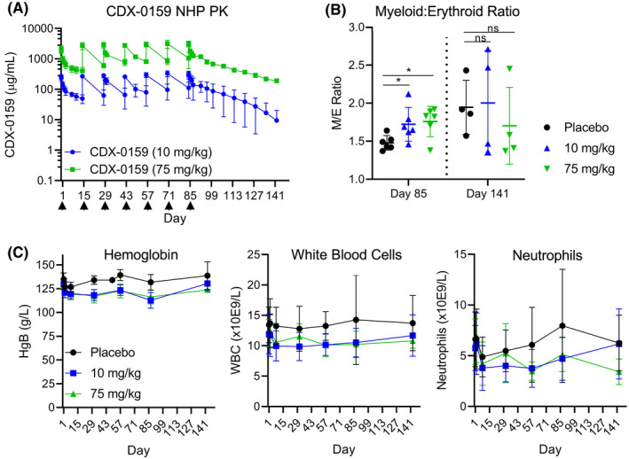
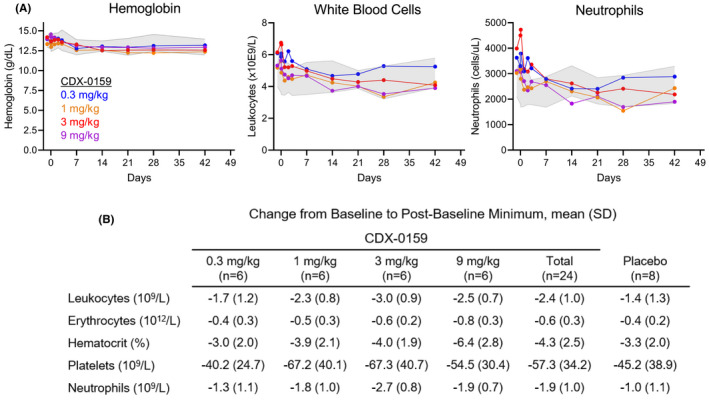
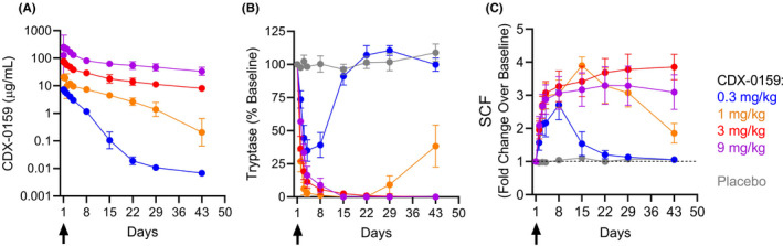
Methods: CDX-0159-mediated KIT inhibition was tested in vitro using KIT-expressing immortalized cells and primary human mast cells. CDX-0159 safety and pharmacokinetics were evaluated in a 13-week good laboratory practice (GLP)-compliant cynomolgus macaque study. A single ascending dose (0.3, 1, 3, and 9 mg/kg), double-blinded placebo-controlled phase 1a human healthy volunteer study (n = 32) was conducted to evaluate the safety, pharmacokinetics, and pharmacodynamics of CDX-0159.

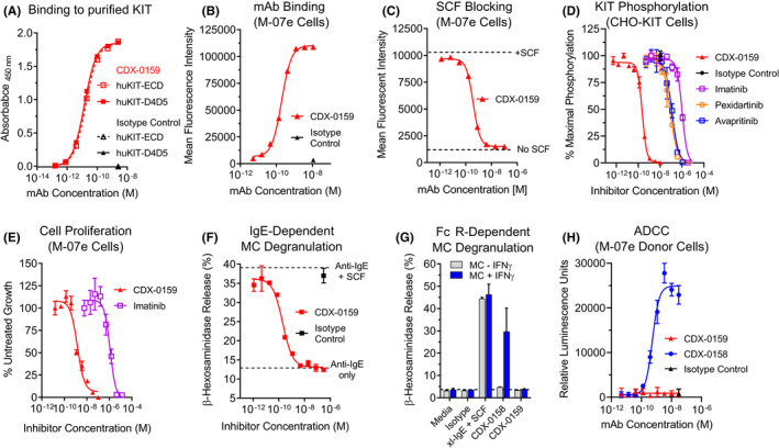
Results: CDX-0159 inhibits SCF-dependent KIT activation in vitro. Fc modifications in CDX-0159 led to elimination of effector function and reduced serum clearance. In cynomolgus macaques, multiple high doses were safely administered without a significant impact on hematology, a potential concern for KIT inhibitors. A single dose of CDX-0159 in healthy human subjects was generally well tolerated and demonstrated long antibody exposure. Importantly, CDX-0159 led to dose-dependent, profound suppression of plasma tryptase, a MC-specific protease associated with tissue MC burden, indicative of systemic MC suppression or ablation.

Conclusion: CDX-0159 administration leads to systemic mast cell ablation and may represent a safe and novel approach to treat mast cell-driven disorders.

Keywords: CDX-0159; KIT; mast cell; monoclonal antibody.


Conflict of interest statement

DA, MBM, LC, JG, AC, LAV, PAM, LJT, TRH, TK, DY, EC, and MHC are full‐time employees of Celldex Therapeutics. SBS is a full‐time employee of Boehringer‐Ingelheim. MM reports grants and/or personal fees from Allakos, Amgen, Aralez, ArgenX, AstraZeneca, Celldex, Centogene, CSL Behring, FAES, Genentech, GIInnovation, Innate Pharma, Kyowa Kirin, Leo Pharma, Lilly, Menarini, Moxie, Novartis, Roche, Sanofi/Regeneron, Third HarmonicBio, UCB, and Uriach. MK is a full‐time employee of Altasciences, which has received research grant/funding (institution) from Actelion Pharmaceuticals, Acurx Pharmaceuticals, Bioxcel Therapeutics, Grifols, Jazz Pharmaceuticals, Novus Therapeutics, Pfizer, DynPort Vaccine Company, Novo Nordisk, FDA/NIH, and ViroDefense. MK holds a leadership role at Altasciences. JG and RG are inventors in patent applications No: 63/140,642 and 63/140,621.

Figures

FIGURE 1
FIGURE 1
CDX‐0159 Inhibits SCF‐dependent KIT Activation and MC Degranulation. (A) CDX‐0159 binds to purified human KIT extracellular domain (huKIT‐ECD) and a fragment comprised of the membrane proximal dimerization domains Ig4 and Ig5 (huKIT‐D4D5) with indistinguishable potency. Purified KIT proteins were immobilized in ELISA plates followed by titration with CDX‐0159. (B) Binding to KIT‐expressing M‐07e cells was demonstrated by flow cytometry, with an EC50 value of 153 ± 22 pM (C). CDX‐0159 completely blocks binding of 10 nM fluorescently labeled SCF to M‐07e cells with a potency of 118 ± 6 pM. (D) Inhibition of SCF‐dependent KIT tyrosine phosphorylation in CHO cells expressing human KIT is demonstrated for CDX‐0159 and KIT‐targeting TKIs imatinib, pexidartinib, and avapritinib. (E) SCF‐dependent proliferation of M‐07E cells is inhibited by CDX‐0159 more potently than with imatinib. (F) IgE‐dependent MC degranulation as measured by β‐hexosaminidase release is significantly enhanced by addition of 100 ng/mL of SCF. CDX‐0159 fully inhibits SCF‐dependent β‐hexosaminidase release. (G) Fc‐silencing mutations in CDX‐0159 abolish FcγR‐dependent MC activation. In MCs pre‐treated with IFNγ to upregulate FcγRI, CDX‐0158 but not CDX‐0159 induces MC β‐hexosaminidase release. Cross‐linked IgE (xl‐IgE) plus SCF is used as a positive control. (H) CDX‐0159 does not elicit measurable ADCC. Fc‐silencing mutations abolish ADCC observed with CDX‐0158 using a reporter assay using Jurkat cells with an NFAT‐luciferase reporter element under the control of FcγRIII as effector cells, and M‐07e as target cells. All experiments were performed at least 3 independent times. Mean values and S.E.M.s are shown
FIGURE 2
FIGURE 2
Repeat dosing of CDX‐0159 in non‐human primates does not induce significant myelosuppression. (A) CDX‐0159 administration every two weeks results in high drug levels and exposure at 10 and 75 mg/kg throughout the dosing period 13weeks and post‐treatment recovery (8 weeks). Arrows denote dosing. (B) In bone marrow smears, CDX‐0159‐induced marginal increases in myeloid/erythroid ratios at the end of treatment which were not seen at the end of the recovery period. Cohort means (n = 6 for on‐treatment and n = 4 for recovery animals) and S.E.M. values are shown. p‐values; ns: not significant; *: < 0.05. (C) Repeat CDX‐0159 administration induces mild rapid decreases in hemoglobin values (left) without further decline despite high and prolonged drug exposure. A similar pattern is observed in total leukocyte count (center) and neutrophils (right), although the total counts exhibit greater intra‐subject variability. Cohort means (n = 10 for on‐treatment and n = 4 for recovery animals) and S.E.M. values are shown. In all cases, no meaningful differences in hematology values between genders were observed
FIGURE 3
FIGURE 3
CDX‐0159 induces mild transient decreases in hemoglobin and neutrophil parameters in healthy human subjects. Mean levels of hemoglobin (A) leukocytes (B) and neutrophils (C) after a single dose of CDX‐0159 in healthy subjects are shown. Gray shaded area represents the 95% confidence interval values from placebo‐treated subjects. (D) Maximal mean post‐baseline values for several hematology parameters are shown
FIGURE 4
FIGURE 4
CDX‐0159 induces systemic MC depletion at saturating doses. (A) The PK analysis showed a dose‐dependent increase in levels of CDX‐0159 as expected. Results indicate a PK profile and volume of distribution consistent with those of a monoclonal antibody through this dosing range with evidence of target mediated clearance below approximately 5 μg/ml. Geometric means and 95% confidence intervals are shown. (B) Dose‐dependent reductions in plasma tryptase are observed after a single i.v. dose of CDX‐0159. At the 1 mg/kg dose or above, tryptase suppression below the level of assay detection (1 ng/ml) is observed in all subjects for at least 3 weeks at 1 mg/kg or 6 weeks for 3 and 9 mg/kg. No notable tryptase modulation was observed in placebo‐treated patients. Tryptase values below the lower limit of quantitation were assigned a value of 0 ng/ml. Dose cohort means and S.E.Ms are reported. (C) Dose‐dependent increases in SCF levels were observed, mirroring the pattern of tryptase suppression. No significant modulation of SCF was observed in placebo‐treated subjects. Subjects with baseline levels below the LLoQ (100 pg/ml) were set to 100 pg/ml for normalization. Dose cohort means and S.E.Ms are reported

References

    1. Varricchi G, Rossi FW, Galdiero MR, et al. Physiological roles of mast cells: collegium internationale allergologicum update 2019. Int Arch Allergy Immunol. 2019;179:247‐261. - PubMed
    1. Dudeck A, Köberle M, Goldmann O, et al. Mast cells as protectors of health. Journal of Allergy and Clinical Immunology. 2019;144:S4‐S18. - PubMed
    1. Dahlin JS, Maurer M, Metcalfe DD, Pejler G, Sagi‐Eisenberg R, Nilsson G. The ingenious mast cell: contemporary insights into mast cell behavior and function. Allergy. 2021;77(1):83‐99. - PubMed
    1. Maurer M, Köberle M, Metz M, Biedermann T. Mast cells: Promoters of health and modulators of disease. Journal of Allergy and Clinical Immunology. 2019;144:S1‐S3. - PubMed
    1. Jönsson F, Mast DM. Mast cells and company. Front Immun. 2012;3. 10.3389/fimmu.2012.00016 - DOI - PMC - PubMed

Publication types

MeSH terms