. 2022 May;12(5):1061-1074.
doi: 10.1002/2211-5463.13383. Epub 2022 Mar 5.

The dynamics of cell death patterns and regeneration during acute liver injury in mice

Affiliations

The dynamics of cell death patterns and regeneration during acute liver injury in mice

Shuai Shao et al. FEBS Open Bio. 2022 May.

Abstract

Acute liver injury is a serious clinical syndrome with multiple causes and unclear pathological process. Here, CCl4 - and D-galactosamine/lipopolysaccharide (D-gal/LPS)-induced acute liver injury was established to explore the cell death patterns and determine whether or not liver regeneration occurred. In CCl4 -induced hepatic injury, three phases, including the early, progressive, and recovery phase, were considered based on alterations of serum transaminases and liver morphology. Moreover, in this model, cytokines exhibited double-peak fluctuations; apoptosis and pyroptosis persisted throughout all phases; autophagy occurred in the early and the progressive phases; and sufficient and timely hepatocyte regeneration was observed only during the recovery phase. All of these phenomena contribute to mild liver injury and subsequent regeneration. Strikingly, only the early and progressive phases were observed in the D-gal/LPS model. Slight pyroptosis occurred in the early phase but diminished in the progressive phase, while apoptosis, reduced autophagy, and slight but subsequently diminished regeneration occurred only during the progressive phase, accompanied by a strong cytokine storm, resulting in severe liver injury with high mortality. Taken together, our work reveals variable modes and dynamics of cell death and regeneration, which lead to different consequences for mild and severe acute liver injury, providing a helpful reference for clinical therapy and prognosis.

Keywords: acute liver injury; cell death; cytokines; hepatocyte; prognosis; regeneration.


Conflict of interest statement

The authors declare no conflict of interest.

Figures

Fig. 1
Fig. 1
CCl4‐ and D‐gal/LPS‐induced acute liver injury models. Mice were injected with either CCl4 (olive oil as control) or D‐gal/LPS (saline as control) to induce acute liver injury and were sacrificed at different time points. Macroscopic appearance and H&E staining of the liver tissues for the CCl4 (A)‐ and D‐gal/LPS (E)‐treated mice; serum ALT (B, F), AST (C, G), and LDH (D, H) were measured. Scale bar, 400 μm for 10× and 100 μm for 40×. The data were expressed as the mean ± SEM for at least triplicate experiments. P values were analyzed by Student’s t‐test. *P < 0.05 vs control.
Fig. 2
Fig. 2
Cytokine expression in CCl4‐induced acute liver injury model mice. The expression of inflammation‐related genes TNF‐α (A), IL‐1β (B), Mcp‐1 (C), IL‐10 (D), and IL‐6 (E) was evaluated by qRT‐PCR. The data were expressed as the mean ± SEM for at least triplicate. P values were analyzed by Student’s t‐test. *P < 0.05 vs control.
Fig. 3
Fig. 3
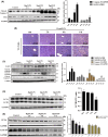
Expression of apoptosis‐, pyroptosis‐, and autophagy‐related genes in liver tissues of CCl4‐induced acute liver injury model mice. (A) The protein level of apoptosis genes in liver tissues was determined by western blot; (B) TUNEL detection of apoptotic gene expression in liver tissue; the protein level of pyroptosis genes in liver tissue (C) and KCs (D) determined by western blot; and (E) the protein level of autophagy‐related gene in liver tissue determined by western blot. GAPDH was used as an internal control. Scale bar, 400 μm for 10× and 100 μm for 40×. The data were expressed as the mean ± SEM for at least triplicate experiments. P values were analyzed by Student’s t‐test. *P < 0.05 vs control.
Fig. 4
Fig. 4
Expression of proliferation‐associated genes in CCl4‐induced acute liver injury model mice. RNA levels of Pcna detected using qRT‐PCR in liver tissue (A) and hepatocytes (E); protein level of PCNA and cyclin D1 detected using western blot in liver tissue (B‐C) and hepatocytes (F, G), GAPDH was used as an internal control; (D) expression of PCNA detected in liver tissue using IHC. Scale bar, 400 μm for 10× and 100 μm for 40×. The data were expressed as the mean ± SEM for at least triplicate experiments. P values were analyzed by Student’s t‐test. *P < 0.05 vs control.
Fig. 5
Fig. 5
Cytokine expression in D‐gal/LPS‐induced acute liver injury model mice. The expression of inflammation‐related genes TNF‐α (A), IL‐1β (B), Mcp‐1 (C), IL‐10 (D), and IL‐6 (E) was evaluated by qRT‐PCR. The data were expressed as the mean ± SEM for at least triplicate experiments. P values were analyzed by Student’s t‐test. *P < 0.05 vs control.
Fig. 6
Fig. 6
Expression of apoptosis, pyroptosis, autophagy, and proliferation in liver tissues of mice in D‐gal/LPS‐induced acute liver injury model mice. (A) The protein level of apoptosis genes in liver tissues was determined by western blot; (B) TUNEL detection of apoptotic gene expression in liver tissue; (C) the protein level of pyroptosis genes in liver tissue determined by western blot; (D) the protein level of autophagy‐related gene in liver tissue determined by western blot; (E) the protein level of regeneration‐related genes in liver tissue determined by western blot. GAPDH was used as an internal control. Scale bar, 400 μm for 10× and 100 μm for 40×. The data were expressed as the mean ± SEM for at least triplicate experiments. P values were analyzed by Student’s t‐test. *P < 0.05 vs control.
Fig. 7
Fig. 7
Schematic diagram illustrating the progress of CCl4‐ and D‐gal/LPS‐induced acute liver injury model mice.

Similar articles

Cited by

References

    1. Stravitz RT, Lee WM. Acute liver failure. Lancet (London, England). 2019;394:869–81.
    1. Luedde T, Kaplowitz N, Schwabe RF. Cell death and cell death responses in liver disease: mechanisms and clinical relevance. Gastroenterology. 2014;147:765–783.e4. - PMC - PubMed
    1. Iorga A, Dara L. Cell death in drug‐induced liver injury. Adv Pharmacol (San Diego, Calif). 2019;85:31–74.
    1. Zhao Y, Shi J, Shao F. Inflammatory caspases: activation and cleavage of gasdermin‐D in vitro and during pyroptosis. Methods Mol Biol (Clifton, NJ). 2018;1714:131–48.
    1. Guo H, Xie M, Zhou C, Zheng M. The relevance of pyroptosis in the pathogenesis of liver diseases. Life Sci. 2019;223:69–73. - PubMed