

Percutaneous intravenous catheter forceps biopsy in right atrial mass: two case reports and literature review
- PMID: 35184743
- PMCID: PMC8859873
- DOI: 10.1186/s12872-022-02507-x
Percutaneous intravenous catheter forceps biopsy in right atrial mass: two case reports and literature review
Abstract
Background: Primary malignant tumors of the heart are rare. Although preoperative histological diagnosis is difficult, it has paramount value in therapeutic strategy development and prognostic estimation. Herein, we reported 2 cases of intracardiac tumors.
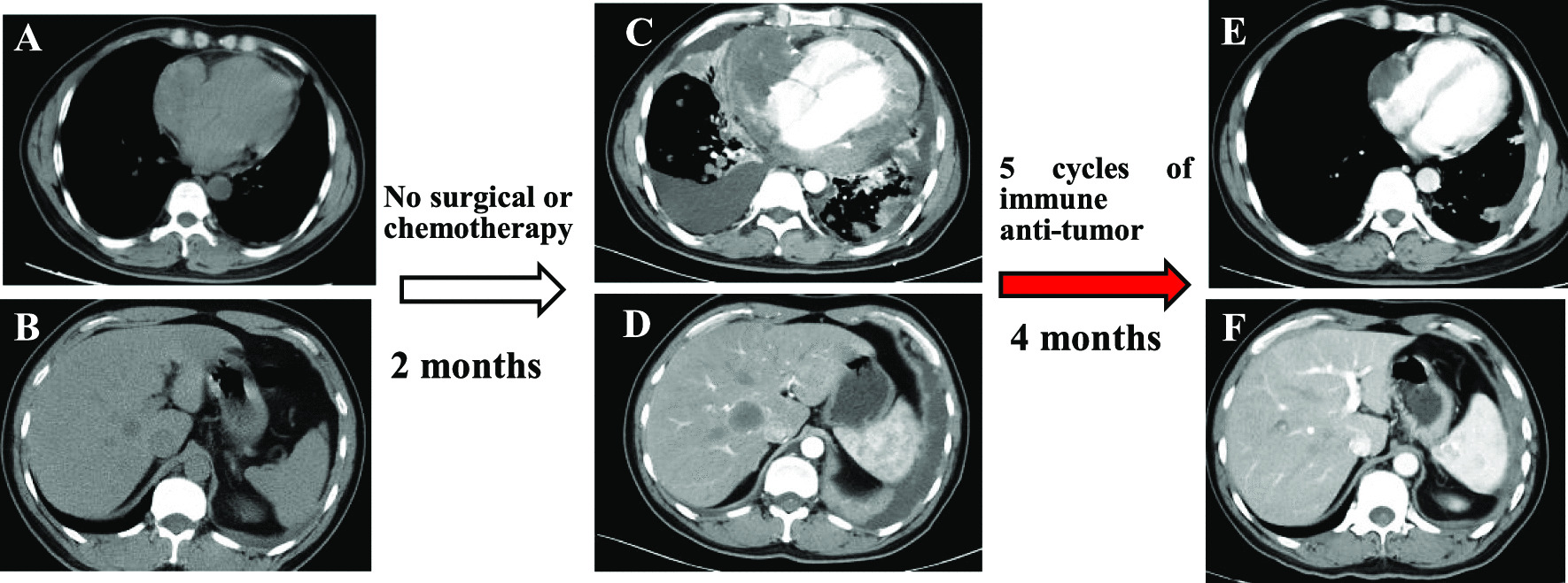
Cases presentation: Both patients presented to the hospital with heart-related symptoms. Echocardiography showed massive masses in the atrium and positron emission tomography-computed tomography (PET/CT) revealed hypermetabolism and invasiveness. One patient cannot take surgery due to extensive metastasis and poor condition. The other patient was primarily diagnosed with lymphoma, and surgery was not recommended. They successfully underwent intravenous atrial biopsy, and histological samples confirmed intimal sarcoma and diffuse large B cell lymphoma. Based on immunohistochemical and molecular assessments, targeted chemotherapy was administered, resulting in clinical and imaging remission at discharge.
Conclusions: Percutaneous intravenous catheter biopsy as a safe invasive test provides an accurate pathological diagnosis after imaging evaluation, and offers a therapeutic direction. Nonmalignant masses and some chemo-radiosensitive malignant tumors in the atrium could have good prognosis after targeted therapy.
Keywords: Cardiac lymphoma; Cardiac sarcoma; Case report; Intimal sarcoma; Intravenous biopsy.
© 2022. The Author(s).
Conflict of interest statement
The authors declare that they have no competing interests.
Figures


References
-
- Butany J, Nair V, Naseemuddin A, Nair GM, Catton C, Yau T. Cardiac tumours: diagnosis and management. Lancet Oncol. 2005;6(4):219–228. - PubMed
-
- Cresti A, Chiavarelli M, Glauber M, Tanganelli P, Scalese M, Cesareo F, Guerrini F, Capati E, Focardi M, Severi S. Incidence rate of primary cardiac tumors: a 14-year population study. J Cardiovasc Med (Hagerstown) 2016;17(1):37–43. - PubMed
-
- Araoz PA, Eklund HE, Welch TJ, Breen JF. CT and MR imaging of primary cardiac malignancies. Radiographics. 1999;19(6):1421–1434. - PubMed
Publication types
MeSH terms
LinkOut - more resources
Full Text Sources
Medical
