. 2022 Feb 21;7(1):51.
doi: 10.1038/s41392-021-00870-3.

Activation of RAS/MAPK pathway confers MCL-1 mediated acquired resistance to BCL-2 inhibitor venetoclax in acute myeloid leukemia

Affiliations

Activation of RAS/MAPK pathway confers MCL-1 mediated acquired resistance to BCL-2 inhibitor venetoclax in acute myeloid leukemia

Qi Zhang et al. Signal Transduct Target Ther. .

Erratum in

  • Correction: Activation of RAS/MAPK pathway confers MCL-1 mediated acquired resistance to BCL-2 inhibitor venetoclax in acute myeloid leukemia.
    Zhang Q, Riley-Gillis B, Han L, Jia Y, Lodi A, Zhang H, Ganesan S, Pan R, Konoplev SN, Sweeney SR, Ryan JA, Jitkova Y, Dunner K Jr, Grosskurth SE, Vijay P, Ghosh S, Lu C, Ma W, Kurtz S, Ruvolo VR, Ma H, Weng CC, Ramage CL, Baran N, Shi C, Cai T, Davis RE, Battula VL, Mi Y, Wang J, DiNardo CD, Andreeff M, Tyner JW, Schimmer A, Letai A, Padua RA, Bueso-Ramos CE, Tiziani S, Leverson J, Popovic R, Konopleva M. Zhang Q, et al. Signal Transduct Target Ther. 2022 Apr 1;7(1):110. doi: 10.1038/s41392-022-00958-4. Signal Transduct Target Ther. 2022. PMID: 35365596 Free PMC article. No abstract available.

Abstract

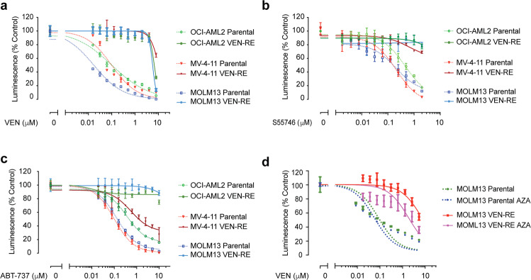
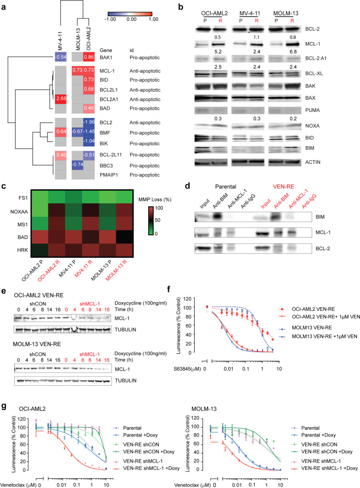
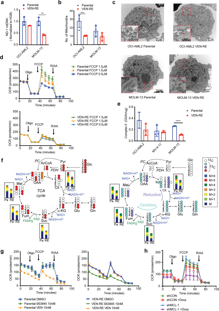
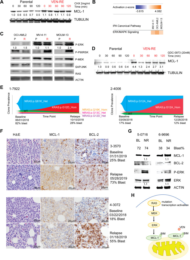
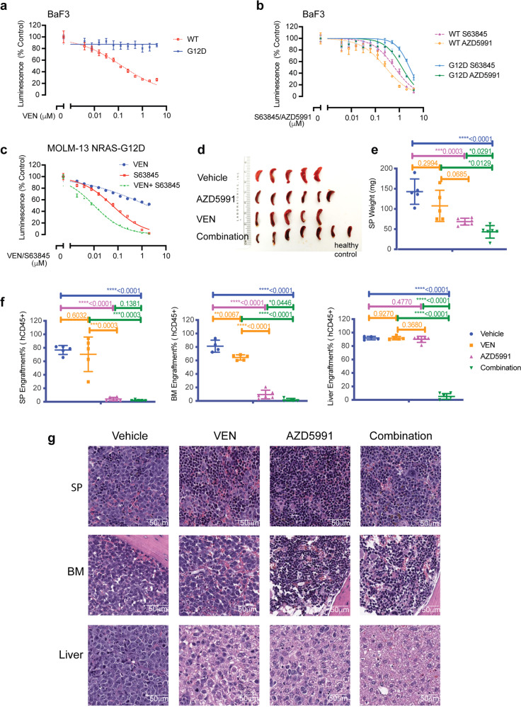
Despite high initial response rates, acute myeloid leukemia (AML) treated with the BCL-2-selective inhibitor venetoclax (VEN) alone or in combinations commonly acquires resistance. We performed gene/protein expression, metabolomic and methylation analyses of isogenic AML cell lines sensitive or resistant to VEN, and identified the activation of RAS/MAPK pathway, leading to increased stability and higher levels of MCL-1 protein, as a major acquired mechanism of VEN resistance. MCL-1 sustained survival and maintained mitochondrial respiration in VEN-RE cells, which had impaired electron transport chain (ETC) complex II activity, and MCL-1 silencing or pharmacologic inhibition restored VEN sensitivity. In support of the importance of RAS/MAPK activation, we found by single-cell DNA sequencing rapid clonal selection of RAS-mutated clones in AML patients treated with VEN-containing regimens. In summary, these findings establish RAS/MAPK/MCL-1 and mitochondrial fitness as key survival mechanisms of VEN-RE AML and provide the rationale for combinatorial strategies effectively targeting these pathways.


Conflict of interest statement

R.A.P. received financial support from AbbVie and Genentech. A.D.S. has received honorarium from Takeda, Novartis, Jazz, and Otsuka Pharmaceuticals and research support from Medivir AB and Takeda. A.D.S. is named as an inventor on a patent application related to the use of DNT cells in AML. A.D.S. owns stock in AbbVie Pharmaceuticals. A.D.S holds the Ronald N. Buick Chair in Oncology Research. MK: Consulting/honorarium: AbbVie, Genentech, F. Hoffman La-Roche, Stemline Therapeutics, Amgen, Forty-Seven, Kisoji; research funding/clinical trials support: AbbVie, Genentech, F. Hoffman La-Roche, Eli Lilly, Cellectis, Calithera, Ablynx, Stemline Therapeutics, Agios, Ascentage, Astra Zeneca, Forty-Seven, Kisoji; stock options/royalties: Reata Pharmaceutical.

Figures

Fig. 1
Fig. 1
Venetoclax-resistant (VEN-RE) cells had hypermethylated genomes. ad The indicated parental cells (dotted lines) and VEN-RE cells (solid lines) were treated with the indicated inhibitors for 48 h, then cell viability was determined by CellTiter-Glo assay. Luminescence reads were normalized to those of DMSO-treated control cells. Data are mean ± SD. a Results of viability assays for cells treated with 0–10 μM VEN. b Results of viability assays for cells treated with 0–2 μM of the BCL-2 inhibitor S55746. c Results of viability assays for cells treated with 0–10 μM of the BCL-2/BCL-XL inhibitor ABT-737. d Viability of MOLM-13 parental and VEN-RE exposed to AZA and subsequently treated with 0–10 μM VEN
Fig. 2
Fig. 2
MCL-1 became the primary antiapoptotic protein in VEN-RE cells. a Heatmap showing results of RNA sequencing analysis of BCL-2 family genes in parental and VEN-RE AML cell lines (FDR ≤ 0.2). Genes in red had higher expression, and genes in blue had ower expression, in VEN-RE cells. The numbers in the cells represent normalized expression levels to those in parental lines. b Western blots showing BCL-2 family protein expression in the indicated parental (P) and VEN-RE (R) cell lines. Numbers below the bands are quantifications normalized to levels in the parental lines. c Heatmap showing results of BH3 profiling of the indicated parental and resistant AML cell lines when exposed to the indicated peptides. Data were normalized to DMSO (as negative control) and FCCP (as positive control). The colors indicate the loss of mitochondrial membrane potential (MMP). d Co-immunoprecipitation of parental and VEN-RE MOLM-13 cells protein lysates pulled down with anti-BIM and anti-MCL-1 to determine the binding of BIM with BCL-2 and MCL-1. IgG served as a negative control. e Western blots showing inducible MCL-1 expression. MCL-1 knockdown was induced with 100 ng/mL doxycycline for 0–8 h in VEN-RE MOLM-13 and for 0–16 h in VEN-RE OCI-AML2 cells transfected with a control shRNA (shCON) or shMCL-1. f VEN-RE OCI-AML2 and MOLM-13 cells were treated with 0-4 μM of the MCL-1 inhibitor S63845 with (solid lines) or without 1 μM VEN (dotted lines) for 48 h, then cell viability was determined by CellTiter-Glo assay. Luminescence reads were normalized to those of DMSO-treated controls. Data represent mean ± SD from triplicate independent experiments. g Parental, VEN-RE, and VEN-RE OCI-AML2 and MOLM-13 cells transfected with a control shRNA (shCON) or a shRNA targeting MCL-1 (shMCL-1) that were induced with (solid lines) or without 100 ng/mL doxycycline (dotted lines) and treated with 0–10 μM VEN for 48 h. Cell viability was determined by CellTiter-Glo assay. Luminescence reads were normalized to DMSO-treated control cells. Data are mean ± SD
Fig. 3
Fig. 3
VEN-RE cells were deficient in mitochondrial respiration. a Mitochondrial ND-1 DNA expression levels were detected by RT-PCR and normalized to HGB levels in the indicated parental and VEN-RE cells. **P < 0.005. b Number of mitochondria in five randomly selected parental and VEN-RE cells in different fields under transmission electron microscopy (TEM). ***P < 0.0005. c TEM images of mitochondrial morphology. Areas marked in red are enlarged in the insets on each panel. Scale bars indicate 2 μm in the larger windows and 500 nm in the insets. d Results of Seahorse assays showing oxygen consumption rate (OCR) with the indicated concentrations of FCCP) in parental (dotted lines) and VEN-RE MOLM-13 cells (solid lines). e Electron transport chain complex II activity in parental and VEN-RE cell lines. ****P < 0.0001. f 13C labeling pattern of TCA cycle intermediates following labeling with U-13C-glucose (left) or 13C5,15N2-glutamine (right). Bar plots show the relative intensity of stable isotope enrichment fractions of TCA cycle intermediates for parental and VEN-RE MOLM-13 cells. The glucose-derived 13C enrichment fraction (total of M + 2, +3, and +4) for fumarate (relative to succinate enrichment levels) shows slightly decreased levels in VEN-RE cells. Cit Citrate, Suc Succinate, Fum Fumarate, Mal Malate. g Results of Seahorse assays showing OCR in parental (dotted lines) and VEN-RE (solid lines) MOLM-13 cells treated with indicated inhibitors for 1 h. h Results of Seahorse assays showing OCR in VEN-RE MOLM-13 cells transfected with shCON (dotted lines) or shMCL-1 (solid lines) that were induced with or without doxycycline (100 ng/mL) for 6 h
Fig. 4
Fig. 4
MAPK pathway activation in VEN-RE cells upregulated MCL-1. a Western blots showing MCL-1 protein levels in parental and VEN-RE MOLM-13 cells treated with 2 mg/mL cycloheximide (CHX) for 0–120 min. b Heatmap showing activation scores for ERK/MAPK signaling in VEN-RE cells based on pathway analysis of the RNA sequencing data. c Western blots showing protein levels of MAPK pathway components in parental (P) and VEN-RE (R) cells. d Western blots showing protein levels of MCL-1 in parental and VEN-RE MOLM-13 cells treated with 20 nM of the MEK inhibitor GDC-0973 (GDC) and 2 mg/mL CHX for 0–120 min. e Fishplots showing clone prevalence at two time points (baseline and relapse) in patient samples 1-7922 and 2-4006. f MCL-1 and BCL-2 expression by immunohistochemistry (IHC) in two paired samples from patients at the time of relapse on VEN clinical trial. Scale bar on the bottom right indicates 20 μm. g Protein expression in two AML patients primary refractory to VEN-based therapy pre- (BL) or post-treatment (no response, NR). h Mechanism of VEN resistance: RAS/MAPK/MCL-1 axis
Fig. 5
Fig. 5
NRAS-G12D mutation confers resistance to VEN and sensitivity to MCL-1 inhibition. a, b The BaF3 cells expressing NRAS-WT (dotted lines) or NRAS-G12D (solid lines) were treated with VEN (a) or indicated MCL-1 inhibitors S63845 or AZD5991 (b) for 24 h, after which cell viability was determined by CellTiter-Glo assay. Luminescence reads were normalized to those of DMSO-treated control cells. Data represent mean ± SD from triplicate independent experiments. c MOLM-13 cells expressing NRAS-G12D were treated with indicated inhibitor for 24 h, then cell viability was determined by CellTiter-Glo assay. Luminescence reads were normalized to those of DMSO-treated control cells. Data are mean ± SD. d Spleen harvested from mice received vehicle, AZD5991, VEN, or the combination of AZD5991 and VEN. One spleen from a healthy mouse is shown for comparison. e Weight of spleens collected from each group. Data are mean ± SD. f Tumor burden shown by %hCD45 from spleens (SP), bone marrows (BM) and livers from each mouse in specified cohort. Data are mean ± SD. g H&E staining of SP, BM, and liver from one representative mouse from each group

References

    1. Dombret H, Gardin C. An update of current treatments for adult acute myeloid leukemia. Blood. 2016;127:53–61. - PMC - PubMed
    1. DiNardo CD, et al. Venetoclax combined with decitabine or azacitidine in treatment-naive, elderly patients with acute myeloid leukemia. Blood. 2019;133:7–17. - PMC - PubMed
    1. DiNardo CD, et al. Safety and preliminary efficacy of venetoclax with decitabine or azacitidine in elderly patients with previously untreated acute myeloid leukaemia: a non-randomised, open-label, phase 1b study. Lancet Oncol. 2018;19:216–228. - PubMed
    1. Maiti A, et al. Outcomes in Molecular Subgroups and Resistance Patterns with Ten-Day Decitabine and Venetoclax (DEC10-VEN) in Acute Myeloid Leukemia. Blood. 2019;134:645–645.
    1. Tahir SK, et al. Potential mechanisms of resistance to venetoclax and strategies to circumvent it. BMC Cancer. 2017;17:399. - PMC - PubMed

Publication types

MeSH terms