. 2022 Feb;74(1):181-192.
doi: 10.1007/s10616-021-00504-0. Epub 2021 Nov 9.

Immortalization of cells derived from domestic dogs through expressing mutant cyclin-dependent kinase 4, cyclin D1, and telomerase reverse transcriptase

Affiliations

Immortalization of cells derived from domestic dogs through expressing mutant cyclin-dependent kinase 4, cyclin D1, and telomerase reverse transcriptase

Izzah Munirah et al. Cytotechnology. 2022 Feb.

Abstract

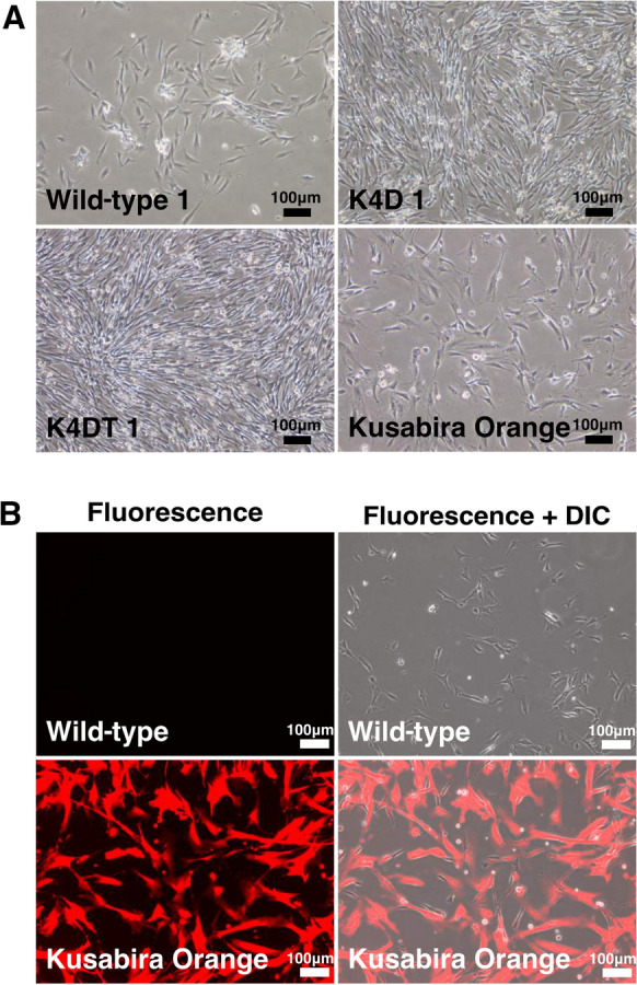
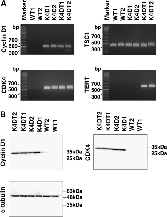
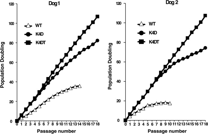
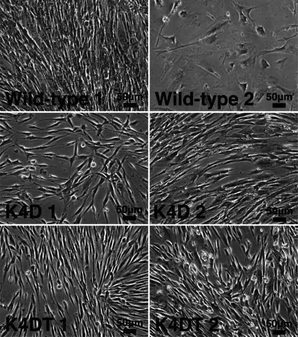
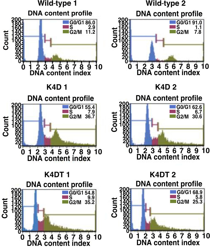
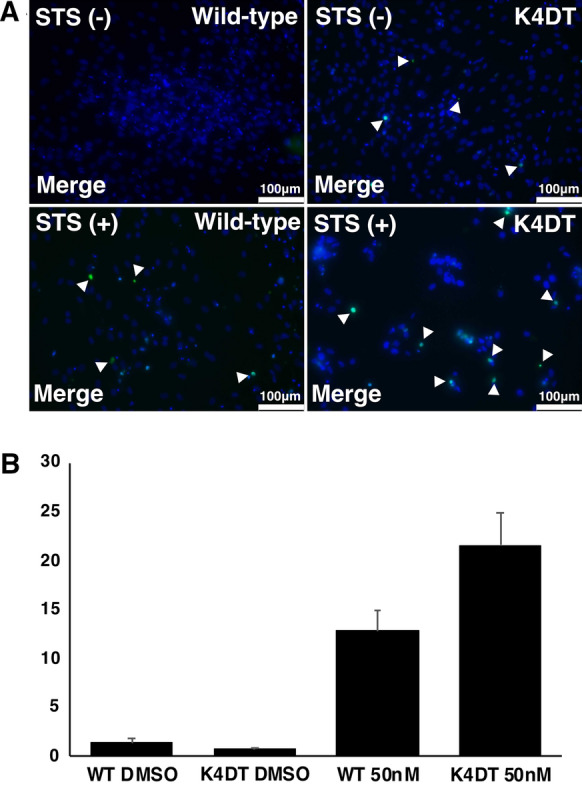
Dog is the first animal that was established as a close partner of human beings. Based on the vast genetic diversity and breeding, dogs exhibit unique genetic evolution and diversity from Chihuahua to St. Bernard. The safety tests of the pharmacological products also included domestic dogs as the test subjects. Although the safety confirmation test of chemicals for human use is important, the welfare of experimental animals requires special consideration. In this study, we cultured domestic dog-derived primary fibroblasts isolated from their muscle tissues. Furthermore, we successfully immortalized them through lentivirus-mediated gene transfer of mutant cyclin-dependent kinase 4 (CDK4), cyclin D1, and telomere reverse transcriptase (TERT). We further demonstrated that the established immortalized domestic dog-derived fibroblasts retained the characteristics of the original parental cells. These cells might act a suitable in vivo model system to replace the implication of animals for evaluating the potential toxicity of pharmacological chemicals.

Supplementary information: The online version contains supplementary material available at 10.1007/s10616-021-00504-0.

Keywords: CDK4; Cyclin D1; Dog; Immortalization; Telomerase reverse transcriptase.


Conflict of interest statement

Conflict of interestThe authors declare that they have no conflict of interest.

Figures

Fig. 1
Fig. 1
Cell morphology and fluorescence detection after the introduction of CDK4, cyclin D1, TERT, and Kusabira Orange (KO) expressions. A Cell morphology of each cell type: wild-type, K4D, K4DT, and KO. B The primary dog cells expressing red-expressing fluorescence (KO). The left panel shows the KO fluorescence detection of lentivirus-infected cells. The right panel shows the merged images of differential interference contrast (DIC) and fluorescence
Fig. 2
Fig. 2
Detection of genomic expression cassette and protein expression after the establishment of immortalized cells. A The genomic expression cassette of cyclin D1, CDK4, and TERT was detected using polymerase chain reaction (PCR). The TSC1 expression cassette was detected as a positive control. B Western blot analysis results for cyclin D1 and CDK4 protein expression. α-tubulin protein expression was detected as a positive control
Fig. 3
Fig. 3
Cell division quantification of established immortalized dog cells. Graphs show the results of the PD value calculated after each sequential passaging to analyze the growth and number of cell divisions. The graph on the left presents the results for dog 1. The graph on the right presents the results for dog 2
Fig. 4
Fig. 4
Cellular senescence detection of wild-type and recombinant cells derived from the Miniature Dachshund dogs. Each cell type was stained with SA-β-gal to detect cellular senescence. Parental cells exhibited bright blue staining, which indicated that the cells were undergoing aging or cellular senescence
Fig. 5
Fig. 5
Cell cycle analysis of wild-type and recombinant dog cells. Histograms showing the cell cycle analysis results: upper panel shows the results of the parental cells, middle panel for K4D cells, and bottom panel for K4DT cells. Each color represents the percentage of cells in each phase of the cell cycle: blue, red, and green represent the G0/G1, S, and G2/M phases, respectively
Fig. 6
Fig. 6
Detection of F-actin distribution by phalloidin fluorescence. Wild-type, K4D, and K4DT cells were stained with phalloidin and DAPI to observe the condition of the F-actin. The top panel shows the staining figures for wild-type cells, the middle panel for K4D cells, and the bottom panel for K4DT cells
Fig. 7
Fig. 7
Detection of apoptosis of wild type and K4DT cells after the exposure to staurosporine by TUNEL assay. The cells were counter-stained by DAPI (blue fluorescence), and positive cells for TUNEL were highlighted with green fluorescence that was marked by arrows (A). Bar graph of TUNEL positive cells, the Y-axis represents the average value of TUNEL-positive cells (B). The average value and standard error from six biological replications were shown
Fig. 8
Fig. 8
Multiple comparisons of CDK4 and cyclin D1 protein sequences between dog and human. Multiple alignments of CDK4 and cyclin D1 protein sequences between dogs and humans were obtained from the UCSC Genome Browser. The red highlighted parts indicate the conserved amino acids between the two sequences. Asterisk (*) marks the 24 R (arginine) of the sequence, which is the core sequence for p16 protein binding in the CDK4 sequence (A). The LXCXE motif (which allows the activation of the cyclin-CDK complex) in cyclin D1 sequence (B) is conserved in both human and dog sequences. The highlighted cyclin box is also conserved in both sequences

Similar articles

Cited by

References

    1. Ananiev J, Tchernev G, Patterson JW, et al. The guardian of genome. Acta Medica Bulg. 2011;38:72–82.
    1. Debacq-Chainiaux F, Erusalimsky JD, Campisi J, Toussaint O. Protocols to detect senescence-associated beta-galactosidase (SA-betagal) activity, a biomarker of senescent cells in culture and in vivo. Nat Protoc. 2009;4:1798–1806. doi: 10.1038/nprot.2009.191. - DOI - PubMed
    1. Donai K, Kuroda K, Guo Y, et al. Establishment of a reporter system to monitor silencing status in induced pluripotent stem cell lines. Anal Biochem. 2013;443:104–112. doi: 10.1016/j.ab.2013.08.014. - DOI - PubMed
    1. Fukuda T, Iino Y, Eitsuka T, et al. Cellular conservation of endangered midget buffalo (Lowland Anoa, Bubalus quarlesi) by establishment of primary cultured cell, and its immortalization with expression of cell cycle regulators. Cytotechnology. 2016;68:1937–1947. doi: 10.1007/s10616-016-0004-0. - DOI - PMC - PubMed
    1. Fukuda T, Gouko R, Eitsuka T, et al. Human-derived corneal epithelial cells expressing cell cycle regulators as a new resource for in vitro ocular toxicity testing. Front Genet. 2019;10:1–10. doi: 10.3389/fgene.2019.00587. - DOI - PMC - PubMed