Review
. 2022 Feb 3:15:805169.
doi: 10.3389/fnmol.2022.805169. eCollection 2022.

Crosstalk Between the NLRP3 Inflammasome/ASC Speck and Amyloid Protein Aggregates Drives Disease Progression in Alzheimer's and Parkinson's Disease

Affiliations
Review

Crosstalk Between the NLRP3 Inflammasome/ASC Speck and Amyloid Protein Aggregates Drives Disease Progression in Alzheimer's and Parkinson's Disease

Jonathan Hulse et al. Front Mol Neurosci. .

Abstract

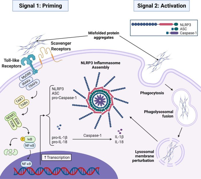
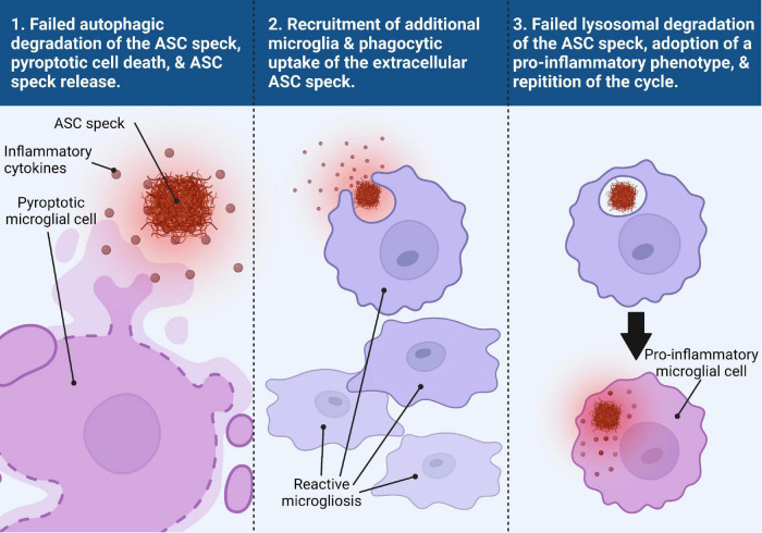
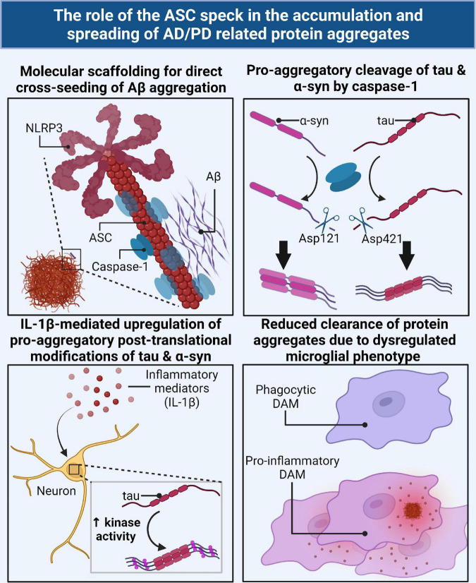
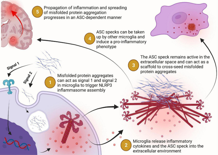
Two key pathological hallmarks of neurodegenerative diseases, including Alzheimer's disease (AD) and Parkinson's disease (PD), are the accumulation of misfolded protein aggregates and the chronic progressive neuroinflammation that they trigger. Numerous original research and reviews have provided a comprehensive understanding of how aggregated proteins (amyloid β, pathological tau, and α-synuclein) contribute to the disease, including driving sterile inflammation, in part, through the aggregation of multi-protein inflammasome complexes and the ASC speck [composed of NOD-, LRR-, and pyrin domain-containing protein 3 (NLRP3), Apoptosis-associated speck-like protein containing a C-terminal caspase activation and recruitment domain (ASC), and inflammatory caspase-1] involved in innate immunity. Here, we provide a unique perspective on the crosstalk between the aggregation-prone proteins involved in AD/PD and the multi-protein inflammasome complex/ASC speck that fuels feed-forward exacerbation of each other, driving neurodegeneration. Failed turnover of protein aggregates (both AD/PD related aggregates and the ASC speck) by protein degradation pathways, prionoid propagation of inflammation by the ASC speck, cross-seeding of protein aggregation by the ASC speck, and pro-aggregatory cleavage of proteins by caspase-1 are some of the mechanisms that exacerbate disease progression. We also review studies that provide this causal framework and highlight how the ASC speck serves as a platform for the propagation and spreading of inflammation and protein aggregation that drives AD and PD.

Keywords: ASC; Alzheimer’s disease; NLRP3; Parkinson’s disease; autophagy-lysosomal degradation; inflammasomes; neuroinflammation; protein aggregation.


Conflict of interest statement

The authors declare that the research was conducted in the absence of any commercial or financial relationships that could be construed as a potential conflict of interest.

Figures

FIGURE 1
FIGURE 1
AD and PD related misfolded protein aggregates (Aβ, tau, and α-syn) initiate signal 1 and signal 2 required for assembly of the NLRP3 inflammasome and subsequent ASC speck. In signal 1, misfolded protein aggregates bind to toll-like and scavenger receptors on the microglial cell surface initiating a MyD88-dependent signaling transduction event resulting in the translocation of NF-κB into the nucleus to initiate the transcription of important inflammasome components and the immature forms of the inflammatory cytokines pro-IL-1β and pro-IL-18. In signal 2, misfolded protein aggregates are taken into the cell via phagocytosis and trafficked to the lysosome where they disrupt the lysosomal membrane and trigger assembly of the inflammasome. Oligomerization of NLRP3 then recruits ASC which oligomerizes with NLRP3 via homotypic interactions between PYD regions of both proteins. Pro-caspase-1 is then recruited to the oligomerized ASC via homotypic interactions between CARD regions. Dimerization of pro-caspase-1 triggers an autolytic cleavage event producing two active caspase-1 enzymes that can mature pro-IL-1β and pro-IL-18 into the mature IL-1β and IL-18. MyD88, myeloid differentiation primary response gene 88; IRAK, IL-1 receptor-associated kinase; TRAF6, TNF receptor associated factor 6; TAK1, transforming growth factor-β-activated kinase-1; TAB, TAK1 binding proteins; NEMO, NF-κB essential modulator; IKKα/β, Inhibitor of NF-κB Kinase; IκB, Inhibitor of NF-κB; NF-κB, nuclear factor-kappaB; IL-1β/18, interleukin-1β/18.
FIGURE 2
FIGURE 2
Trans-microglial propagation of inflammation via ASC speck. Upon activation and assembly of the ASC speck from the NLRP3 inflammasome in response to misfolded protein aggregates, the microglial cell fails to degrade the ASC speck via autophagic clearance mechanisms (panel 1). Persistence of the ASC speck can result in either a non-pyroptotic exocytosis of the ASC speck or pyroptotic cell death, resulting in the release of inflammatory cytokines and the ASC speck into the extracellular space (panel 1). The extracellular ASC speck remains functionally active and the presence of inflammatory cytokines helps to recruit additional reactive microglia to the site of inflammation (panel 2). The ASC speck can then be taken up by another microglial cell where it can be degraded by the lysosomes (panel 3). Failed degradation of the ASC speck and possible lysosomal damage induces the microglial cell to adopt a pro-inflammatory phenotype (panel 3). This microglial cell can then undergo the same process as the previous microglial cell, perpetuating a viscous inflammatory cycle.
FIGURE 3
FIGURE 3
The ASC speck directly and indirectly contributes to the accumulation and spreading of AD/PD related misfolded protein aggregates via several mechanisms. The ASC speck can directly cross-seed the aggregation of Aβ by serving as a molecular scaffold for aggregation (top left). Caspase-1, the effector component of the inflammasome/ASC speck, can directly cleave tau and α-syn monomers into a pro-aggregatory form for seeding nucleation (top right). IL-1β, the downstream product of the ASC speck, can act as a cellular signal to upregulate key enzymes for the pro-aggregatory PTMs of tau and α-syn (bottom left). The ASC speck contributes to the dysregulation of protective disease-associated microglia (DAMs) and subsequent skewed pro-inflammatory phenotype resulting in a decrease in phagocytic clearance of AD/PD related misfolded protein aggregates (bottom right).
FIGURE 4
FIGURE 4
Misfolded protein aggregates bind to toll-like or scavenger receptors which triggers the expression of NLRP3 and caspase-1 to prime the cell for inflammasome assembly in a MyD88-NFκB signaling dependent manner. Extracellular misfolded protein aggregates may be internalized by the phagolysosomal pathway but can escape lysosomal degradation and trigger assembly of the NLRP3 inflammasome (step 1). The NLRP3 inflammasome catalyzes the maturation of interleukin-1β and interleukin-18 which can be transported extracellularly or released into the extracellular environment during pyroptosis for inflammatory signaling (step 2). Assembly of the NLRP3 inflammasome may progress to the formation of the ASC speck, a supramolecular complex with enhanced inflammatory signaling capacity, which may then be released into the extracellular environment following pyroptosis or some other non-pyroptic mechanism (step 2). Once in the extracellular space, the ASC speck remains functionally active for inflammatory cytokine maturation, pro-aggregatory modifications of other proteins, and cross-seeding of neuronally derived misfolded protein aggregation (step 3). The ASC speck can also propagate inflammation from one microglial cell to another by inducing a pro- inflammatory phenotype in cells that phagocytose the ASC speck but fail to degrade it (step 4). Together, these activities of the ASC speck implicate its role in the propagation of inflammation and spreading of protein aggregation in a vicious cycle that contributes to the chronic progressive nature of Alzheimer’s disease and Parkinson’s disease (step 5).

References

    1. Afonina I. S., Zhong Z., Karin M., Beyaert R. (2017). Limiting inflammation—the negative regulation of NF-κB and the NLRP3 inflammasome. Nat. Immunol. 18 861–869. 10.1038/ni.3772 - DOI - PubMed
    1. Arendt T., Stieler J. T., Holzer M. (2016). Tau and tauopathies. Brain Res. Bull. 126 238–292. 10.1016/j.brainresbull.2016.08.018 - DOI - PubMed
    1. Asai H., Ikezu S., Tsunoda S., Medalla M., Luebke J., Haydar T., et al. (2015). Depletion of microglia and inhibition of exosome synthesis halt tau propagation. Nat. Neurosci. 18 1584–1593. 10.1038/nn.4132 - DOI - PMC - PubMed
    1. Baroja-Mazo A., Martín-Sánchez F., Gomez A. I., Martínez C. M., Amores-Iniesta J., Compan V., et al. (2014). The NLRP3 inflammasome is released as a particulate danger signal that amplifies the inflammatory response. Nat. Immunol. 15 738–748. 10.1038/ni.2919 - DOI - PubMed
    1. Barron M., Gartlon J., Dawson L. A., Atkinson P. J., Pardon M.-C. (2017). A state of delirium: deciphering the effect of inflammation on tau pathology in Alzheimer’s disease. Exp. Gerontol. 94 103–107. 10.1016/j.exger.2016.12.006 - DOI - PMC - PubMed