Review
. 2022 Feb 3:16:799753.
doi: 10.3389/fncel.2022.799753. eCollection 2022.

Modes of Brain Cell Death Following Intracerebral Hemorrhage

Affiliations
Review

Modes of Brain Cell Death Following Intracerebral Hemorrhage

Yan Zhang et al. Front Cell Neurosci. .

Abstract

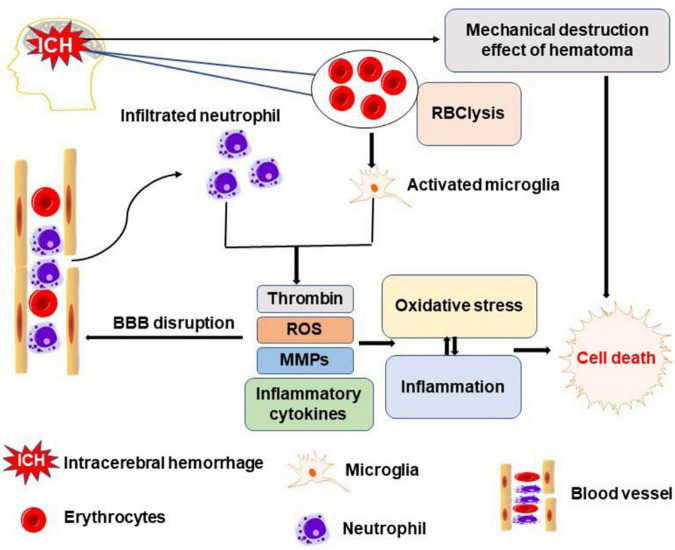
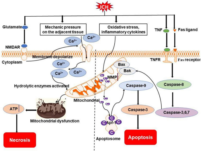
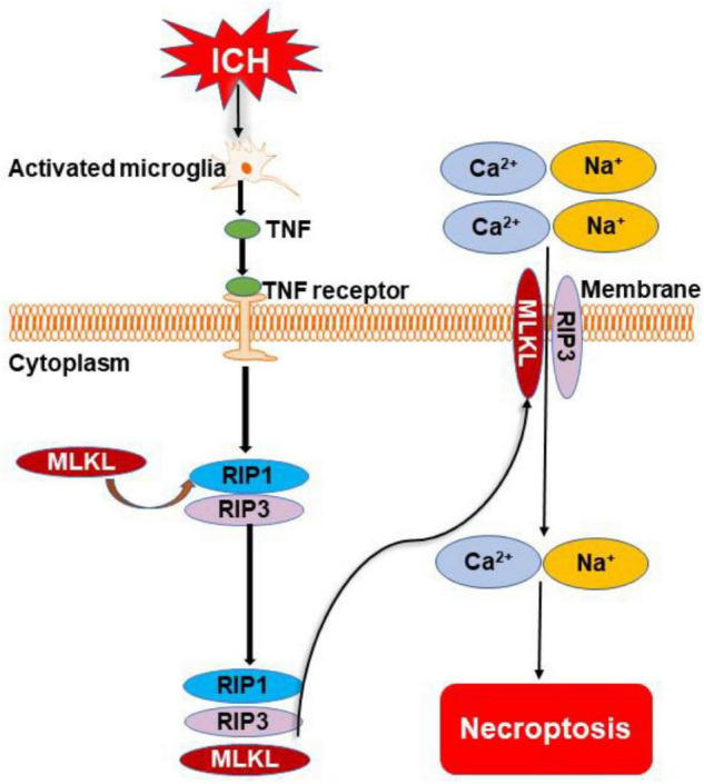
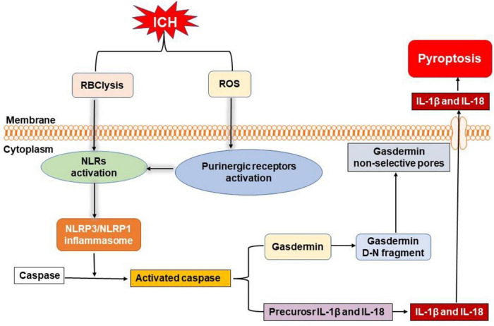
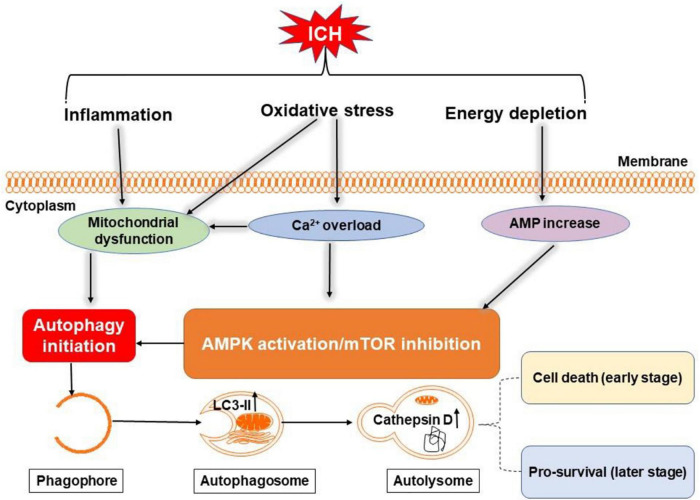
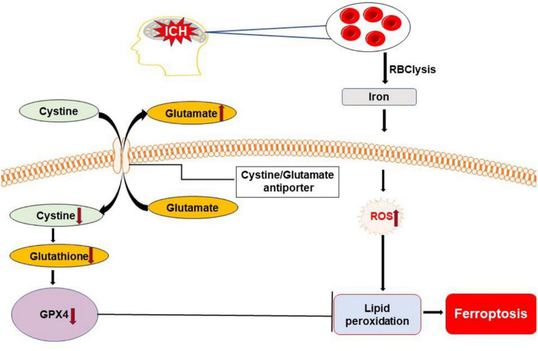
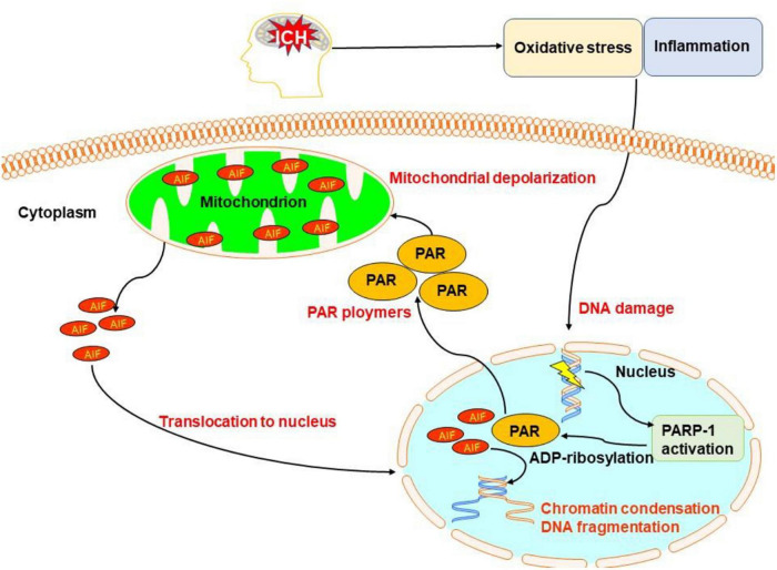
Intracerebral hemorrhage (ICH) is a devastating form of stroke with high rates of mortality and morbidity. It induces cell death that is responsible for neurological deficits postinjury. There are no therapies that effectively mitigate cell death to treat ICH. This review aims to summarize our knowledge of ICH-induced cell death with a focus on apoptosis and necrosis. We also discuss the involvement of ICH in recently described modes of cell death including necroptosis, pyroptosis, ferroptosis, autophagy, and parthanatos. We summarize treatment strategies to mitigate brain injury based on particular cell death pathways after ICH.

Keywords: apoptosis; autophagy; cell death; ferroptosis; intracerebral hemorrhage; necrosis; parthanatos.


Conflict of interest statement

The authors declare that the research was conducted in the absence of any commercial or financial relationships that could be construed as a potential conflict of interest.

Figures

FIGURE 1
FIGURE 1
Schematic representation of major pathways leading to brain cell death after intracerebral hemorrhage (ICH). Mechanical compression of brain tissue by the hematoma directly leads to brain cell death. The degradation products of erythrocytes activate microglia which, together with invading neutrophils, release toxic substances, such as thrombin, reactive oxygen species (ROS), matrix metalloproteinases (MMPs), and inflammatory cytokines. These events of neuroinflammation and oxidative stress culminate in neuronal and glial cell death, vasogenic edema, and breakdown of the blood-brain barrier.
FIGURE 2
FIGURE 2
Overview of apoptosis and necrosis pathways following ICH. Oxidative stress, inflammatory cytokines (e.g., TNF), and Fas/Fas ligand after ICH may activate intrinsic caspase-dependent pathways to induce the emergence of mitochondrial membrane permeability (MMP). Cytochrome c is then released from mitochondria to activate caspases to initiate the process of cell death. Mechanical compression by the hematoma on adjacent tissue and activation of NMDAR by excessive glutamate after ICH can result in an influx of calcium, which causes mitochondrial dysfunction. Ultimately, cells go to die due to insufficient ATP produced by mitochondria. NMDAR, N-methyl-D-aspartate receptor; TNF, tumor necrosis factor; TNFR, TNF receptor; Bak, Bcl-2 homologous antagonist killer; Bax, Bcl-2 associated X protein; Ca2+, calcium ion; C, cytochrome c; Apaf-1, apoptotic protease-activating factor 1.
FIGURE 3
FIGURE 3
Overview of necroptosis pathways following ICH. Microglia are activated after ICH. They release tumor necrosis factor (TNF) to initiate necroptosis by binding to TNF receptor. Phosphorylation of receptor-interacting protein 1 (RIP1) to develop necrosome also occurs. The necrosome cooperates with receptor-interacting protein 3 (RIP3) for the recruitment of mixed lineage kinase domain-like protein (MLKL). The complex of MLKL and RIP3 transfers to the cell membrane and forms a channel to cause the inward flow of Ca2+ and Na+. Finally, the cell dies through the necroptotic pathway.
FIGURE 4
FIGURE 4
Overview of pyroptosis pathways following ICH. Nucleotide-binding oligomerization domain-like receptors (NLRs) can be activated by the degradation products of erythrocytes (such as hemoglobin, heme, and iron) and activated purinergic receptors via ROS, leading to the formation of inflammasome. Then, NLR pyrin domain-containing 3 (NLRP3) and NLRP1 inflammasome activate caspase to initiate the cleavage and activation of interleukin-1β (IL-1β) and interleukin-18 (IL-18). Moreover, active caspase can also cleave gasdermin to form gasdermin D-N fragment, which causes non-selective pores in membrane, inducing the release of mature IL-1β and IL-18 to elicit neuroinflammation and cell death after ICH.
FIGURE 5
FIGURE 5
Overview of autophagy pathways following ICH. Oxidative stress, inflammation, and energy depletion after ICH may induce autophagy by activating adenosine monophosphate-activated protein kinase (AMPK) or inhibiting mammalian target of rapamycin (mTOR) and damaging mitochondria. During autophagy, light chain 3-II (LC3-II) in autophagosome and cathepsin D in lysosomes are increased. Excessive autophagy may be detrimental in the early stage of ICH, but maybe neuroprotective in the later stages through clearance of cellular debris.
FIGURE 6
FIGURE 6
Overview of ferroptosis pathways following ICH. Dysfunction of the cystine/glutamate antiporter after ICH leads to decreased synthesis of glutathione (GSH) and activity of glutathione peroxidase 4 (GPX4). Iron released from lysed erythrocytes can produce highly toxic hydroxyl radicals to attack DNA, proteins, and lipid membranes. The deficiency of GPX4 combined with the presence of toxic iron leads to the accumulation of lipid peroxides and the execution of ferroptosis. ROS, reactive oxygen species.
FIGURE 7
FIGURE 7
Overview of pathanatos pathways following ICH. Oxidative stress and inflammation after ICH damages DNA, leading to the activation of poly(ADP-ribose) polymerase-1 (PARP-1), which catalyzes the excessive synthesis of PAR intracellularly. PAR directly interacts with the C-terminus of membrane-bound apoptosis-inducing factor and triggers the release of apoptosis-inducing factor (AIF) from the mitochondria. AIF then begins its journey toward the nucleus and activates caspases causing chromatin condensation and DNA fragmentation.

References

    1. Bai Q., Sheng Z., Liu Y., Zhang R., Yong V. W., Xue M. (2020a). Intracerebral haemorrhage: from clinical settings to animal models. Stroke Vasc. Neurol. 5 388–395. 10.1136/svn-2020-000334 - DOI - PMC - PubMed
    1. Bai Q., Xue M., Yong V. W. (2020b). Microglia and macrophage phenotypes in intracerebral haemorrhage injury: therapeutic opportunities. Brain 143 1297–1314. 10.1093/brain/awz393 - DOI - PubMed
    1. Bao X., Wu G., Hu S., Huang F. (2008). Poly(Adp-Ribose) polymerase activation and brain edema formation by hemoglobin after intracerebral hemorrhage in rats. Acta Neurochir. Suppl. 105 23–27. 10.1007/978-3-211-09469-3_5 - DOI - PubMed
    1. Blaser H., Dostert C., Mak T. W., Brenner D. (2016). Tnf and Ros crosstalk in inflammation. Trends Cell Biol. 26 249–261. 10.1016/j.tcb.2015.12.002 - DOI - PubMed
    1. Bobinger T., Burkardt P., Huttner H. B., Manaenko A. (2018). Programmed cell death after intracerebral hemorrhage. Curr. Neuropharmacol. 16 1267–1281. 10.2174/1570159X15666170602112851 - DOI - PMC - PubMed

LinkOut - more resources