

Habenular Involvement in Response to Subcallosal Cingulate Deep Brain Stimulation for Depression
- PMID: 35185654
- PMCID: PMC8854862
- DOI: 10.3389/fpsyt.2022.810777
Habenular Involvement in Response to Subcallosal Cingulate Deep Brain Stimulation for Depression
Abstract
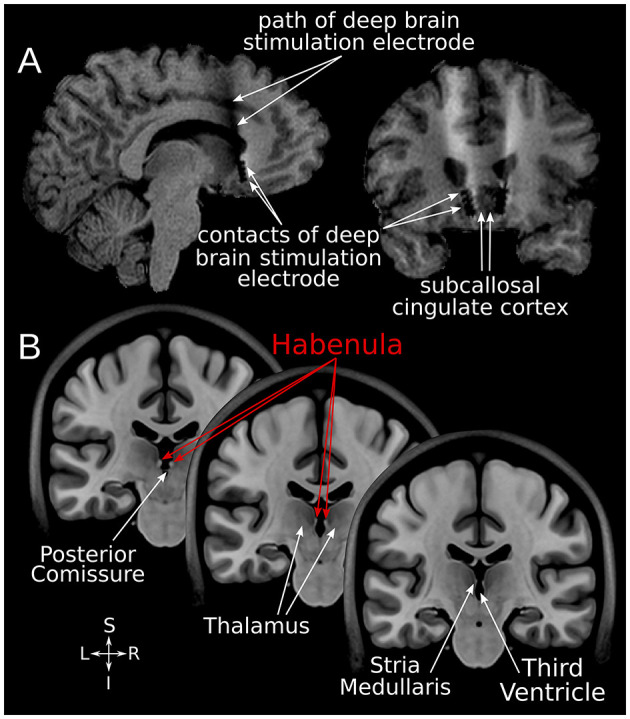
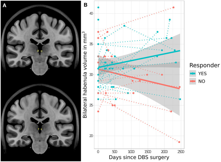
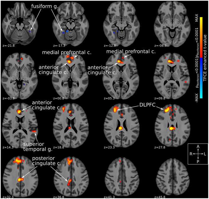
The habenula (Hb) is a small, evolutionarily conserved epithalamic structure implicated in functions such as reward and mood regulation. Prior imaging work suggests that Hb's structural and functional properties may relate to treatment response in depression and other mood disorders. We used multimodal MRI techniques to investigate the potential involvement of Hb in response to subcallosal cingulate area deep brain stimulation (SCC-DBS) for treatment-resistant mood disorders. Using an automated segmentation technique, we compared Hb volume at baseline and at a subsequent post-operative timepoint (4.4 ± 3.0 years after surgery) in a cohort of 32 patients who received SCC-DBS. Clinical response to treatment (≥50% decrease in HAMD-17 from baseline to 12 months post-operation) was significantly associated with longitudinal Hb volume change: responders tended to have increased Hb volume over time, while non-responders showed decreased Hb volume (t = 2.4, p = 0.021). We additionally used functional MRI (fMRI) in a subcohort of SCC-DBS patients (n = 12) to investigate immediate within-patient changes in Hb functional connectivity associated with SCC-DBS stimulation. Active DBS was significantly associated with increased Hb connectivity to several prefrontal and corticolimbic regions (TFCE-adjusted p Bonferroni < 0.0001), many of which have been previously implicated in the neurocircuitry of depression. Taken together, our results suggest that Hb may play an important role in the antidepressant effect of SCC-DBS.
Keywords: deep brain stimulation; depression; habenula; neuroimaging; neuromodulation; treatment biomarker.
Copyright © 2022 Elias, Germann, Loh, Boutet, Pancholi, Beyn, Bhat, Woodside, Giacobbe, Kennedy and Lozano.
Conflict of interest statement
AML is the co-founder of Functional Neuromodulation (a DBS-related company), is a consultant for Boston Scientific, Medtronic, and Abbott (companies that produce DBS hardware). PG and SK are consultants for Abbott. GE, AB, and AML have intellectual property in the field of DBS. The remaining authors declare that the research was conducted in the absence of any commercial or financial relationships that could be construed as a potential conflict of interest.
Figures
Similar articles
-
Defining critical white matter pathways mediating successful subcallosal cingulate deep brain stimulation for treatment-resistant depression.Biol Psychiatry. 2014 Dec 15;76(12):963-9. doi: 10.1016/j.biopsych.2014.03.029. Epub 2014 Apr 13. Biol Psychiatry. 2014. PMID: 24832866 Free PMC article.
-
Field potential 1/f activity in the subcallosal cingulate region as a candidate signal for monitoring deep brain stimulation for treatment-resistant depression.J Neurophysiol. 2019 Sep 1;122(3):1023-1035. doi: 10.1152/jn.00875.2018. Epub 2019 Jul 17. J Neurophysiol. 2019. PMID: 31314668 Free PMC article. Clinical Trial.
-
Neuroanatomical predictors of response to subcallosal cingulate deep brain stimulation for treatment-resistant depression.J Psychiatry Neurosci. 2020 Jan 1;45(1):45-54. doi: 10.1503/jpn.180207. J Psychiatry Neurosci. 2020. PMID: 31525860 Free PMC article.
-
Subcallosal Cingulate Cortex Deep Brain Stimulation for Treatment-Resistant Depression: A Systematic Review.Front Neurol. 2022 Apr 1;13:780481. doi: 10.3389/fneur.2022.780481. eCollection 2022. Front Neurol. 2022. PMID: 35432155 Free PMC article. Review.
-
A Decade of Progress in Deep Brain Stimulation of the Subcallosal Cingulate for the Treatment of Depression.J Clin Med. 2020 Oct 12;9(10):3260. doi: 10.3390/jcm9103260. J Clin Med. 2020. PMID: 33053848 Free PMC article. Review.
Cited by
-
Electroconvulsive therapy-induced volumetric brain changes converge on a common causal circuit in depression.Mol Psychiatry. 2024 Feb;29(2):229-237. doi: 10.1038/s41380-023-02318-2. Epub 2023 Nov 20. Mol Psychiatry. 2024. PMID: 37985787 Free PMC article.
-
Deep Brain Stimulation (DBS) in Treatment-Resistant Depression (TRD): Hope and Concern.Adv Exp Med Biol. 2024;1456:161-186. doi: 10.1007/978-981-97-4402-2_9. Adv Exp Med Biol. 2024. PMID: 39261429 Review.
-
Habenular volume changes after venlafaxine treatment in patients with major depression.Psychiatry Clin Neurosci. 2024 Aug;78(8):468-472. doi: 10.1111/pcn.13684. Epub 2024 Jun 12. Psychiatry Clin Neurosci. 2024. PMID: 38867362 Free PMC article.
-
The disappointment centre of the brain gets exciting: a systematic review of habenula dysfunction in depression.Transl Psychiatry. 2024 Dec 19;14(1):499. doi: 10.1038/s41398-024-03199-x. Transl Psychiatry. 2024. PMID: 39702626 Free PMC article.
-
The habenula in mood disorders: A systematic review of human studies.Mol Psychiatry. 2025 Aug 1. doi: 10.1038/s41380-025-03105-x. Online ahead of print. Mol Psychiatry. 2025. PMID: 40750674
References
LinkOut - more resources
Full Text Sources
Research Materials