. 2022 Feb 4:13:762580.
doi: 10.3389/fimmu.2022.762580. eCollection 2022.

Indole-3-Acetic Acid Alters Intestinal Microbiota and Alleviates Ankylosing Spondylitis in Mice

Affiliations

Indole-3-Acetic Acid Alters Intestinal Microbiota and Alleviates Ankylosing Spondylitis in Mice

Jun Shen et al. Front Immunol. .

Abstract

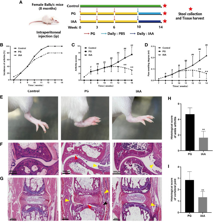
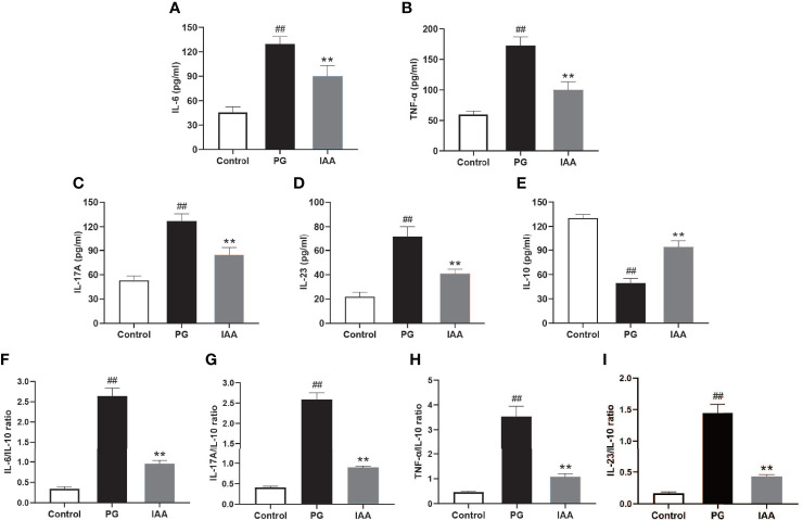
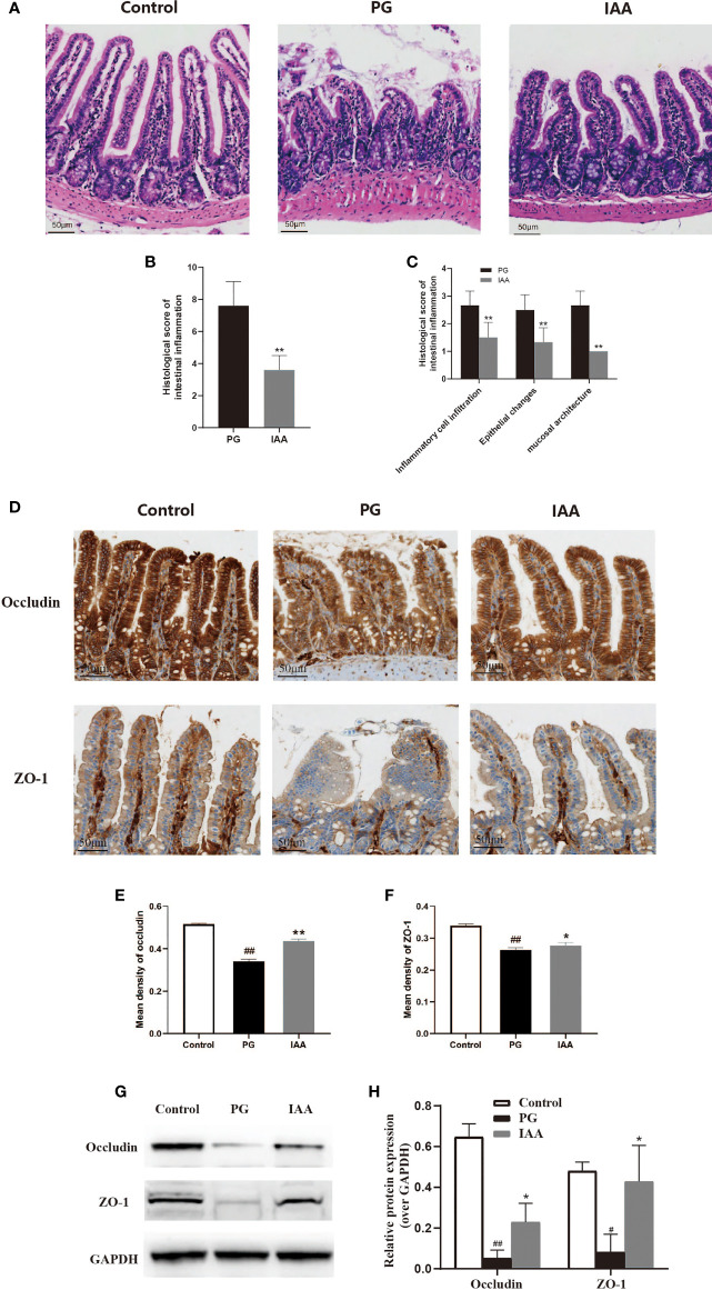
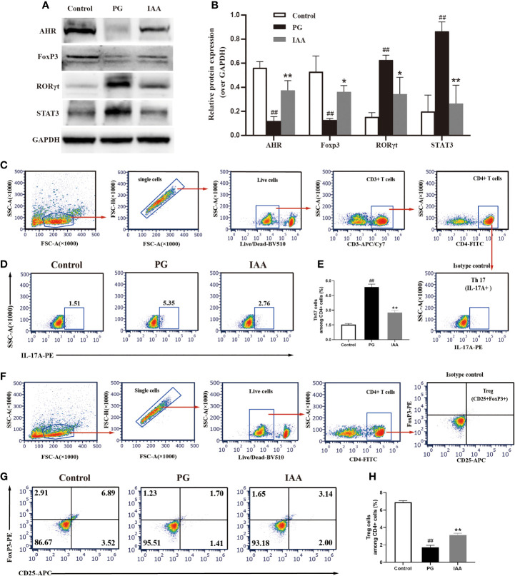
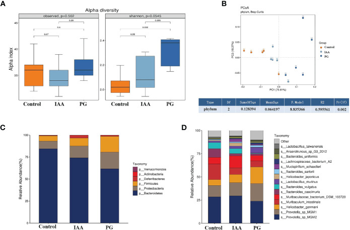
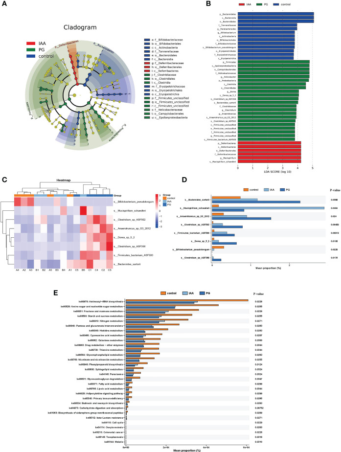
Ankylosing spondylitis (AS) is a systemic, chronic, and inflammatory autoimmune disease associated with the disorder of intestinal microbiota. Unfortunately, effective therapies for AS are lacking. Recent evidence has indicated that indole-3-acetic acid (IAA), an important microbial tryptophan metabolite, can modulate intestinal homeostasis and suppress inflammatory responses. However, reports have not examined the in vivo protective effects of IAA against AS. In this study, we investigated the protective effects and underlying mechanisms through which IAA acts against AS. We constructed a proteoglycan (PG)-induced AS mouse model and administered IAA (50 mg/kg body weight) by intraperitoneal injection daily for 4 weeks. The effects of IAA on AS mice were evaluated by examining disease severity, intestinal barrier function, aryl hydrocarbon receptor (AhR) pathway, T-helper 17 (Th17)/T regulatory (Treg) balance, and inflammatory cytokine levels. The intestinal microbiota compositions were profiled through whole-genome sequencing. We observed that IAA decreased the incidence and severity of AS in mice, inhibited the production of pro-inflammatory cytokines (tumor necrosis factor α [TNF-α], interleukin [IL]-6, IL-17A, and IL-23), promoted the production of the anti-inflammatory cytokine IL-10, and reduced the ratios of pro-/anti- inflammatory cytokines. IAA ameliorated pathological changes in the ileum and improved intestinal mucosal barrier function. IAA also activated the AhR pathway, upregulated the transcription factor forehead box protein P3 (FoxP3) and increased Treg cells, and downregulated the transcription factors retinoic acid receptor-related orphan receptor gamma t (RORγt) and signal transducer and activator of transcription 3 (STAT3) and decreased Th17 cells. Furthermore, IAA altered the composition of the intestinal microbiota composition by increasing Bacteroides and decreasing Proteobacteria and Firmicutes, in addition to increasing the abundances of Bifidobacterium pseudolongum and Mucispirillum schaedleri. In conclusion, IAA exerted several protective effects against PG-induced AS in mice, which was mediated by the restoration of balance among the intestinal microbial community, activating the AhR pathway, and inhibiting inflammation. IAA might represent a novel therapeutic approach for AS.

Keywords: ankylosing spondylitis; aryl hydrocarbon receptor; indole-3-acetic acid; inflammatory response; intestinal microbiota.


Conflict of interest statement

The authors declare that the research was conducted in the absence of any commercial or financial relationships that could be construed as a potential conflict of interest.

Figures

Figure 1
Figure 1
IAA attenuates disease progression and severity in PG-induced AS mice. (A) Schematic diagram for study design. Incidence of arthritis (B), arthritis index score (C), and degree of hind feet swelling (D) in three groups of mice (n = 10). (E) Representative pictures of the hind feet in three groups of mice at the end of week 14. (F) Representative pictures of ankle joint sections in three groups of mice (HE staining, scale bar = 300 µm). PG group was characterized by inflammatory cell infiltration in the synovium of the joint capsule (yellow arrow), cartilage degeneration, and bone erosion on the articular surface (red arrow). (G) Representative pictures of vertebral joint sections in three groups of mice (HE staining, scale bar = 300 µm). PG group was characterized by inflammatory cell infiltration in the ligaments around the intervertebral disc (yellow arrow), partial destruction of intervertebral disc structure (red arrow), and new chondrocytes (black arrow). (H) Histologic scores for ankle joint sections in the PG and IAA groups (n = 6). (I) Histologic scores of vertebral joint sections in the PG and IAA groups (n = 6). Data are expressed as the mean ± standard deviation (SD); # p < 0.05, ## p < 0.01 vs. control; **p < 0.01 vs. PG alone. IAA, indole-3 acetic acid; PG, proteoglycan; AS, ankylosing spondylitis; HE, hematoxylin and eosin.
Figure 2
Figure 2
IAA inhibits pro-inflammatory cytokines expressions, elevates anti-inflammatory cytokine expression, and reduces the ratios of pro-/anti- inflammatory cytokines in AS mice. Ankylosing spondylitis (AS) mice were administered intraperitoneal injections of indole-3 acetic acid (IAA; 50 mg/kg/day) for 4 weeks. The serum levels of the pro-inflammatory cytokines interleukin (IL)-6, tumor necrosis factor (TNF)-α, IL-17A, and IL-23, and the anti-inflammatory cytokine IL-10 were detected by enzyme-linked immunosorbent assay (ELISA). (A) IL-6, (B) TNF-α, (C) IL-17A, (D) IL-23, (E) IL-10, (F) IL-6/IL-10, (G) IL-17A/IL-10, (H) TNF-α/IL-10, (I) IL-23/IL-10. (n = 6). Data are expressed as the mean ± standard deviation (SD); ## p < 0.01 vs. control; **p < 0.01 vs. PG alone.
Figure 3
Figure 3
IAA ameliorates the pathological morphology of ileum tissue and improves intestinal mucosal barrier function in AS mice. (A) Representative pictures of ileum sections from three groups of mice at the end of week 14. (HE staining, scale bar = 50 µm). (B) Histologic scores for intestinal inflammation in the proteoglycan (PG) and indole-3 acetic acid (IAA) group (n = 6). (C) Histologic score of intestinal inflammation was divided into three categories: inflammatory cell infiltrates, epithelial changes, and mucosal architecture (n = 6). (D) The proteins of ZO-1 and occludin in ileum tissue from three groups of mice were detected by immunohistochemistry, as shown in representative pictures from one of three independent experiments (scale bar = 50 µm). (E) ZO-1 and (F) occludin staining intensities were quantified as the mean optical density (n = 6). (G) The tight junction protein (ZO-1 and occludin) levels in ileum tissue from three groups of mice were analyzed by western blot analysis, as shown in representative blots from one of three independent experiments. (H) Relative protein expression of ZO-1 and occludin using densitometric analysis, with GAPDH as a loading control (n = 3). Data are expressed as the mean ± standard deviation (SD); # p < 0.05, ## p < 0.01 vs. control; *p < 0.05, **p < 0.01 vs. PG alone. HE, hematoxylin and eosin; ZO-1, zonula occludens-1; GAPDH, glyceraldehyde 3-phosphate dehydrogenase.
Figure 4
Figure 4
IAA activates AhR signaling pathway and restores Th17/Treg balance in AS mice. (A) The protein levels of aryl hydrocarbon receptor (AhR) and Th17/Treg-related transcription factors, such as forkhead box protein P3 (FoxP3), signal transducer and activator of transcription 3 (STAT3), and retinoic acid receptor–related orphan receptor gamma (RORγt) in ileum tissue from three groups of mice were analyzed by western blot analysis, as shown in representative blots from one of three independent experiments. (B) Relative protein expression of AhR, FoxP3, STAT3, and RORγt using densitometric analysis, with glyceraldehyde 3-phosphate dehydrogenase (GAPDH) as a loading control (n=3). (C-H) Flow cytometric analysis of the frequencies of Th17 and Treg cells in ileum lamina propria from three groups of mice. (C) Gating strategy used for analysis of Th17 cells. Arrows indicate that Th17 cells were sequentially gated from single cells, live cells, CD3+ cells and CD4+ cells. Gated CD4+ cells were analyzed for expression of IL-17A. Representative plots of IL-17A expression (D) and percentage of IL-17A+ cells (E) among CD4+ T cells (n=6). (F) Gating strategy used for analysis of Treg cells. Arrows indicate that Treg cells were sequentially gated from single cells, live cells, and CD4+ cells. Gated CD4+ cells were analyzed for expression of CD25 and Foxp3. Representative plots of CD25 and Foxp3 expression (G) and percentage of CD25+Foxp3+ T cells (H) among CD4+ T cells (n = 6). Data are expressed as the mean ± standard deviation (SD); ## p < 0.01 vs. control; *p < 0.05, **p < 0.01 vs. PG alone. IAA, indole-3 acetic acid; Th17, T-helper 17; Treg, T regulatory; AS, ankylosing spondylitis.
Figure 5
Figure 5
IAA modulates the diversity and composition of the intestinal microbiota in AS mice. (A) Alpha diversity in three groups was assessed by observed species and Shannon index. (B) Beta diversity at the phylum level in three groups was assessed by principal coordinate analysis (PCoA) of Bray–Curtis dissimilarity paired with permutational multivariate analysis of variance (PERMANOVA). Relative abundance analyses of intestinal microbiota composition at the phylum (C) and species (D) levels in three groups. (n = 3). IAA, indole-3 acetic acid; AS, ankylosing spondylitis.
Figure 6
Figure 6
Comparison of the taxa and functional profiles of intestinal microbiota in three groups. The linear discriminant analysis effect size (LEfSe) analysis of the intestinal microbiota at the species level in three groups was presented on the cladogram (A) and bar plot (B) with linear discriminant analysis (LDA) score ≥ 3 and p < 0.05. (C) Heat map of 8 significantly different taxon abundances at the species level in three groups. (D) Box plot generated by rank-sum test (Kruskal–Wallis test) of relative abundance among the intestinal microbiota at the species level in three groups. (E) The differential analysis of functional predictions in three groups was performed based on level 3 of the Kyoto Encyclopedia of Genes and Genomes (KEGG) functional categories.

References

    1. Babaie F, Hasankhani M, Mohammadi H, Safarzadeh E, Rezaiemanesh A, Salimi R, et al. . The Role of Gut Microbiota and IL-23/IL-17 Pathway in Ankylosing Spondylitis Immunopathogenesis: New Insights and Updates. Immunol Lett (2018) 196:52–62. doi: 10.1016/j.imlet.2018.01.014 - DOI - PubMed
    1. Rosine N, Miceli-Richard C. Innate Cells: The Alternative Source of IL-17 in Axial and Peripheral Spondyloarthritis? Front Immunol (2020) 11:553742. doi: 10.3389/fimmu.2020.553742 - DOI - PMC - PubMed
    1. Venken K, Jacques P, Mortier C, Labadia ME, Decruy T, Coudenys J, et al. . Rorγt Inhibition Selectively Targets IL-17 Producing iNKT and γδ-T Cells Enriched in Spondyloarthritis Patients. Nat Commun (2019) 10(1):9. doi: 10.1038/s41467-018-07911-6 - DOI - PMC - PubMed
    1. Zhai J, Rong J, Li Q, Gu J. Immunogenetic Study in Chinese Population With Ankylosing Spondylitis: Are There Specific Genes Recently Disclosed? JC Immunol (2013) 2013:419357. doi: 10.1155/2013/419357 - DOI - PMC - PubMed
    1. Feldtkeller E, Khan MA, van der Heijde D, van der Linden S, Braun J. Age at Disease Onset and Diagnosis Delay in HLA-B27 Negative vs. Positive Patients With Ankylosing Spondylitis. Rheumatol Int (2003) 23(2):61–6. doi: 10.1007/s00296-002-0237-4 - DOI - PubMed

Publication types

MeSH terms