Regulation of Peripheral Inflammation by a Non-Viable, Non-Colonizing Strain of Commensal Bacteria
- PMID: 35185874
- PMCID: PMC8847375
- DOI: 10.3389/fimmu.2022.768076
Regulation of Peripheral Inflammation by a Non-Viable, Non-Colonizing Strain of Commensal Bacteria
Abstract
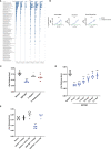
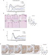
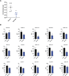
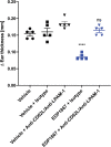
The gastrointestinal tract represents one of the largest body surfaces that is exposed to the outside world. It is the only mucosal surface that is required to simultaneously recognize and defend against pathogens, while allowing nutrients containing foreign antigens to be tolerated and absorbed. It differentiates between these foreign substances through a complex system of pattern recognition receptors expressed on the surface of the intestinal epithelial cells as well as the underlying immune cells. These immune cells actively sample and evaluate microbes and other particles that pass through the lumen of the gut. This local sensing system is part of a broader distributed signaling system that is connected to the rest of the body through the enteric nervous system, the immune system, and the metabolic system. While local tissue homeostasis is maintained by commensal bacteria that colonize the gut, colonization itself may not be required for the activation of distributed signaling networks that can result in modulation of peripheral inflammation. Herein, we describe the ability of a gut-restricted strain of commensal bacteria to drive systemic anti-inflammatory effects in a manner that does not rely upon its ability to colonize the gastrointestinal tract or alter the mucosal microbiome. Orally administered EDP1867, a gamma-irradiated strain of Veillonella parvula, rapidly transits through the murine gut without colonization or alteration of the background microbiome flora. In murine models of inflammatory disease including delayed-type hypersensitivity (DTH), atopic dermatitis, psoriasis, and experimental autoimmune encephalomyelitis (EAE), treatment with EDP1867 resulted in significant reduction in inflammation and immunopathology. Ex vivo cytokine analyses revealed that EDP1867 treatment diminished production of pro-inflammatory cytokines involved in inflammatory cascades. Furthermore, blockade of lymphocyte migration to the gut-associated lymphoid tissues impaired the ability of EDP1867 to resolve peripheral inflammation, supporting the hypothesis that circulating immune cells are responsible for promulgating the signals from the gut to peripheral tissues. Finally, we show that adoptively transferred T cells from EDP1867-treated mice inhibit inflammation induced in recipient mice. These results demonstrate that an orally-delivered, non-viable strain of commensal bacteria can mediate potent anti-inflammatory effects in peripheral tissues through transient occupancy of the gastrointestinal tract, and support the development of non-living bacterial strains for therapeutic applications.
Keywords: CNS; Th1; Th17; Th2; bacteria; gastrointestinal tract; inflammation.
Copyright © 2022 Ramani, Cormack, Cartwright, Alami, Parameswaran, Abdou, Wang, Hilliard-Barth, Argueta, Raghunathan, Caffry, Davitt, Romano, Ng, Kravitz, Rommel, Sizova, Kiran, Pradeep, Ponichtera, Ganguly, Bodmer and Itano.
Conflict of interest statement
All authors are employees and shareholders of Evelo Biosciences, which is sponsoring the clinical development of EDP1867.
Figures


Similar articles
-
Development, validation and implementation of an in vitro model for the study of metabolic and immune function in normal and inflamed human colonic epithelium.Dan Med J. 2015 Jan;62(1):B4973. Dan Med J. 2015. PMID: 25557335 Review.
-
Commensal bacteria (normal microflora), mucosal immunity and chronic inflammatory and autoimmune diseases.Immunol Lett. 2004 May 15;93(2-3):97-108. doi: 10.1016/j.imlet.2004.02.005. Immunol Lett. 2004. PMID: 15158604 Review.
-
The Probiotic Compound VSL#3 Modulates Mucosal, Peripheral, and Systemic Immunity Following Murine Broad-Spectrum Antibiotic Treatment.Front Cell Infect Microbiol. 2017 May 5;7:167. doi: 10.3389/fcimb.2017.00167. eCollection 2017. Front Cell Infect Microbiol. 2017. PMID: 28529928 Free PMC article.
-
Increased Expression of DUOX2 Is an Epithelial Response to Mucosal Dysbiosis Required for Immune Homeostasis in Mouse Intestine.Gastroenterology. 2015 Dec;149(7):1849-59. doi: 10.1053/j.gastro.2015.07.062. Epub 2015 Aug 7. Gastroenterology. 2015. PMID: 26261005 Free PMC article.
-
Fecal microbiota transplantation prevents Candida albicans from colonizing the gastrointestinal tract.Microbiol Immunol. 2019 May;63(5):155-163. doi: 10.1111/1348-0421.12680. Epub 2019 May 15. Microbiol Immunol. 2019. PMID: 30919462
Cited by
-
Harnessing the small intestinal axis to resolve systemic inflammation.Front Immunol. 2022 Nov 15;13:1060607. doi: 10.3389/fimmu.2022.1060607. eCollection 2022. Front Immunol. 2022. PMID: 36458009 Free PMC article.
-
Study of the Relationship between Mucosal Immunity and Commensal Microbiota: A Bibliometric Analysis.Nutrients. 2023 May 20;15(10):2398. doi: 10.3390/nu15102398. Nutrients. 2023. PMID: 37242281 Free PMC article. Review.
-
Probiotics combined with prebiotics alleviated seasonal allergic rhinitis by altering the composition and metabolic function of intestinal microbiota: a prospective, randomized, double-blind, placebo-controlled clinical trial.Front Immunol. 2024 Nov 1;15:1439830. doi: 10.3389/fimmu.2024.1439830. eCollection 2024. Front Immunol. 2024. PMID: 39555052 Free PMC article. Clinical Trial.
-
A dynamic atlas of immunocyte migration from the gut.Sci Immunol. 2024 Jan 5;9(91):eadi0672. doi: 10.1126/sciimmunol.adi0672. Epub 2024 Jan 5. Sci Immunol. 2024. PMID: 38181094 Free PMC article.
-
Betaine alleviates deficits in motor behavior, neurotoxic effects, and neuroinflammatory responses in a rat model of demyelination.Toxicol Rep. 2025 Feb 28;14:101974. doi: 10.1016/j.toxrep.2025.101974. eCollection 2025 Jun. Toxicol Rep. 2025. PMID: 40129881 Free PMC article.
References
MeSH terms
Substances
LinkOut - more resources
Full Text Sources
Other Literature Sources
Medical

