. 2022 Feb 3:13:790043.
doi: 10.3389/fimmu.2022.790043. eCollection 2022.

Neutrophils Contribute to ER Stress in Lung Epithelial Cells in the Pristane-Induced Diffuse Alveolar Hemorrhage Mouse Model

Affiliations

Neutrophils Contribute to ER Stress in Lung Epithelial Cells in the Pristane-Induced Diffuse Alveolar Hemorrhage Mouse Model

Gantsetseg Tumurkhuu et al. Front Immunol. .

Abstract

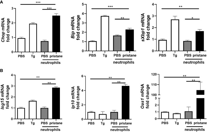
Diffuse alveolar hemorrhage (DAH), although rare, is a life-threatening complication of systemic lupus erythematosus (SLE). Little is known about the pathophysiology of DAH in humans, although increasingly neutrophils, NETosis and inflammatory monocytes have been shown to play an important role in the pristane-induced model of SLE which develops lung hemorrhage and recapitulates many of the pathologic features of human DAH. Using this experimental model, we asked whether endoplasmic reticulum (ER) stress played a role in driving the pathology of pulmonary hemorrhage and what role infiltrating neutrophils had in this process. Analysis of lung tissue from pristane-treated mice showed genes associated with ER stress and NETosis were increased in a time-dependent manner and reflected the timing of CD11b+Ly6G+ neutrophil accumulation in the lung. Using precision cut lung slices from untreated mice we observed that neutrophils isolated from the peritoneal cavity of pristane-treated mice could directly induce the expression of genes associated with ER stress, namely Chop and Bip. Mice which had myeloid-specific deletion of PAD4 were generated and treated with pristane to assess the involvement of PAD4 and PAD4-dependent NET formation in pristane-induced lung inflammation. Specific deletion of PAD4 in myeloid cells resulted in decreased expression of ER stress genes in the pristane model, with accompanying reduction in IFN-driven genes and pathology. Lastly, coculture experiments of human neutrophils and human lung epithelial cell line (BEAS-2b) showed neutrophils from SLE patients induced significantly more ER stress and interferon-stimulated genes in epithelial cells compared to healthy control neutrophils. These results support a pathogenic role of neutrophils and NETs in lung injury during pristane-induced DAH through the induction of ER stress response and suggest that overactivation of neutrophils in SLE and NETosis may underlie development of DAH.

Keywords: ER stress; NEtosis; diffuse alveolar hemorrhage (DAH); lung inflammation; neutrophil (PMN); systemic lupus erythematosus (SLE).


Conflict of interest statement

The authors declare that the research was conducted in the absence of any commercial or financial relationships that could be construed as a potential conflict of interest.

Figures

Figure 1
Figure 1
Increased levels of ER stress markers in lung of pristane mice associate with neutrophil infiltration. (A) Time course analysis of CD11b+Ly6G+ neutrophil infiltration in whole lung of C57BL/6 mice (n=4-6 mice per group), after pristane treatment determined by FACS. (B) Time course analysis of ER stress gene expression in the whole lung from indicated days after pristane treatment as measured by qPCR. (C) ER stress gene expression in the lung day 7 after pristane treatment as measured by qPCR. (D) Representative images immunostaining for CHOP (green), E-Cadherin (red), and DAPI (blue). Values are the mean ± SD. Statistical significance was determined by one-way ANOVA test. *p < 0.05, **p < 0.01 and ***p ≤ 0.001.
Figure 2
Figure 2
Neutrophils from pristane-treated mice induce higher expression of ER stress and ISGs in co-culture with precision cut lung slices (PCLS). PCLS from untreated mice were cultured (1 slice/well) and stimulated with thapsigargin (Tg) as an ER stress positive control or cocultured with peritoneal neutrophils (1x10⁶/well) from PBS- or pristane-treated mice. Expression of (A) ER stress markers and (B) ISGs were determined from total cells by qPCR. Values are the mean ± SD. and are representative 3 independent experiments. Statistical significance was determined by one-way ANOVA test. *p < 0.05, **p < 0.01 and ***p ≤ 0.001.
Figure 3
Figure 3
Lung inflammation and ER stress genes are driven by PAD4-expressing neutrophils 7 days post pristane treatment. (A) Representative haematoxylin and eosin (H&E)-stained lung sections (x200) of PBS- or pristane-treated PAD4fl/fl or LysMCrePAD4fl/fl mice. Quantification of lung damage was determined from 10 randomly selected images. (B) Expression of ER stress genes and ISGs were determined by qPCR of whole lung. (C) PCLS were prepared and cocultured with neutrophils as described in Figure 2 , and PAD4fl/fl or LysMCrePAD4fl/fl peritoneal neutrophils were isolated after 7 days of PBS or pristane treatment. Values were normalized to neutrophils isolated from PBS-treated mice of their respective genotype. ER stress genes and ISGs were determined by qPCR of whole cells. Values are the mean ± SD. In each case data is representative of 2 individual experiments with per group. Statistical significance was determined by one-way ANOVA test. * p < 0.05, ** p < 0.01, and *** p ≤ 0.001. ns, not significant.
Figure 4
Figure 4
Active SLE patient neutrophils are inflammatory and induce ER stress in human BEAS-2b bronchial epithelial cells. (A) The expression levels of ISGs in neutrophils isolated from the whole blood of inactive SLE (IAP) or moderate to active SLE patients (SLEDAI > 4, AP) as determined by qPCR (n=3 for each group). (B) Neutrophils from healthy control and active SLE patient (n=3 per group) whole blood were isolated and cultured alone or directly with BEAS-2b cells at a 1:4 ratio and incubated overnight. The expression levels of ER stress genes were determined by qPCR of whole cells. Values are the mean ± SD. Statistical significance was determined by one-way ANOVA test. *p < 0.05, **p < 0.01, and ***p ≤ 0.001.

References

    1. Kamen DL, Strange C. Pulmonary Manifestations of Systemic Lupus Erythematosus. Clinics Chest Med (2010) 31(3):479–88. doi: 10.1016/j.ccm.2010.05.001 - DOI - PubMed
    1. Agmon-Levin N, Selmi C. The Autoimmune Side of Heart and Lung Diseases. Clin Rev Allergy Immunol (2013) 44(1):1–5. doi: 10.1007/s12016-012-8335-x - DOI - PubMed
    1. Amarnani R, Yeoh SA, Denneny EK, Wincup C. Lupus and the Lungs: The Assessment and Management of Pulmonary Manifestations of Systemic Lupus Erythematosus. Front Med (Lausanne) (2020) 7:610257. doi: 10.3389/fmed.2020.610257 - DOI - PMC - PubMed
    1. Garcia-Romo GS, Caielli S, Vega B, Connolly J, Allantaz F, Xu Z, et al. . Netting Neutrophils Are Major Inducers of Type I IFN Production in Pediatric Systemic Lupus Erythematosus. Sci Trans Med (2011) 3(73):73ra20. doi: 10.1126/scitranslmed.3001201 - DOI - PMC - PubMed
    1. Lande R, Ganguly D, Facchinetti V, Frasca L, Conrad C, Gregorio J, et al. . Neutrophils Activate Plasmacytoid Dendritic Cells by Releasing Self-DNA-Peptide Complexes in Systemic Lupus Erythematosus. Sci Trans Med (2011) 3(73):73ra19. doi: 10.1126/scitranslmed.3001180 - DOI - PMC - PubMed

Publication types

MeSH terms