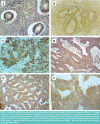
Evaluation of immunohistochemical expression of stem cell markers (NANOG and CD133) in normal, hyperplastic, and malignant endometrium
- PMID: 35186145
- PMCID: PMC8852636
- DOI: 10.25122/jml-2021-0206
Evaluation of immunohistochemical expression of stem cell markers (NANOG and CD133) in normal, hyperplastic, and malignant endometrium
Abstract
Cancer stem cells (CSC) are a potential cause for recurrence, metastasis, and resistance of tumors to different therapeutic modalities like hormonal radiotherapy and chemotherapy. We investigated two CSC markers (NANOG and CD 133) in normal, hyperplastic endometrium and endometrial carcinoma. A total of 93 formalin-fixed paraffin-embedded tissue blocks were used for immunohistochemical expression of NANOG and CD133 markers. NANOG expression was detected in 88.37% of endometrial carcinoma cases compared to 15% of the normal proliferative endometrium and 60% of hyperplasia cases. In endometrial carcinoma, high NANOG expression was significantly correlated with high grade, deep myometrial invasion, lymph node metastasis, and high stage with p-values (0.009, 0.005, 0.014, and 0.003, respectively). CD133 was positive in 76.74% of endometrial carcinoma cases, and it showed a significant correlation with deep myometrial invasion, positive lymph node, positive lymphovascular invasion, and high stage (p-values 0.003, 0.001, 0.003, and 0.013, respectively). Normal endometrium showed less expression of CD133 (only 5%) than hyperplasia and endometrial carcinoma with a statistically highly significant difference (p less than 0.0001). Hyperplastic cases with atypia expressed higher CD133 than those without atypia (6 out of 12 versus 3 out of 18). However, this difference was not statistically significant (p-value 0.111). The cancer stem cell markers NANOG and CD 133 are expressed in a high percentage in endometrial carcinoma compared to normal and hyperplasia and their expression is positively correlated with the aggressive behavior of the tumor. High expression of these two markers in apparently normal tissue around the tumor and in hyperplastic conditions with atypia suggests the possibility to use NANOG and CD133 expression as a diagnostic marker distinguishing dysplasia from reactive atypia. Therefore, inhibition of these markers can be a promising method to stop the progression of early cancers.
Keywords: CD 133; NANOG; cancer stem cell markers; endometrial carcinoma; endometrial hyperplasia; immunohistochemical; normal endometrium.
©2022 JOURNAL of MEDICINE and LIFE.
Figures

Similar articles
-
Immunohistochemical analysis of c-myc, c-jun and estrogen receptor in normal, hyperplastic and neoplastic endometrium.Pathol Oncol Res. 2005;11(1):32-9. doi: 10.1007/BF03032403. Epub 2005 Mar 31. Pathol Oncol Res. 2005. PMID: 15800680
-
Computerized image analysis of p53 and proliferating cell nuclear antigen expression in benign, hyperplastic, and malignant endometrium.Arch Pathol Lab Med. 2001 Jul;125(7):872-9. doi: 10.5858/2001-125-0872-CIAOPA. Arch Pathol Lab Med. 2001. PMID: 11419970
-
Enhanced CD24 expression in endometrial carcinoma and its expression pattern in normal and hyperplastic endometrium.Histol Histopathol. 2009 Mar;24(3):309-16. doi: 10.14670/HH-24.309. Histol Histopathol. 2009. PMID: 19130400
-
Melanoma Cell Adhesion Molecule (CD 146) in Endometrial Physiology and Disorder.Adv Exp Med Biol. 2025;1474:131-148. doi: 10.1007/5584_2024_826. Adv Exp Med Biol. 2025. PMID: 39400880 Review.
-
Autophagy Involvement in Non-Neoplastic and Neoplastic Endometrial Pathology: The State of the Art with a Focus on Carcinoma.Int J Mol Sci. 2024 Nov 12;25(22):12118. doi: 10.3390/ijms252212118. Int J Mol Sci. 2024. PMID: 39596186 Free PMC article. Review.
Cited by
-
Characteristics of Cancer Stem Cells and Their Potential Role in Endometrial Cancer.Cancers (Basel). 2024 Mar 7;16(6):1083. doi: 10.3390/cancers16061083. Cancers (Basel). 2024. PMID: 38539419 Free PMC article. Review.
-
Inhibiting MEK1 R189 citrullination enhances the chemosensitivity of docetaxel to multiple tumour cells.Philos Trans R Soc Lond B Biol Sci. 2023 Nov 20;378(1890):20220246. doi: 10.1098/rstb.2022.0246. Epub 2023 Oct 2. Philos Trans R Soc Lond B Biol Sci. 2023. PMID: 37778380 Free PMC article.
References
MeSH terms
Substances
LinkOut - more resources
Full Text Sources
Research Materials
