. 2022 Feb 9:2022:4691576.
doi: 10.1155/2022/4691576. eCollection 2022.

Chemical Distance Measurement and System Pharmacology Approach Uncover the Novel Protective Effects of Biotransformed Ginsenoside C-Mc against UVB-Irradiated Photoaging

Affiliations

Chemical Distance Measurement and System Pharmacology Approach Uncover the Novel Protective Effects of Biotransformed Ginsenoside C-Mc against UVB-Irradiated Photoaging

Xiao-Yi Liu et al. Oxid Med Cell Longev. .

Abstract

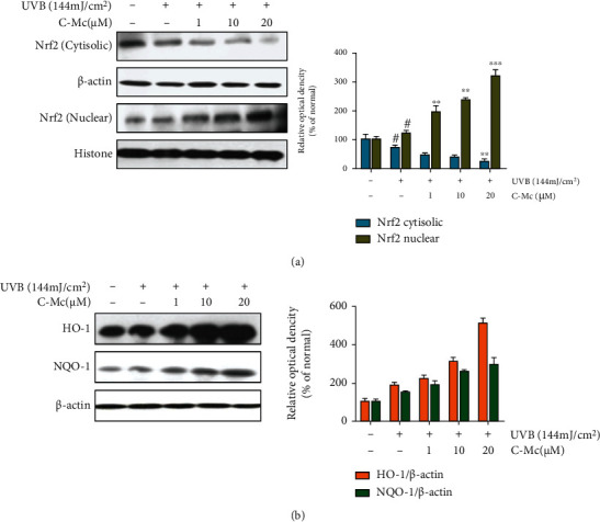
Long-term exposure to ultraviolet light induces photoaging and may eventually increase the risk of skin carcinogenesis. Rare minor ginsenosides isolating from traditional medicine Panax (ginseng) have shown biomedical efficacy as antioxidation and antiphotodamage agents. However, due to the difficulty of component extraction and wide variety of ginsenoside, the identification of active antiphotoaging ginsenoside remains a huge challenge. In this study, we proposed a novel in silico approach to identify potential compound against photoaging from 82 ginsenosides. Specifically, we calculated the shortest distance between unknown and known antiphotoaging ginsenoside set in the chemical space and applied chemical structure similarity assessment, drug-likeness screening, and ADMET evaluation for the candidates. We highlighted three rare minor ginsenosides (C-Mc, Mx, and F2) that possess high potential as antiphotoaging agents. Among them, C-Mc deriving from American ginseng (Panax quinquefolius L.) was validated by wet-lab experimental assays and showed significant antioxidant and cytoprotective activity against UVB-induced photodamage in human dermal fibroblasts. Furthermore, system pharmacology analysis was conducted to explore the therapeutic targets and molecular mechanisms through integrating global drug-target network, high quality photoaging-related gene profile from multiomics data, and skin tissue-specific expression protein network. In combination with in vitro assays, we found that C-Mc suppressed MMP production through regulating the MAPK/AP-1/NF-κB pathway and expedited collagen synthesis via the TGF-β/Smad pathway, as well as enhanced the expression of Nrf2/ARE to hold a balance of endogenous oxidation. Overall, this study offers an effective drug discovery framework combining in silico prediction and in vitro validation, uncovering that ginsenoside C-Mc has potential antiphotoaging properties and might be a novel natural agent for use in oral drug, skincare products, or functional food.


Conflict of interest statement

The authors declare that there is no conflict of interest regarding the publication of this paper.

Figures

Figure 1
Figure 1
Schematic diagram illustrating in silico methodology and in vitro validation for identification of active ginsenosides against photoaging. (a) In silico identification of antiphotoaging candidates from 82 ginsenosides. (b) In vitro validation of antiphotoaging effects for the promising candidate ginsenoside C-Mc. (c) System pharmacology-based exploration of the potential therapeutic targets, biological process, signal pathway, and regulatory function. (d) In vitro assay for elucidating the mechanism actions of ginsenoside C-Mc against photoaging.
Figure 2
Figure 2
Preliminary identification of potential antiphotoaging ginsenosides through shortest distance measurement among compounds in the PCA space. (a) Calculation of the rational boundary constraint of the space of PCA. (b) Eliminating the improper compounds outside the boundary constraint of the space. (c) Shortest distance calculation between known and unknown antiphotoaging ginsenosides.
Figure 3
Figure 3
Pairwise chemical structure similarity analysis between known and unknown antiphotoaging ginsenosides. (a) Heatmap illustrating the similarity analysis results of the 16 known active ginsenosides and the 22 unknown ginsenosides. (b) Chemical structures of the top 10 ginsenosides with average similarity index higher than 0.7.
Figure 4
Figure 4
Free radical scavenging ability and cell viability of ginsenoside C-Mc. (a) Compound structure and physicochemical properties of ginsenoside C-Mc. The proper range of the chemical physicochemical properties is given by ADMETlab 2.0 [34]. (b–d) ROS production. (e) Cell viability. (f) LDH release. (g) GSH secretion. NHDFs were irradiated or nonirradiated with 144 mJ/cm2 UVB, followed by treatment with the indicated of ginsenoside C-Mc (1, 10, and 20 μM). All data are shown as the mean ± SD of three independent experiments. # and ∗ indicate significant differences from the nonirradiated control and UVB-irradiated control groups. #p < 0.05, ##p < 0.01, and ###p < 0.001 contrast with the nonirradiated control. p < 0.05, ∗∗p < 0.01, and ∗∗∗p < 0.001 contrast with the UVB-irradiated control. p < 0.05, ∗∗p < 0.01, and ∗∗∗p < 0.001.
Figure 5
Figure 5
Global drug-target network of ginsenoside C-Mc interacted with photoaging-related gene network and skin tissue-specific expression protein network. Gene symbol names of the overlapped targets of C-Mc are displayed. The detailed information of the networks is provided in Supplementary Table S3-5.
Figure 6
Figure 6
Gene enrichment analysis of the potential targets of ginsenoside C-Mc overlapped with photoaging and skin-specific genes. (a) Gene Ontology (GO) biological process enrichment and WikiPathway annotation; (b) Sankey diagram illustrating the relationship among the photoaging-relevant enriched pathways/processes, corresponding proteins, and biological function of C-Mc against photoaging.
Figure 7
Figure 7
C-Mc prevented secretion of MMP-1 and MMP-3 and inhibited MAPK/AP-1 signaling pathway in UVB-irradiated NHDFs. Production of (a) MMP-1 and (b) MMP-3 under non-UVB irradiation and UVB-irradiated conditions; (c) MMP-1 and (d) MMP-3 mRNA expression. An equimolar quantity of mRNA was quantified compared to GAPDH. Cells were incubated in absence or presence of ginsenoside C-Mc at the present concentration after exposure to UVB radiation (144 mJ/cm2). (e) The protein levels of p38, ERK, and JNK in UVB-irradiated NHDFs measured by Western blot analysis. NHDFs were irradiated or nonirradiated with UVB, followed by treated with ginsenoside C-Mc for 1 h (MAPK). The signal intensities for phosphorylation levels of p38, ERK, and JNK. (f) The protein levels of c-Fos and c-Jun in UVB-irradiated NHDFs measured by Western blot analysis. NHDFs were irradiated or nonirradiated with UVB, followed by treatment with C-Mc for 4 h (AP-1). The signal intensities for phosphorylation levels of c-Fos and c-Jun. Values shown are the mean ± SD. # and ∗ indicate significant differences from the nonirradiated control and UVB-irradiated control groups. #p < 0.05, ##p < 0.01, and ###p < 0.001 contrast with the nonirradiated control. p < 0.05, ∗∗p < 0.01, and ∗∗∗p < 0.001 contrast with the UVB-irradiated control.
Figure 8
Figure 8
Ginsenoside C-Mc inhibited UVB-induced inflammatory cytokine secretion and alleviated UVB-induced NF-κB/IκB-α expression. Production of (a) IL-6 and (b) VEGF under non-UVB irradiation and UVB-irradiated conditions; (c) IL-6, (d) iNOS, and (e) TNF-α mRNA expression. An equimolar quantity of mRNA was quantified compared to GAPDH. Cells were incubated in absence or presence of ginsenoside C-Mc at the present concentration after exposure to UVB radiation (144 mJ/cm2). (f) Molecular docking simulation for C-Mc binding to NF-κB. (g) The protein levels of NF-κB and IκB-α detected by Western blot. Values shown are the mean ± SD. # and ∗ indicate significant differences from the nonirradiated control and UVB-irradiated control groups. #p < 0.05, ##p < 0.01, and ###p < 0.001 contrast with the nonirradiated control. p < 0.05, ∗∗p < 0.01, and ∗∗∗p < 0.001 contrast with the UVB-irradiated control.
Figure 9
Figure 9
C-Mc increased in TGF-β1 and procollagen type I secretion and regulated TGF-β/Smad pathway in UVB-irradiated NHDFs. Production of (a) procollagen type I and (b) TGF-β1 under non-UVB irradiation and UVB-irradiated conditions. (c) Procollagen type I mRNA expression. An equimolar quantity of mRNA was quantified compared to GAPDH. Cells were incubated in absence or presence of ginsenoside C-Mc at the present concentration after exposure to UVB radiation (144 mJ/cm2). (d) The protein levels of Smad2/3, Smad7, and TGF-β1 in UVB-irradiated NHDFs were measured by Western blot analysis. The NHDFs were irradiated or nonirradiated with UVB, followed by treated with ginsenoside C-Mc for 1.5 h. The signal intensities for phosphorylation levels of Smad2/3, Smad7, and TGF-β1 were tested by Western blotting. Values shown are the mean ± SD. # and ∗ indicate significant differences from the nonirradiated control and UVB-irradiated control groups. #p < 0.05, ##p < 0.01, and ###p < 0.001 contrast with the nonirradiated control. p < 0.05, ∗∗p < 0.01, and ∗∗∗p < 0.001 contrast with the UVB-irradiated control.
Figure 10
Figure 10
Effects of ginsenoside C-Mc on Nrf2, HO-1, and NQO-1 expression in UVB-irradiated NHDFs. (a) The protein levels of Nrf2 in UVB-irradiated NHDFs were measured by Western blot analysis. The signal intensities for phosphorylation levels of Nrf2. The protein levels of Nrf2 in UVB-irradiated NHDFs were measured by Western blot analysis. (b) The signal intensities for phosphorylation levels of HO-1 and NQO-1. The NHDFs were irradiated or nonirradiated with UVB, followed by treated with ginsenoside C-Mc for 3 h. The results were shown as the mean ± SD of at least three independent experiments. #p < 0.05, ##p < 0.01, and ###p < 0.001 contrast with the nonirradiated control. p < 0.05, ∗∗p < 0.01, and ∗∗∗p < 0.001 contrast with the UVB-irradiated control.
Figure 11
Figure 11
Schematic summary illustrating amelioration of ginsenoside C-Mc on photoaging in UVB-irradiated NHDFs via modulating oxidation stress, inflammation, matrix metalloproteinase secretion, collagen degradation, and synthesis.

References

    1. Han A., Chien A. L., Kang S. J. Photoaging. Photoaging. Dermatologic clinics . 2014;32(3):291–299. doi: 10.1016/j.det.2014.03.015. - DOI - PubMed
    1. Cavinato M., Jansen-Dürr P. Molecular mechanisms of UVB-induced senescence of dermal fibroblasts and its relevance for photoaging of the human skin. Experimental Gerontology . 2017;94:78–82. doi: 10.1016/j.exger.2017.01.009. - DOI - PubMed
    1. Weihermann A. C., Lorencini M., Brohem C. A., De Carvalho C. M. Elastin structure and its involvement in skin photoageing. International journal of cosmetic science . 2017;39(3):241–247. doi: 10.1111/ics.12372. - DOI - PubMed
    1. Kammeyer A., Luiten R. M. Oxidation events and skin aging. Ageing research reviews . 2015;21 - PubMed
    1. Cheong Y., Kim C., Kim M. B., Hwang J. K. The anti-photoaging and moisturizing effects of Bouea macrophylla extract in UVB-irradiated hairless mice. Food Science and Biotechnology . 2018;27(1):147–157. doi: 10.1007/s10068-017-0276-y. - DOI - PMC - PubMed