. 2022 Mar 8:27:1225-1234.
doi: 10.1016/j.omtn.2022.02.008. Epub 2022 Feb 13.

RIG-I-induced innate antiviral immunity protects mice from lethal SARS-CoV-2 infection

Affiliations

RIG-I-induced innate antiviral immunity protects mice from lethal SARS-CoV-2 infection

Samira Marx et al. Mol Ther Nucleic Acids. .

Abstract

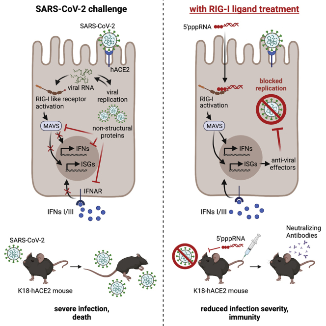
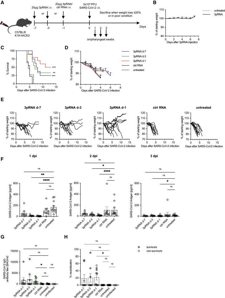
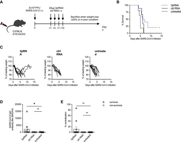
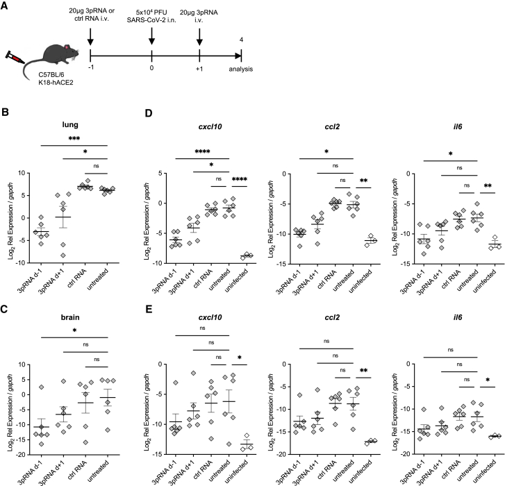
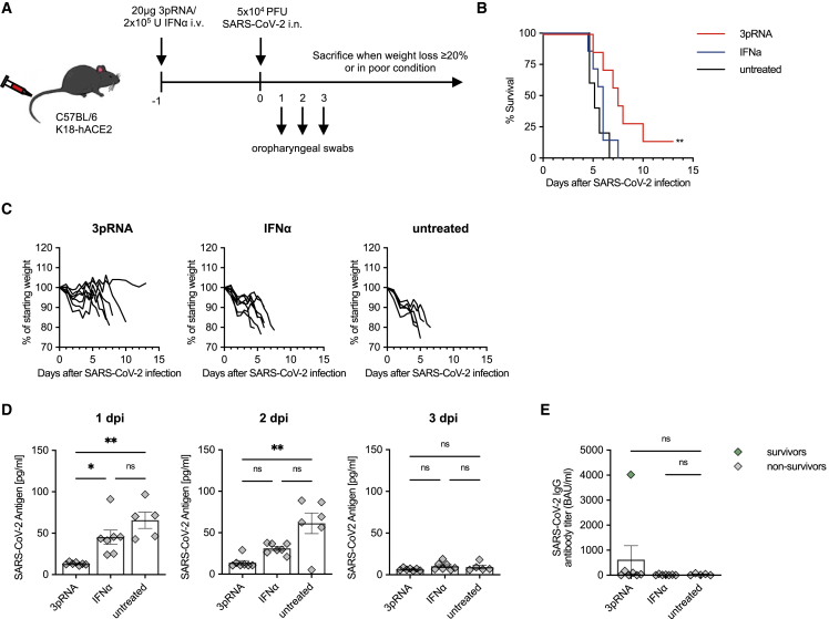
The SARS-CoV-2 pandemic has underscored the need for rapidly usable prophylactic and antiviral treatments against emerging viruses. The targeted stimulation of antiviral innate immune receptors can trigger a broad antiviral response that also acts against new, unknown viruses. Here, we used the K18-hACE2 mouse model of COVID-19 to examine whether activation of the antiviral RNA receptor RIG-I protects mice from lethal SARS-CoV-2 infection and reduces disease severity. We found that prophylactic, systemic treatment of mice with the specific RIG-I ligand 3pRNA, but not type I interferon, 1-7 days before viral challenge, improved survival of mice by up to 50%. Survival was also improved with therapeutic 3pRNA treatment starting 1 day after viral challenge. This improved outcome was associated with lower viral load in oropharyngeal swabs and in the lungs and brains of 3pRNA-treated mice. Moreover, 3pRNA-treated mice exhibited reduced lung inflammation and developed a SARS-CoV-2-specific neutralizing antibody response. These results demonstrate that systemic RIG-I activation by therapeutic RNA oligonucleotide agonists is a promising strategy to convey effective, short-term antiviral protection against SARS-CoV-2 infection, and it has great potential as a broad-spectrum approach to constrain the spread of newly emerging viruses until virus-specific therapies and vaccines become available.

Keywords: COVID-19; K18-hACE2 mouse model; MT: Oligonucleotides: Therapies and Applications; RIG-I; SARS-CoV-2; antiviral immunity; coronavirus; emerging viruses; innate immunity; nucleic acid immunity; pandemic; type I IFN.


Conflict of interest statement

M.S. and G.H. are inventors on a patent on RIG-I ligands.

Figures

None
Graphical abstract
Figure 1
Figure 1
Prophylactic RIG-I stimulation protects mice from lethal SARS-CoV-2 infection (A) Experimental setup. K18-hACE2wt/tg mice were i.v. injected with 20 μg 3pRNA or control RNA complexed to in vivo jetPEI on the indicated days. On day 0 (d-0), mice were infected intranasally with 5 × 104 PFU SARS-CoV-2 virus. Oropharyngeal swabs were obtained on d-1 to d-3 post-infection (dpi). Disease development and survival were monitored up to twice daily until 13 dpi. (B) Weight development of 3pRNA-treated animals. Plotted are the means ± SEMs of 2 independent experiments (3pRNA n = 8, untreated n = 11). (C–E) Kaplan-Meier curve (C) and (D) weight loss (pooled) of SARS-CoV-2-infected animals. (E) Individual weight loss over time of each SARS-CoV-2 infected mouse until reaching the endpoint criteria or 13 dpi. (F) SARS-CoV-2 antigen ELISA of oropharyngeal swab material on 1 dpi to 3 dpi. (G) Quantification of anti-SARS-CoV-2-specific IgG antibody titers in sera of SARS-CoV-2-infected animals collected at their individual time of death or 13 dpi. (H) Percentage of inhibition of SARS-CoV-2 S1/RBD-hACE2 interaction by sera of SARS-CoV-2-infected animals collected at their individual time of death or 13 dpi. (C, D, F–H) Plotted are the means ± SEMs (3pRNA d-7 n = 8, 3pRNA d-3 n = 9, 3pRNA d-1 n = 10, ctrl RNA d-1 n = 9, untreated n = 11). The data are pooled from 2 independent experiments. The statistical significance was calculated by log rank Mantel-Cox test (B) and the non-parametric Kruskal-Wallis test with Dunn’s multiple testing (F and G). ∗p < 0.05, ∗∗p < 0.01, ∗∗∗p < 0.001, ∗∗∗∗p < 0.0001.
Figure 2
Figure 2
Therapeutic intervention with RIG-I ligand confers an intermediate level of protection against lethal SARS-CoV-2 infection in mice (A) Experimental setup. K18-hACE2wt/tg mice were infected intranasally with 5 × 104 PFU SARS-CoV-2. One day post-infection, mice were i.v. injected with 20 μg 3pRNA or control RNA complexed to in vivo jetPEI. Injection of 3pRNA or control RNA was repeated on 4, 7, and 10 dpi. Disease development and survival were monitored up to twice daily until 13 dpi. (B and C) Kaplan-Meier curve of SARS-CoV-2-infected animals (B). (C) Individual weight loss development of each mouse until they reached the endpoint criteria or 13 dpi. (D) Quantification of anti-SARS-CoV-2-specific IgG antibody titers in the sera of SARS-CoV-2-infected animals collected on their individual time point of death or 13 dpi. (E) Percentage of inhibition of SARS-CoV-2 S1/RBD-hACE2 interaction by the sera of SARS-CoV-2-infected animals collected on their individual time point of death or 13 dpi. Plotted are the means ± SEMs (3pRNA n = 10, ctrl RNA n = 9, untreated n = 11). The data are pooled from 2 independent experiments. The statistical significance was calculated by log rank Mantel-Cox test (B) and non-parametric Kruskal-Wallis test with Dunn’s multiple testing (D and E). ∗p < 0.05, ∗∗p < 0.01, ∗∗∗p < 0.001, ∗∗∗∗p < 0.0001.
Figure 3
Figure 3
Reduced viral burden and inflammation in SARS-CoV-2-infected lungs upon RIG-I ligand treatment (A) Experimental setup. K18-hACE2wt/tg mice were i.v. injected with 20 μg 3pRNA or control RNA complexed to in vivo jetPEI 1 day before (3pRNA d-1) or after (3pRNA d+1) infection. Mice were infected intranasally with 5 × 104 PFU SARS-CoV-2 and sacrificed 4 dpi. (B and C) SARS-CoV-2 viral burden in the lungs (B) and in the brain (C) at 4 dpi. (D and E) Cytokine expression in the lungs (D) and brain (E) at 4 dpi. For (B)–(E), expression was quantified by qPCR relative to murine gapdh expression. Plotted are the means ± SEMs (n = 6, uninfected n = 3). The statistical significance was calculated by 1-way ANOVA (Welch) with Dunnett’s T3 multiple testing, when the data were lognormally distributed; otherwise, a non-parametric Kruskal-Wallis test with Dunn’s multiple testing was applied. ∗p < 0.05, ∗∗p < 0.01, ∗∗∗p < 0.001, ∗∗∗∗p < 0.0001.
Figure 4
Figure 4
RIG-I stimulation is superior to type I interferon (IFN) at protecting mice from lethal SARS-CoV-2 infections (A) Experimental setup. K18-hACE2wt/tg mice were i.v. injected with 20 μg 3pRNA complexed to in vivo jetPEI or 2 × 105 U recombinant universal IFN-α (A/D) 1 day before infection. Mice were infected intranasally with 5 × 104 PFU SARS-CoV-2. Oropharyngeal swabs were obtained on 1 to 3 dpi. Disease development and survival were monitored up to twice daily until 13 dpi. (B) Kaplan-Meier curve of SARS-CoV-2-infected animals. (C) Individual weight loss development of each mouse until its individual time of death or 13 dpi. (D) SARS-CoV-2 antigen ELISA in oropharyngeal swab material on 1 to 3 dpi. (E) Quantification of anti-SARS-CoV-2-specific IgG antibody titers in the sera of SARS-CoV-2-infected animals. Plotted are the means ± SEMs (3pRNA and IFN-α n = 7, untreated n = 5). The statistical significance was calculated by log rank Mantel-Cox test (B) and non-parametric Kruskal-Wallis test with Dunn’s multiple testing (D and E). ∗p < 0.05, ∗∗p < 0.01, ∗∗∗p < 0.001, ∗∗∗∗p < 0.0001.

References

    1. Zhou P., Yang X.-L., Wang X.-G., Hu B., Zhang L., Zhang W., Si H.-R., Zhu Y., Li B., Huang C.-L., et al. A pneumonia outbreak associated with a new coronavirus of probable bat origin. Nature. 2020;579:270–273. - PMC - PubMed
    1. Zhu N., Zhang D., Wang W., Li X., Yang B., Song J., Zhao X., Huang B., Shi W., Lu R., et al. A novel coronavirus from patients with pneumonia in China, 2019. N. Engl. J. Med. 2020;382:727–733. - PMC - PubMed
    1. Chen X., Laurent S., Onur O.A., Kleineberg N.N., Fink G.R., Schweitzer F., Warnke C. A systematic review of neurological symptoms and complications of COVID-19. J. Neurol. 2021;268:392–402. - PMC - PubMed
    1. Gupta A., Madhavan M.V., Sehgal K., Nair N., Mahajan S., Sehrawat T.S., Bikdeli B., Ahluwalia N., Ausiello J.C., Wan E.Y., et al. Extrapulmonary manifestations of COVID-19. Nat. Med. 2020;26:1017–1032. - PMC - PubMed
    1. Alwan N.A. The road to addressing Long Covid. Science. 2021;373:491–493. - PubMed

LinkOut - more resources