. 2022 Feb 3:8:768419.
doi: 10.3389/fcvm.2021.768419. eCollection 2021.

EP-PINNs: Cardiac Electrophysiology Characterisation Using Physics-Informed Neural Networks

Affiliations

EP-PINNs: Cardiac Electrophysiology Characterisation Using Physics-Informed Neural Networks

Clara Herrero Martin et al. Front Cardiovasc Med. .

Abstract

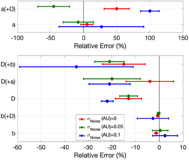
Accurately inferring underlying electrophysiological (EP) tissue properties from action potential recordings is expected to be clinically useful in the diagnosis and treatment of arrhythmias such as atrial fibrillation. It is, however, notoriously difficult to perform. We present EP-PINNs (Physics Informed Neural Networks), a novel tool for accurate action potential simulation and EP parameter estimation from sparse amounts of EP data. We demonstrate, using 1D and 2D in silico data, how EP-PINNs are able to reconstruct the spatio-temporal evolution of action potentials, whilst predicting parameters related to action potential duration (APD), excitability and diffusion coefficients. EP-PINNs are additionally able to identify heterogeneities in EP properties, making them potentially useful for the detection of fibrosis and other localised pathology linked to arrhythmias. Finally, we show EP-PINNs effectiveness on biological in vitro preparations, by characterising the effect of anti-arrhythmic drugs on APD using optical mapping data. EP-PINNs are a promising clinical tool for the characterisation and potential treatment guidance of arrhythmias.

Keywords: Physics Informed Neural Network (PINN); arrhythmia (any); artificial intelligence; atrial fibrillation; biophysical modelling; cardiac electrophysiology; optical mapping; parameter estimation.


Conflict of interest statement

The authors declare that the research was conducted in the absence of any commercial or financial relationships that could be construed as a potential conflict of interest.

Figures

Figure 1
Figure 1
Numerical solutions to the Aliev-Panfilov monodomain system for: (a) Planar wave. (b) Centrifugal wave. (c) Spiral wave. (d) Centrifugal wave in the presence of a square heterogeneity in D. (e) Spiral wave in the presence of a square heterogeneity in D.
Figure 2
Figure 2
Network architectures and training schemes for EP-PINNs used in this study. Neurons are represented by σ. In forward simulations only network parameters θ were estimated, whereas in the inverse setting one or more EP parameters (λ) were also calculated. (A) Architecture used in all forward simulations and all inverse simulations (1D and 2D) with homogeneous EP parameters. (B) Architecture used in all 2D forward simulations and all inverse simulations where D was a spatially-varying field. (C) Used training schemes. 1 was used in in silico 1D problems, whereas training scheme 2 was used in 2D problems and for optical mapping experimental data. The number of NN neurons and layers varied across different experiments, as did the learning rates (lr) and number of iterations (iter) in training scheme 2. Further details about the architecture and parameterisation of the NNs and training schemes can be found in Supplementary Table 2.
Figure 3
Figure 3
Effect of the number of sampled experimental points (A) and experimental noise (B) on EP-PINNs V estimates. The error in estimation is measured using the root mean square error (RMSE, see Equation 7) in forward mode in 1D. Representative V(t) plots sampled at a random spatial location are shown as insets for some of the probed conditions.
Figure 4
Figure 4
EP-PINNs 2D forward solutions to the Aliev-Panfilov monodomain system for the same conditions as the GT data depicted in Figure 1. (a) Planar wave. (b) Centrifugal wave. (c) Spiral wave. (d) Centrifugal wave in the presence of a square heterogeneity in D. (e) Spiral wave in the presence of a square heterogeneity in D.
Figure 5
Figure 5
Error in global EP parameter estimates by EP-PINNs in the inverse setting in 1D in the presence of different amounts of experimental noise. We show the relative error for a, b and D, when estimated separately and in pairs.
Figure 6
Figure 6
1D inverse EP-PINNs solution for a detailed canine left atrial model in the conditions of: (A) no remodelling. (B) moderate AF remodelling. (C) severe AF remodelling. Representative V(t) plots are shown throughout, accompanied by the models 90% APD and EP-PINNS estimates for b, a parameter inversely proportional to APD.
Figure 7
Figure 7
EP-PINNs inverse solution in homogeneous conditions in 2D. (a) Relative error for global estimates of: a and D and b and D, when estimated separately or simultaneously. (b–d) Corresponding representative V maps for: planar wave (b), centrifugal wave (c), and spiral wave (d). Compare (b–d) to the corresponding GT in Figures 1a-c and the forward solutions in Figures 4a–c.
Figure 8
Figure 8
EP-PINNs 2D inverse solution in the presence of heterogeneities in D. Maps showing representative V and D estimates for: (a,c) centrifugal wave and (b,d) spiral wave. (e) Error for global estimates of a and b and RMSE for estimates of D across the 2D domain, for all estimated parameter combinations. Compare (a,b) to the corresponding GT in Figures 1d,e and the forward solutions in Figures 4d,e.
Figure 9
Figure 9
1D inverse EP-PINNs solution for experimental optical mapping data, in the presence of (A) ICaL channel blocker nifedipine and (B) IKr channel blocker E-4031. (Δb refers to the change in model parameter b in the presence of the drug when compared to the baseline value. Each AP was acquired in separate datasets and juxtaposed in the figure to allow visual comparisons.

Comment in

References

    1. Hindricks G, Potpara T, Dagres N, Arbelo E, Bax JJ, Blomström-Lundqvist C, et al. . 2020. ESC Guidelines for the diagnosis and management of atrial fibrillation developed in collaboration with the European Association for Cardio-Thoracic Surgery (EACTS). Eur Heart J. (2020). 42:373–498. 10.1093/eurheartj/ehaa945 - DOI - PubMed
    1. Ganesan AN, Shipp NJ, Brooks AG, Kuklik P, Lau DH, Lim H, et al. . Long-term outcomes of catheter ablation of atrial fibrillation: a systematic review and meta-analysis. J Am Heart Assoc. (2013) 2:e004549. 10.1161/JAHA.112.004549 - DOI - PMC - PubMed
    1. Nattel S, Dobrev D. Controversies about atrial fibrillation mechanisms. Circ Res. (2017) 120:1396–9. 10.1161/CIRCRESAHA.116.310489 - DOI - PubMed
    1. Calkins H, Kuck KH, Cappato R, Brugada J, John Camm A, Chen SA, et al. . 2012 HRS/EHRA/ECAS expert consensus statement on catheter and surgical ablation of atrial fibrillation. J Intervent Cardiac Electrophysiol. (2012). 14:171–257. - PubMed
    1. Clayton RH, Bernus O, Cherry E, Dierckx H, Fenton F, Mirabella L, et al. . Models of cardiac tissue electrophysiology: progress, challenges and open questions. Prog Biophys Mol Biol. (2011) 104:22–48. 10.1016/j.pbiomolbio.2010.05.008 - DOI - PubMed