Review
. 2022 Jan 25;8(1):61-70.
eCollection 2022 Feb 25.

Hypoxia-preconditioning of human adipose-derived stem cells enhances cellular proliferation and angiogenesis: A systematic review

Affiliations
Review

Hypoxia-preconditioning of human adipose-derived stem cells enhances cellular proliferation and angiogenesis: A systematic review

John P Garcia et al. J Clin Transl Res. .

Abstract

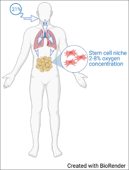
Background: Human adipose-derived stem cells (hADSCs) have gained attention lately because of their ease of harvesting and ability to be substantially multiplied in laboratory cultures. Stem cells are usually cultured under atmospheric conditions; however, preconditioning stem cells under hypoxic conditions seems beneficial.

Aim: This systematic review aims to investigate the effect of hypoxia preconditioning and its impact on the proliferation and angiogenic capacity of the hADSCs.

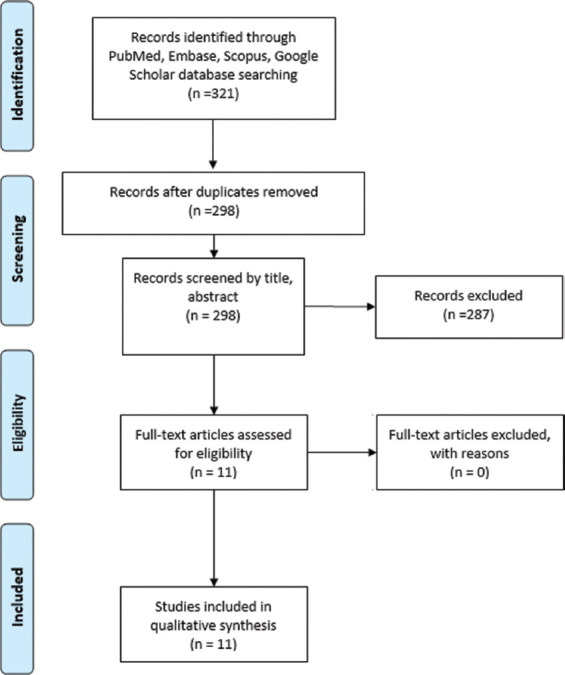
Methods: We performed a systematic review by searching PubMed, Scopus, Embase, and Google Scholar databases from all years through March 22, 2021, following Preferred Reporting Items for Systematic Reviews and Meta-Analyses guidelines. Medical Subject Headings terms "adipose-derived stem cell," "Hypoxia," "cell proliferation," and "angiogenesis" guided our search. Only articles written in English using experimental models comparing a preconditioned group against a control group of hADSCs with data on proliferation and angiogenic capacity were included.

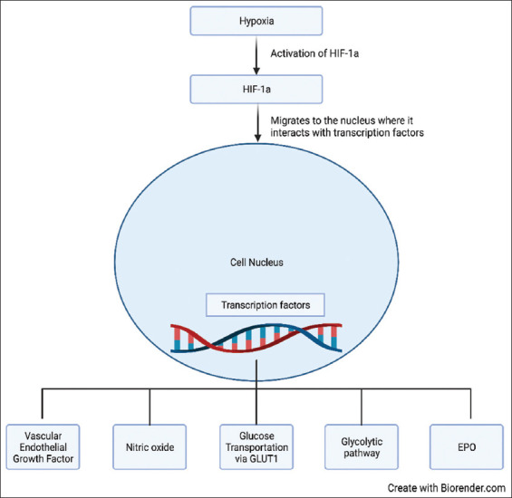
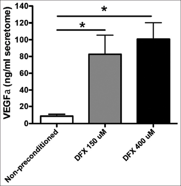
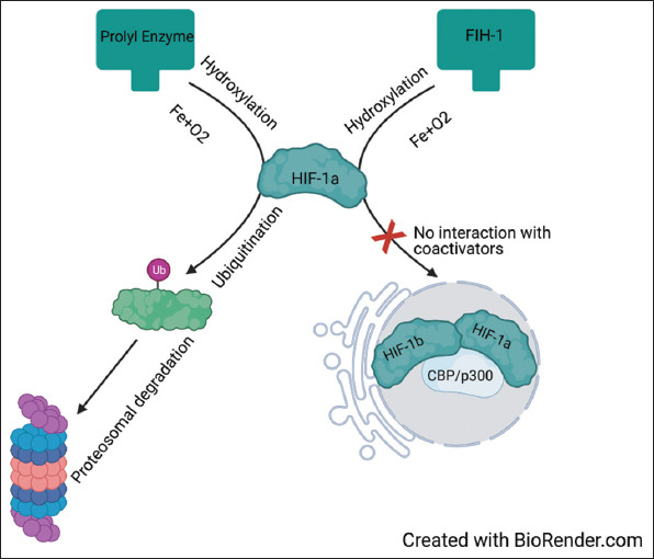
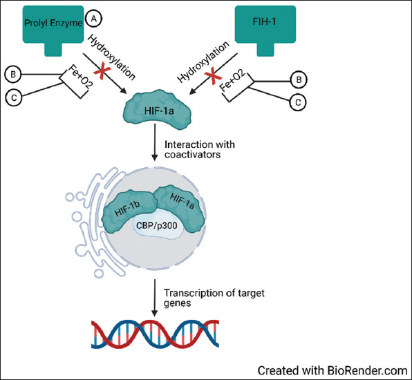
Results: Our search yielded a total of 321 articles. 11 articles met our inclusion criteria and were ultimately included in this review. Two studies induced hypoxia using hypoxia-inducible factor-1 alpha stabilizing agents, while nine reached hypoxia by changing oxygen tension conditions around the cells. Four articles conducted in-vivo studies to correlate their in-vitro findings, which proved to be consistent. Although 1 article indicated cell proliferation inhibition with hypoxia preconditioning, the remaining 10 found enhanced proliferation in preconditioned groups compared to controls. All articles showed an enhanced angiogenic capacity of hADSCs after hypoxia preconditioning.

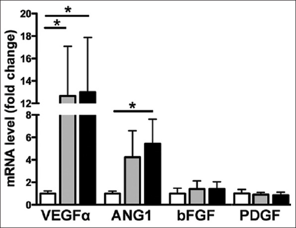
Conclusion: In this review, we found evidence to support hypoxia preconditioning of hADSCs before implantation. Benefits include enhanced cell proliferation with a faster population doubling rate and increased secretion of multiple angiogenic growth factors, enhancing angiogenesis capacity.

Relevance for patients: Although regenerative therapy is a promising field of study and treatment in medicine, much is still unknown. The potential for angiogenic therapeutics with stem cells is high, but more so, if we discover ways to enhance their natural angiogenic properties. Procedures and pathologies alike require the assistance of angiogenic treatments to improve outcome, such is the case with skin grafts, muscle flaps, skin flaps, or myocardial infarction to mention a few. Enhanced angiogenic properties of stem cells may pave the way for better outcomes and results for patients.

Keywords: adipose-derived stem cells; angiogenesis; cell hypoxia; cell proliferation; growth factors; human stem cells; regenerative medicine.


Conflict of interest statement

The authors declare no conflict of interest

Figures

Figure 1
Figure 1. Oxygen concentration variability. Illustrates the difference in oxygen concentration in ambient air and as it reaches body tissues.
Figure 2
Figure 2. Hypoxia regulated genes. When a cell encounters hypoxic conditions, hypoxia inducible factor-1a is activated, this leads to its interaction with transcription factors within the nucleus which further leads to activation of downstream genes which help cells endure hypoxic conditions.
Figure 3
Figure 3. Study selection flow chart. Flow chart describing the study selection process according to Preferred Reporting Items for Systematic Reviews and Meta-Analyses guidelines.
Figure 4
Figure 4. Risk of bias graph created with RevMan 5.3 following the Risk of Bias In Non-randomized Studies of Interventions -I guidelines of the Cochrane Library. Green indicates a low risk of bias, yellow indicates an unclear risk of bias, and red indicates a high risk of bias.
Figure 5
Figure 5. Risk of bias summary created with RevMan 5.3 following the Risk of Bias In Non-randomized Studies of Interventions-I guidelines of the Cochrane Library. A low risk of bias is indicated by green color, yellow indicates an unclear risk of bias, and red indicates a high risk of bias.
Figure 6
Figure 6. Deferoxamine preconditioning increases the expression levels of pro-angiogenic factors. Total RNA was obtained from MSCs exposed to 150 μM DFX, 400 μMDFX, or the vehicle for 48 h and subjected to quantitative reverse transcriptase ± PCR analysis. White bars represent non-preconditioned MSCs, gray bars represent MSCs preconditioned with 150 μM DFX, and black bars represent MSCs preconditioned with 400μMDFX. Data are shown as mean ± SEM. n = 4 per experimental group (biological repeats). Experiments were repeated 3 times at the technical level. *P<0.05. MSCs: Mesenchymal stem cells; DFX: Deferoxamine; PCR: Polymerase chain reaction; VEGFa: Vascular endothelial growth factor-a; ANG-1: Angiopoietin-1; bFGF: Basic fibroblast growth factor; PDGF: Platelet-derived growth factor; SEM: Standard error of the mean.
Figure 7
Figure 7. Deferoxamine preconditioning increases the secretion of pro-angiogenic factors. The secretomes obtained from MSCs were exposed to 150 μMDFX, 400 μMDFX, or the vehicle for 48 h. Quantification of VEGFα in MSC secretomes is shown. Data is presented as mean ± SEM. N = 4 per experimental group (biological repeats). Experiments were repeated 3 times at the technical level. *P<0.05. MSC: Mesenchymal stem cells; DFX: Deferoxamine; VEGFa: Vascular endothelial growth factor-a; SEM: standard mean error.
Figure 8
Figure 8. HIF-1a degradation in normoxia. HIF-1a is primarily degraded through two pathways. The first pathway utilizes prolyl hydroxylase enzymes to ubiquitinate HIF-1a for proteasomal degradation. The second pathway acts by hydroxylation of asparagine residues by FIH-1, preventing HIF-1a from interacting with co-activators (HIF-1b and CBP/p300) inside the nucleus. HIF-1a, hypoxia-inducible factor-1 alpha; FIH, factor inhibiting HIF-1; HIF-1b, hypoxia-inducible factor-1; CBP, CREB-binding protein; p300, E1A binding protein p300.
Figure 9
Figure 9. HIF-1a activation by hypoxia and stabilizing agents. (A) DMOG is a prolyl hydroxylase enzyme inhibitor, thus preventing ubiquitination and proteasomal degradation of HIF-1a. (B) Oxygen and Fe are required for hydroxylation. However, hypoxia decreases oxygen and Fe availability for these reactions, reducing HIF-1a degradation. (C) Deferoxamine is a Fe chelator, thus decreasing Fe availability for hydroxylation, decreasing HIF-1a degradation. As HIF-1 is accumulating, it translocates into the nucleus, where it interacts with its co-activators (HIF-1b and CBP/p300), engaging in transcriptional activity of downstream genes. HIF-1a, hypoxia-inducible factor-1 alpha; CBP, CREB-binding protein; p300, E1A binding protein p300; Fe, iron; DMOG, dimethyloxalylglycine.

References

    1. Mazini L, Rochette L, Admou B, Amal S, Malka G. Hopes and Limits of Adipose-Derived Stem Cells (ADSCs) and Mesenchymal Stem Cells (MSCs) in Wound Healing. Int J Mol Sci. 2020;21:1306. - PMC - PubMed
    1. Hatzistergos KE, Quevedo H, Oskouei BN, Hu Q, Feigenbaum GS, Margitich IS, et al. Bone Marrow Mesenchymal Stem Cells Stimulate Cardiac Stem Cell Proliferation and Differentiation. Circ Res. 2010;107:913–22. - PMC - PubMed
    1. Ntege EH, Sunami H, Shimizu Y. Advances in Regenerative Therapy:A Review of the Literature and Future Directions. Regen Ther. 2020;14:136–53. - PMC - PubMed
    1. Caplan AI. Mesenchymal stem cells in regenerative medicine. In: Atala A, Lanza R, Mikos AG, Nerem R, editors. Principles of Regenerative Medicine. 3rd ed. Boston: Academic Press; 2019. pp. 219–27. Ch. 15.
    1. Zhao L, Johnson T, Liu D. Therapeutic angiogenesis of adipose-derived stem cells for ischemic diseases. Stem Cell Res Ther. 2017;8:125. - PMC - PubMed

LinkOut - more resources