

Mitophagy, Ferritinophagy and Ferroptosis in Retinal Pigment Epithelial Cells Under High Glucose Conditions: Implications for Diabetic Retinopathy and Age-Related Retinal Diseases
- PMID: 35187384
- PMCID: PMC8856657
Mitophagy, Ferritinophagy and Ferroptosis in Retinal Pigment Epithelial Cells Under High Glucose Conditions: Implications for Diabetic Retinopathy and Age-Related Retinal Diseases
Abstract
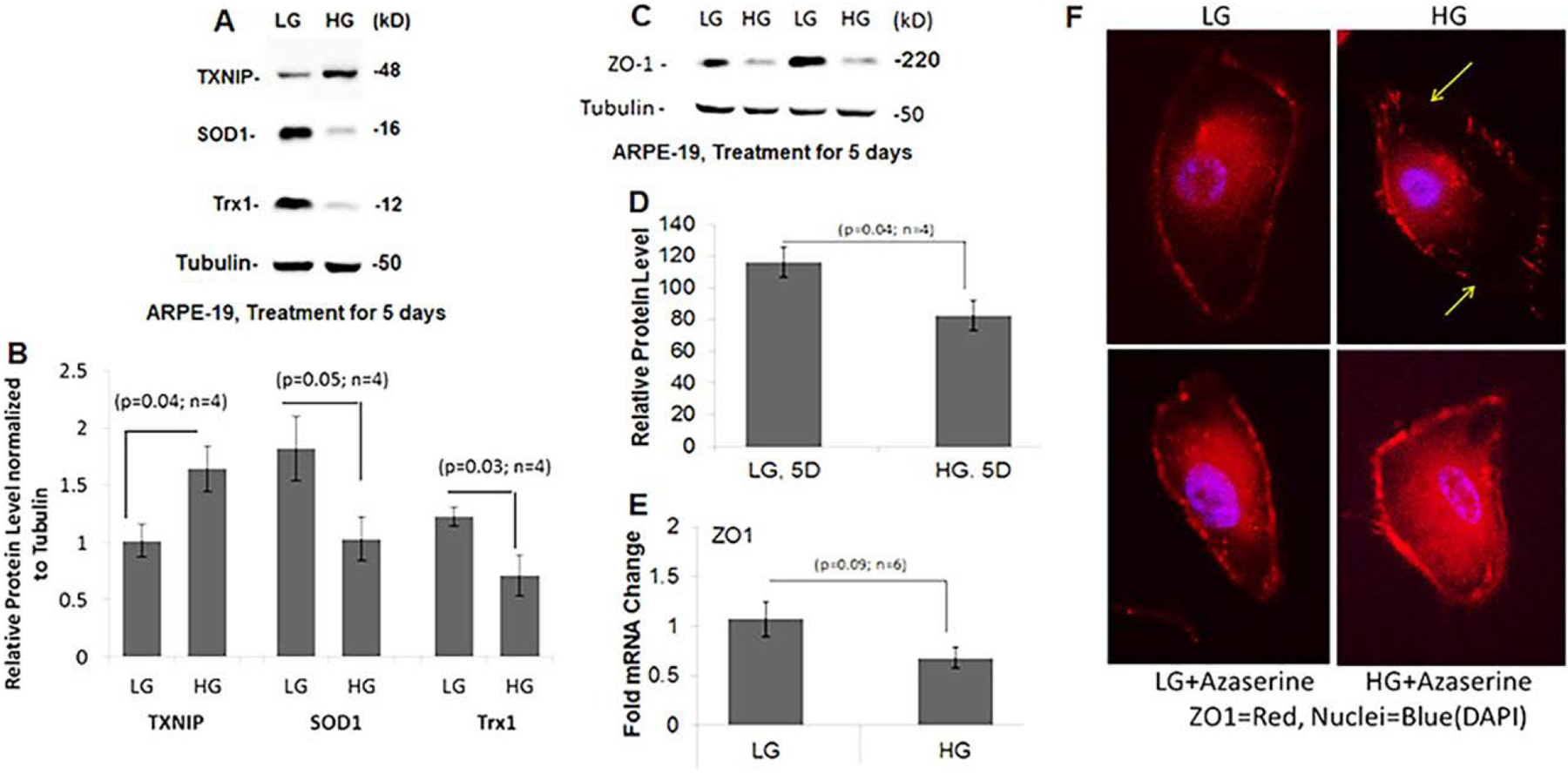
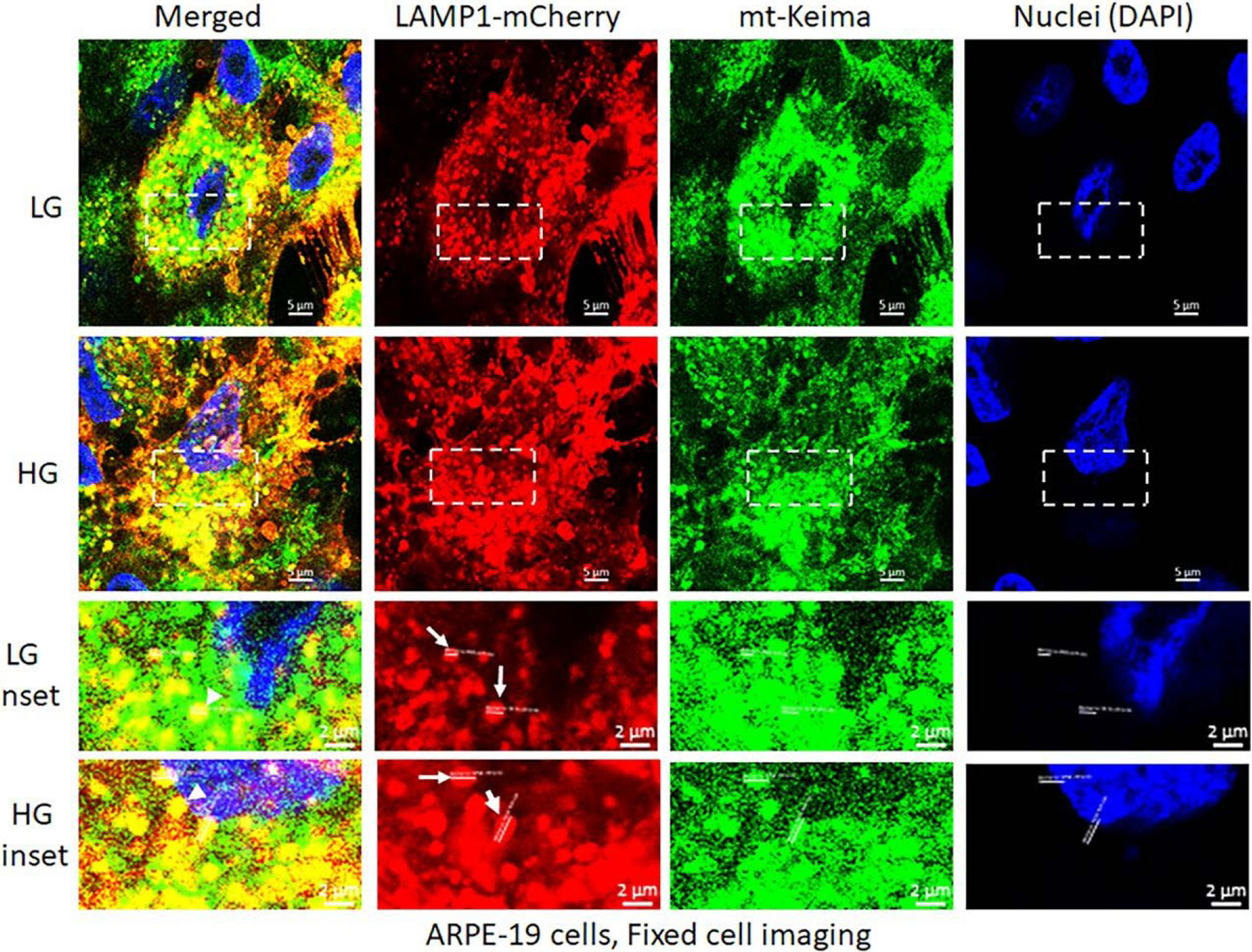
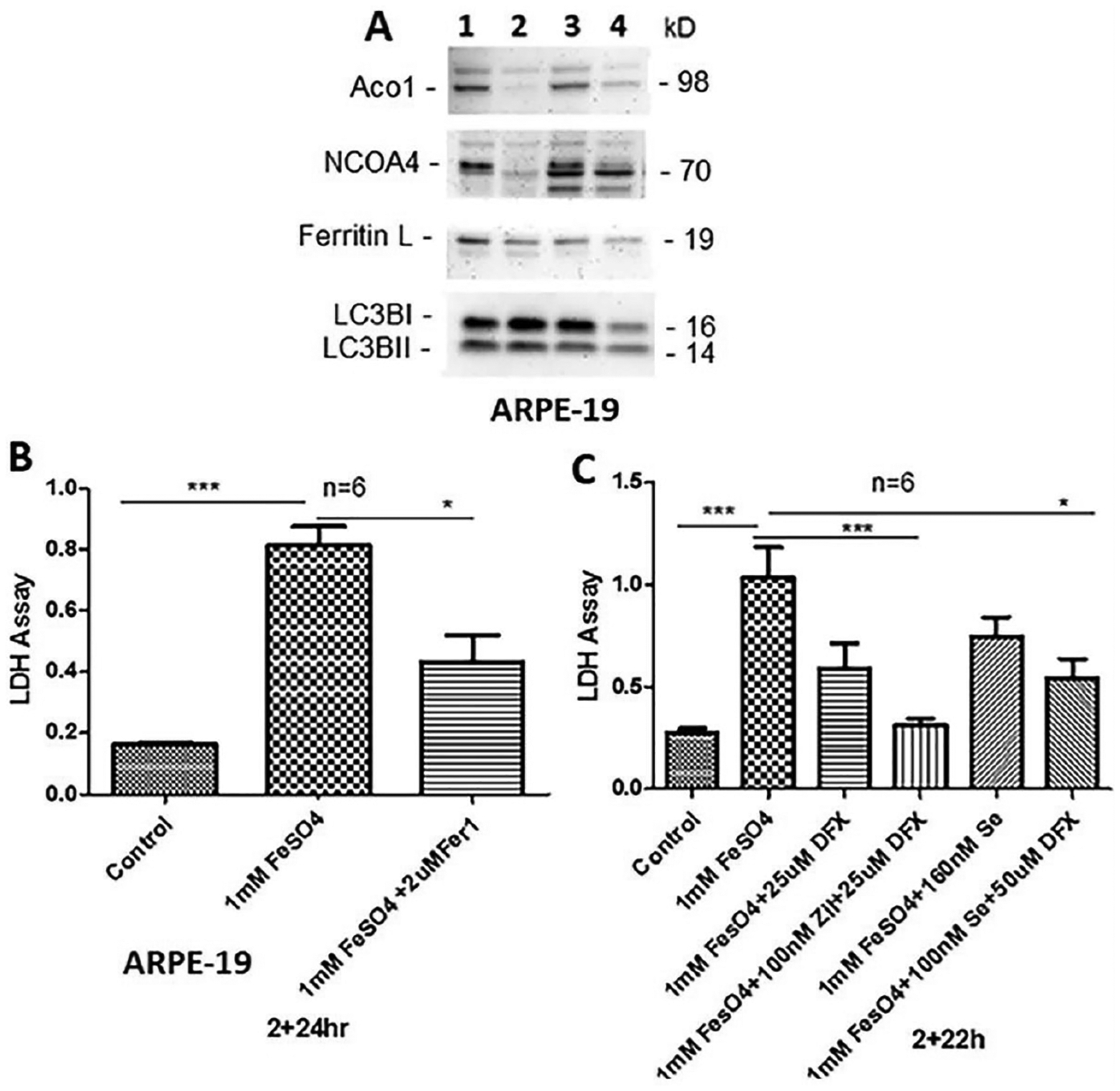
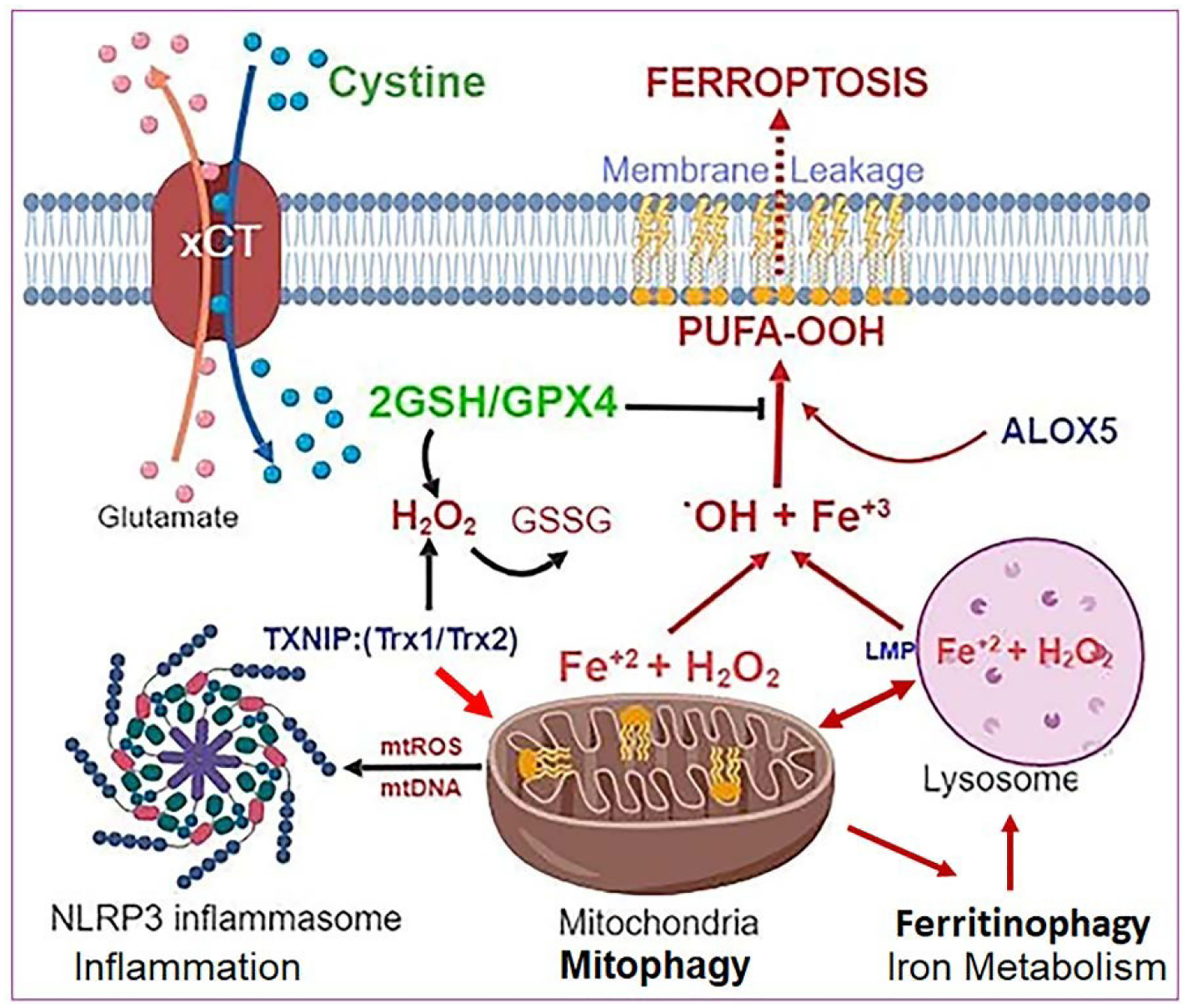
Diabetic retinopathy (DR) is a devastating disease leading to blindness among majority of working adults around the globe. Nonetheless, an effective treatment or cure for the disease is still to be achieved. This is because the cellular and molecular mechanisms of DR are complex and not fully understood yet. In this article, we describe how high glucose induced TXNIP upregulation and associated redox stress may cause mitochondrial dysfunction, mitophagy, ferritinophagy (iron release by autophagy) and lysosome destabilization. Labile irons react with hydrogen peroxide (H2O2) to generate hydroxyl radicals (.OH) by the Fenton reaction and cause membrane phospholipid peroxidation due to reduction in glutathione (GSH) level and glutathione peroxidase 4 (GPX4) activity, which cause ferroptosis, a recently identified non-apoptotic cell death mechanism. We used in this study a retinal pigment epithelial cell line, ARPE- 19 and exposed it to high glucose in in vitro cultures to highlight some of the intricacies of these cellular processes, which may be relevant to the pathogenesis of DR and age-related retinal neurodegenerative diseases, such as age-related macular degeneration, AMD.
Keywords: Diabetic retinopathy; Ferritinophagy; Ferroptosis; Mitophagy; Oxidative stress; RPE; TXNIP.
Figures
Similar articles
-
Auranofin Mediates Mitochondrial Dysregulation and Inflammatory Cell Death in Human Retinal Pigment Epithelial Cells: Implications of Retinal Neurodegenerative Diseases.Front Neurosci. 2019 Oct 10;13:1065. doi: 10.3389/fnins.2019.01065. eCollection 2019. Front Neurosci. 2019. PMID: 31649499 Free PMC article.
-
Astragaloside-IV alleviates high glucose-induced ferroptosis in retinal pigment epithelial cells by disrupting the expression of miR-138-5p/Sirt1/Nrf2.Bioengineered. 2022 Apr;13(4):8240-8254. doi: 10.1080/21655979.2022.2049471. Bioengineered. 2022. PMID: 35302431 Free PMC article.
-
Interferon-γ induces retinal pigment epithelial cell Ferroptosis by a JAK1-2/STAT1/SLC7A11 signaling pathway in Age-related Macular Degeneration.FEBS J. 2022 Apr;289(7):1968-1983. doi: 10.1111/febs.16272. Epub 2021 Nov 22. FEBS J. 2022. PMID: 34741776
-
Iron Accumulation and Lipid Peroxidation in the Aging Retina: Implication of Ferroptosis in Age-Related Macular Degeneration.Aging Dis. 2021 Apr 1;12(2):529-551. doi: 10.14336/AD.2020.0912. eCollection 2021 Apr. Aging Dis. 2021. PMID: 33815881 Free PMC article. Review.
-
Ferroptosis: An Energetic Villain of Age-Related Macular Degeneration.Biomedicines. 2025 Apr 17;13(4):986. doi: 10.3390/biomedicines13040986. Biomedicines. 2025. PMID: 40299661 Free PMC article. Review.
Cited by
-
Transcriptional patterns of human retinal pigment epithelial cells under protracted high glucose.Mol Biol Rep. 2024 Apr 4;51(1):477. doi: 10.1007/s11033-024-09479-5. Mol Biol Rep. 2024. PMID: 38573426
-
Mitophagy and Ferroptosis in Sepsis-Induced ALI/ARDS: Molecular Mechanisms, Interactions and Therapeutic Prospects of Medicinal Plants.J Inflamm Res. 2024 Oct 29;17:7819-7835. doi: 10.2147/JIR.S488655. eCollection 2024. J Inflamm Res. 2024. PMID: 39494205 Free PMC article. Review.
-
Comprehensive analysis of ferritinophagy-related genes and immune infiltration landscape in diabetic retinopathy.Front Endocrinol (Lausanne). 2023 Jul 14;14:1177488. doi: 10.3389/fendo.2023.1177488. eCollection 2023. Front Endocrinol (Lausanne). 2023. PMID: 37522124 Free PMC article.
-
Mitophagy-related regulated cell death: molecular mechanisms and disease implications.Cell Death Dis. 2024 Jul 16;15(7):505. doi: 10.1038/s41419-024-06804-5. Cell Death Dis. 2024. PMID: 39013891 Free PMC article. Review.
-
Impaired Removal of the Damaged Mitochondria in the Metabolic Memory Phenomenon Associated with Continued Progression of Diabetic Retinopathy.Mol Neurobiol. 2024 Jan;61(1):188-199. doi: 10.1007/s12035-023-03534-1. Epub 2023 Aug 18. Mol Neurobiol. 2024. PMID: 37596436 Free PMC article.
References
-
- Frank M, Duvezin Caubet S, Koob S, Occhipinti A, Jagasia R, et al. (2012) Mitophagy is triggered by mild oxidative stress in a mitochondrial fission dependent manner. Biochim Biophys Acta 1823(12): 2297–2310. - PubMed
Grants and funding
LinkOut - more resources
Full Text Sources