. 2022 Jan 27;3(2):zqac001.
doi: 10.1093/function/zqac001. eCollection 2022.

ATP Synthase K+- and H+-fluxes Drive ATP Synthesis and Enable Mitochondrial K+-"Uniporter" Function: II. Ion and ATP Synthase Flux Regulation

Affiliations

ATP Synthase K+- and H+-fluxes Drive ATP Synthesis and Enable Mitochondrial K+-"Uniporter" Function: II. Ion and ATP Synthase Flux Regulation

Magdalena Juhaszova et al. Function (Oxf). .

Abstract

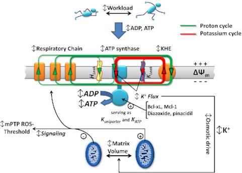
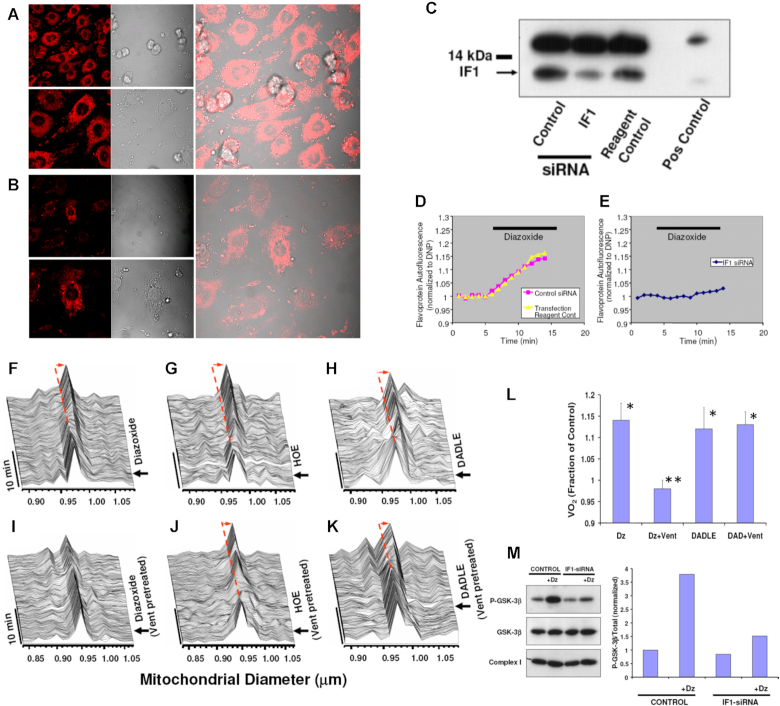
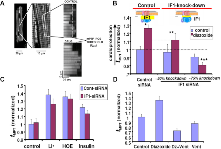
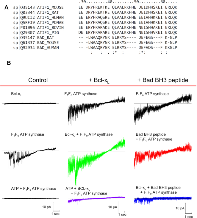
We demonstrated that ATP synthase serves the functions of a primary mitochondrial K+ "uniporter," i.e., the primary way for K+ to enter mitochondria. This K+ entry is proportional to ATP synthesis, regulating matrix volume and energy supply-vs-demand matching. We show that ATP synthase can be upregulated by endogenous survival-related proteins via IF1. We identified a conserved BH3-like domain of IF1 which overlaps its "minimal inhibitory domain" that binds to the β-subunit of F1. Bcl-xL and Mcl-1 possess a BH3-binding-groove that can engage IF1 and exert effects, requiring this interaction, comparable to diazoxide to augment ATP synthase's H+ and K+ flux and ATP synthesis. Bcl-xL and Mcl-1, but not Bcl-2, serve as endogenous regulatory ligands of ATP synthase via interaction with IF1 at this BH3-like domain, to increase its chemo-mechanical efficiency, enabling its function as the recruitable mitochondrial KATP-channel that can limit ischemia-reperfusion injury. Using Bayesian phylogenetic analysis to examine potential bacterial IF1-progenitors, we found that IF1 is likely an ancient (∼2 Gya) Bcl-family member that evolved from primordial bacteria resident in eukaryotes, corresponding to their putative emergence as symbiotic mitochondria, and functioning to prevent their parasitic ATP consumption inside the host cell.

Keywords: ATP synthase regulation; ATPase Inhibitory Factor-1 (IF₁); Bcl-2 family proteins; mitochondrial permeability transition pore; mitochondrial potassium transport; volume regulation.


Figures

Graphical Abstract
Graphical Abstract
ATP Synthase K+- and H+-fluxes Drive ATP Synthesis and Enable Mitochondrial K+-“Uniporter” Function: II. Ion and ATP Synthase Flux Regulation.
Figure 1.
Figure 1.
Knockdown of IF1 expression in neonatal cardiomyocytes using RNA interference. (A) IF1 immunocytochemical labeling of control, and (B) IF1 siRNA treated cells. (C) Western blot analysis of control vs siRNA treated samples; positive control corresponds to adult rat heart. (D,E) FP autofluorescence (normalized to dinitrophenol, DNP) as marker of mKATP activity. (D) Dz induced FP oxidation in control, and (E) No effect of Dz was observed in IF1 siRNA-treated cells. (FK) In situ monitoring of the amplitude and kinetics of regulatory mitochondrial swelling (resulting from increased mitochondrial K+ influx and/or retention) in intact cardiomyocytes, based on Fourier analysis of laser linescan transmittance imaging. (F) KCO, Dz; (G) The NHE-1 inhibitor, HOE, and (H) the δ-opioid peptide, DADLE, induced mitochondrial swelling. (I) The Fo inhibitor, Vent, blocked Dz-induced mitochondrial swelling, while it had no effect on swelling induced by (J) HOE or (K) DADLE. Arrow indicates the time point of drug addition. (L) Mitochondrial respiration (indexed by oxygen consumption, VO2 with respect to Dz and Vent treatment as in (D) in myocytes. (M) mKATP (Dz)-protection signaling via GSK-3β requires IF1 (see text for details). While the KCO, Dz, causes a robust increase in P-GSK-3β in control cells, this was largely prevented in IF1-siRNA treated cells.
Figure 2.
Figure 2.
Measurements of the mitochondrial permeability transition ROS threshold (tMPT, the index of cardioprotection) in myocytes. (A) typical positive tMPT effect of a drug is illustrated vs Control. (B) tMPT decreases in proportion to the degree of IF1 knock-down, compared to control cells. (C) GSK-3β-dependent protection signaling which does not require mKATP activated K+ flux (i.e., Li+, HOE, insulin) is unaffected by IF1-knock-down. (D) Block of Fo by Vent prevents mKATP (Dz)-mediated cardioprotection. * P< 0.05 vs paired Control; **, ***P = ns vs paired Control.
Figure 3.
Figure 3.
Regulation of F1Fo current and activity by Dz, Bcl-2 family peptides and proteins mediated by IF1. (A) AA alignment of the 26-residue BAD BH3 peptide and F1Fo inhibitory factor IF1 from mouse, rat, bovine, pig, monkey and human. The consistency of the alignment is indicated in the last row (asterisk shows complete conservation) (see also Figure S4). BH3 peptide L12 aligns with L42 in full length IF1. (B) Effect of Bcl-xL (20 nM) and BH3 peptide (20 nM) on the voltage ramp (from -60 mV to +60 mV) evoked F1Fo currents. Column headers about the red line denote three experimental groups; Bcl-xl and BH3 peptide middle and right columns, respectively; control currents (left column). Bottom traces (left and middle) correspond to 2 mM ATP inhibited F1Fo current.
Figure 4.
Figure 4.
Current-time integral of voltage ramp evoked F1Fo currents (A) Augmentation of the current-time integral by Bcl-xL and Dz (30 µM) is reversed by a 26 AA peptide consisting of the BH3-domain of Bad. Control peptide with a single AA substitution L12A has no effect. (B) Reconstitution of F1Fo from IF1–/– mice. Addition of IF1 (100 nM) restores the stimulatory effect of Dz, Bcl-xL and Mcl-1 on the current-time integral of voltage ramp evoked F1Fo currents from IF1–/– mice. The order of addition of IF1 and Dz, Bcl-xL or Mcl-1 is varied among the various groups as indicated. (C) F1Fo activity (ATP synthesis) driven by a H+ or (D) a K+ gradient in PL. (E) ATP production/consumption kinetics (chemiluminescence traces) in a K+ gradient in PL. (F) Dose-response of ATP inhibition of F1Fo (H+) currents and Dz and Bcl-xL activated F1Fo currents: x-axis-(linear) ATP concentration used for inhibition of F1Fo currents, y-axis (log) normalized current-time integral of F1Fo currents. Dz and Bcl-xL produced a parallel shift in the F1Fo activity vs control resulting in the energy of an additional ∼2.8 mM ATP being required to provide sufficient counter-torque to limit F1Fo to the same level of function as under control conditions. The relative change in efficiency was calculated as the ratio of the free energy of ATP hydrolysis during activation by Dz and Bcl-xL over that under basal conditions. * P<0.05, ** P< 0.02.
Figure 5.
Figure 5.
Scheme of the H+ and K+ transport across the inner mitochondrial membrane. All the energy available for work and to drive ionic movements derives from the original H+ gradient established by proton pumps in the respiratory chain (see also). A central point is the obligatory preservation of charge and mass balance under the steady state circuits. Panel A, displays the “original view of cation flux cycles” in which the H+ gradient is being harnessed by F1Fo directly to make ATP, whereas a certain amount of K+ enters the matrix through an ordinary K+ channel mechanism (a “mKATP-uniporter” channel), driven by ΔΨ, and extruded via KHE utilizing the energy remaining in the fraction of the H+ gradient not directly harnessed by F1Fo. The equivalent energy of this fraction being used to extrude K+, and a large fraction of that non-ATP-producing energy would essentially be dissipated as heat in the constant cycle of K+ recirculation. Panel B displays the new mechanism in which the same amount of energy available in the original H+ gradient is entirely available to produce ATP, simply by having the mKATP-uniporter mechanism reside inside, and as natural part of, F1Fo with the traffic of H+ or K+ contributing its energy to producing ATP. The remainder of the H+ gradient energy is now utilized to remove all the K+ that entered via F1Fo. However, the gain is that more ATP is produced for the same input energy by not wasting some of that energy on maintaining what was originally thought to be a separate K+ cycle that does not/cannot generate any ATP. This way is a better, tightly coupled system of energy supply-demand matching through the K+ cycle utilizing F1Fo because the matrix influx of K+ is truly directly proportional to ATP synthesis. Any transient increase in F1Fo activity will thus lead to transient K+ accumulation. This will lead to the attraction of a counter-ion and change of the osmotic drive yielding a “volume-activation of respiration” response which previously has been documented in detail. The scheme depicted in (C) integrates the implications of modestly enhancing the chemo-mechanical efficiency of F1Fo (by KCO's or Bcl-xL/Mcl-1). For the driving energy of the same H+ gradient the F1Fo flux increases, enabling increased respiration and a directly increased K+ flux cycle (yielding an increased volume signal) and enhanced ATP generation (C) vs the basal conditions (B).
Figure 6.
Figure 6.
Interaction of F1 ATPase with IF1 or a BH3 modeled peptide. (A) Ribbon model of the crystal structure of bovine F1 (PDB ID 4Z1M) emphasizing two of the three β subunits (cyan and magenta) and the γ subunit (blue) in interaction with the long α-helix of the inhibitor protein IF1 (orange). The IF1 domain (residues 18–51 are shown in cyan; residues 23–70 from PDB ID 1GMJ are shown in orange) interacts with the β subunit marked in yellow. (B) Surface representation of subunits β (yellow), γ (blue) and α (magenta) of F1 ATPase interacting with IF1 peptide. (C) As panel (B) with the α2-helix peptide containing BH3 domain from the BAD protein (PDB ID 1G5J, as it binds Bcl-xL) in ribbon representation at the IF1 groove in F1 showing the aliphatic side chain of Leu 42 in IF1 and Leu 12 in BH3-BAD peptide (corresponding to Leu114 in human BAD). (D) Same as (C) at an approximately 90° orientation. (E) Phylogenetic tree of the BH3 extended peptides (35 residues) from Bcl-2 proteins and IF1 across eukaryotes. Sequence alignment was computed with Clustal Omega and the tree and divergence times (in Myr) were calculated by MEGA 6.0 (for further details, see Supplemental Information; Figures S4 and S5, and Table S1).

Comment in

References

    1. Juhaszova M, Kobrinsky E, Zorov DBet al. . ATP synthase K+- and H+-flux drive ATP synthesis and enable mitochondrial K+-uniporter function: I. Characterization of ion fluxes. Function. 2021. 10.1093/function/zqab065. - DOI - PMC - PubMed
    1. Garlid KD, Dos Santos P, Xie ZJ, Costa AD, Paucek P. Mitochondrial potassium transport: the role of the mitochondrial ATP-sensitive K(+) channel in cardiac function and cardioprotection. Biochim Biophys Acta. 2003;1606(1-3):1–21. - PubMed
    1. Juhaszova M, Wang S, Zorov DBet al. . The identity and regulation of the mitochondrial permeability transition pore: where the known meets the unknown. Ann NY Acad Sci. 2008;1123(1):197–212.. http://www.ncbi.nlm.nih.gov/entrez/query.fcgi?cmd=Retrieve&db=PubMed&dop.... - PubMed
    1. Juhaszova M, Zorov DB, Kim SHet al. . Glycogen synthase kinase-3beta mediates convergence of protection signaling to inhibit the mitochondrial permeability transition pore. J Clin Invest. 2004;113(11):1535–1549.. http://www.ncbi.nlm.nih.gov/entrez/query.fcgi?cmd=Retrieve&db=PubMed&dop.... - PMC - PubMed
    1. Zorov DB, Juhaszova M, Yaniv Y, Nuss HB, Wang S, Sollott SJ.. Regulation and pharmacology of the mitochondrial permeability transition pore. Cardiovasc Res. 2009;83(2):213–225.. http://www.ncbi.nlm.nih.gov/entrez/query.fcgi?cmd=Retrieve&db=PubMed&dop.... - PMC - PubMed

Publication types

MeSH terms

Substances

LinkOut - more resources