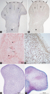
Global DNA methylation and chondrogenesis of rat limb buds in a three-dimensional organ culture system
- PMID: 35188093
- PMCID: PMC9392980
- DOI: 10.17305/bjbms.2021.6584
Global DNA methylation and chondrogenesis of rat limb buds in a three-dimensional organ culture system
Abstract
Although DNA methylation epigenetically regulates development, data on global DNA methylation during development of limb buds (LBs) are scarce. We aimed to investigate the global DNA methylation developmental dynamics in rat LBs cultivated in a serum-supplemented (SS) and in chemically defined serum- and protein-free (SF) three-dimensional organ culture. Fischer rat front- and hind-LBs at 13th and 14th gestation days (GD) were cultivated at the air-liquid interface in Eagle's Minimal Essential Medium (MEM) or MEM with 50% rat serum for 14 days, as SF and SS conditions, respectively. The methylation of repetitive DNA sequences (SINE rat ID elements) was assessed by pyrosequencing. Development was evaluated by light microscopy and extracellular matrix glycosaminoglycans staining by Safranin O. Upon isolation, weak Safranin O staining was present only in more developed GD14 front-LBs. Chondrogenesis proceeded well in all cultures towards day 14, except in the SF-cultivated GD13 hind-LBs, where Safranin O staining was almost absent on day 3. That was associated with a higher percentage of DNA methylation than in SF-cultivated GD13 front-LBs on day three. In SF-cultivated front-LBs, a significant methylation increase between the 3rd and 14th day was detected. In SS-cultivated GD13 front-LBs, methylation increased significantly on day three and then decreased. In older GD14 SS-cultivated LBs, there was no increase of DNA methylation, but they were significantly hypomethylated relative to the SS-cultivated GD13 at days 3 and 14. We confirmed that the global DNA methylation increase is associated with less developed limb organ primordia that strive towards differentiation in vitro, which is of importance for regenerative medicine strategies.
Conflict of interest statement
Conflicts of interest: The authors declare no conflicts of interest.
Figures


Similar articles
-
Transmembranous and enchondral osteogenesis in transplants of rat limb buds cultivated in serum- and protein-free culture medium.Anat Histol Embryol. 2022 Sep;51(5):592-601. doi: 10.1111/ahe.12835. Epub 2022 Jul 11. Anat Histol Embryol. 2022. PMID: 35815632 Free PMC article.
-
Epigenetic drug 5-azacytidine impairs proliferation of rat limb buds in an organotypic model-system in vitro.Croat Med J. 2013 Oct 28;54(5):489-95. doi: 10.3325/cmj.2013.54.489. Croat Med J. 2013. PMID: 24170728 Free PMC article.
-
Differentiation of rat neural tissue in a serum-free embryo culture model followed by in vivo transplantation.Croat Med J. 2001 Dec;42(6):611-7. Croat Med J. 2001. PMID: 11740842
-
Inhibition of prostaglandin synthesis reduces cyclic AMP levels and inhibits chondrogenesis in cultured chick limb mesenchyme.Methods Cell Sci. 2000 Mar;22(1):9-16. doi: 10.1023/a:1009824106368. Methods Cell Sci. 2000. PMID: 10650328 Review.
-
The tissues and regulatory pattern of limb chondrogenesis.Dev Biol. 2020 Jul 15;463(2):124-134. doi: 10.1016/j.ydbio.2020.04.009. Epub 2020 May 15. Dev Biol. 2020. PMID: 32417169 Review.
Cited by
-
Predictive biomarkers for embryotoxicity: a machine learning approach to mitigating multicollinearity in RNA-Seq.Arch Toxicol. 2024 Dec;98(12):4093-4105. doi: 10.1007/s00204-024-03852-w. Epub 2024 Sep 6. Arch Toxicol. 2024. PMID: 39242367
-
Transmembranous and enchondral osteogenesis in transplants of rat limb buds cultivated in serum- and protein-free culture medium.Anat Histol Embryol. 2022 Sep;51(5):592-601. doi: 10.1111/ahe.12835. Epub 2022 Jul 11. Anat Histol Embryol. 2022. PMID: 35815632 Free PMC article.
References
-
- Hill RE, Lettice LA. Limb Development. In: Baldock R, Bard J, Davidson DR, Morriss-Kay G, editors. Kaufman's Atlas of Mouse Development Supplement Coronal Images. Massachusetts, United States: Academic Press; 2016. pp. 193–205.
-
- Barter MJ, Tselepi M, Gõmez R, Woods S, Hui W, Smith GR, et al. Genome-wide microRNA and gene analysis of mesenchymal stem cell chondrogenesis identifies an essential role and multiple targets for miR-140-5p. Stem Cells. 2015;33(11):3266–80. https://doi.org/10.1002/stem.2093. - PMC - PubMed
-
- Greenberg MVC, Bourc'his D. The diverse roles of DNA methylation in mammalian development and disease. Nat Rev Mol Cell Biol. 2019;20(10):590–607. https://doi.org/10.1038/s41580-019-0159-6. - PubMed
-
- Murrell A, Hurd PJ, Wood IC. Epigenetic mechanisms in development and disease. Biochem Soc Trans. 2013;41(3):697–9. https://doi.org/10.1042/BST20130051. - PubMed
-
- Barter MJ, Bui C, Cheung K, Falk J, Gómez R, Skelton AJ, et al. DNA hypomethylation during MSC chondrogenesis occurs predominantly at enhancer regions. Sci Rep. 2020;10(1):1169. https://doi.org/10.1038/s41598-020-58093-5. - PMC - PubMed

