Review
. 2022 Feb 21;79(3):144.
doi: 10.1007/s00018-022-04184-7.

Defective perlecan-associated basement membrane regeneration and altered modulation of transforming growth factor beta in corneal fibrosis

Affiliations
Review

Defective perlecan-associated basement membrane regeneration and altered modulation of transforming growth factor beta in corneal fibrosis

Steven E Wilson. Cell Mol Life Sci. .

Abstract

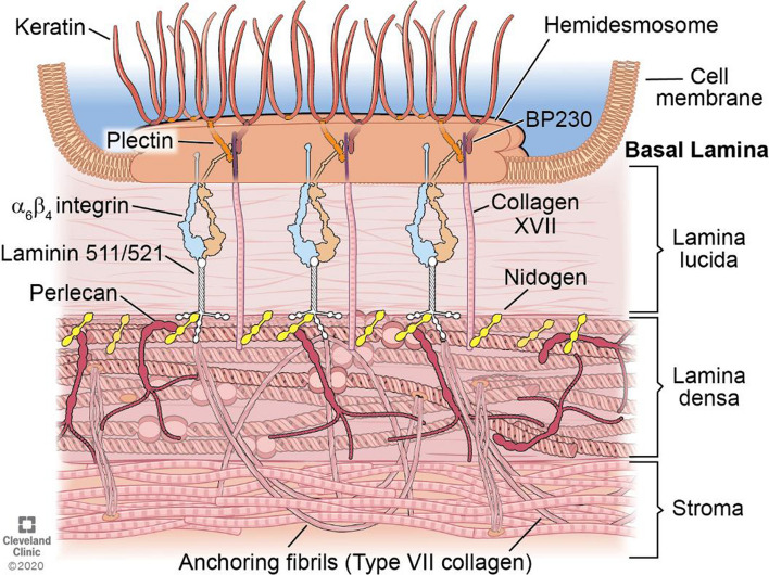
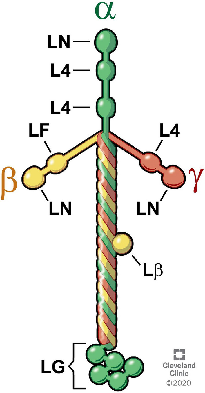
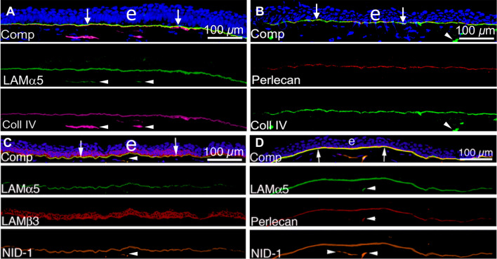
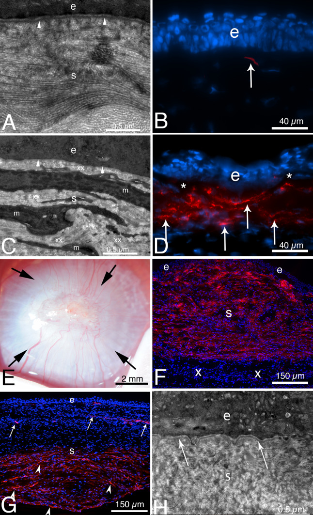
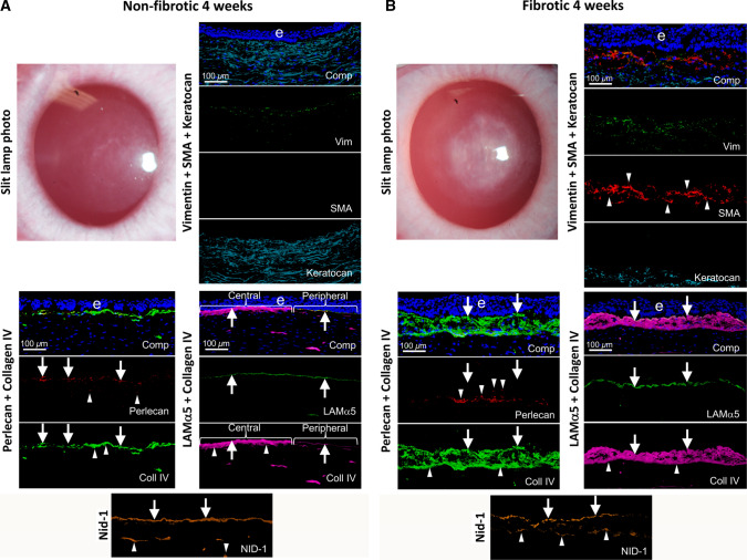
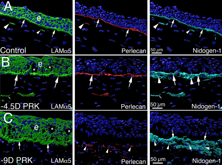
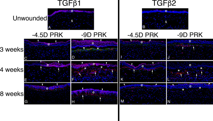
In the cornea, the epithelial basement membrane (EBM) and corneal endothelial Descemet's basement membrane (DBM) critically regulate the localization, availability and, therefore, the functions of transforming growth factor (TGF)β1, TGFβ2, and platelet-derived growth factors (PDGF) that modulate myofibroblast development. Defective regeneration of the EBM, and notably diminished perlecan incorporation, occurs via several mechanisms and results in excessive and prolonged penetration of pro-fibrotic growth factors into the stroma. These growth factors drive mature myofibroblast development from both corneal fibroblasts and bone marrow-derived fibrocytes, and then the persistence of these myofibroblasts and the disordered collagens and other matrix materials they produce to generate stromal scarring fibrosis. Corneal stromal fibrosis often resolves completely if the inciting factor is removed and the BM regenerates. Similar defects in BM regeneration are likely associated with the development of fibrosis in other organs where perlecan has a critical role in the modulation of signaling by TGFβ1 and TGFβ2. Other BM components, such as collagen type IV and collagen type XIII, are also critical regulators of TGF beta (and other growth factors) in the cornea and other organs. After injury, BM components are dynamically secreted and assembled through the cooperation of neighboring cells-for example, the epithelial cells and keratocytes for the corneal EBM and corneal endothelial cells and keratocytes for the corneal DBM. One of the most critical functions of these reassembled BMs in all organs is to modulate the pro-fibrotic effects of TGFβs, PDGFs and other growth factors between tissues that comprise the organ.

Keywords: Basement membrane assembly; Collagen type IV; Cornea; Descemet’s membrane; Dystroglycan; Epithelial barrier function; Epithelial basement membrane; HGF; Integrins; KGF; Laminins; Nidogens; PDGF; Perlecan; Regeneration; TGF beta.


Conflict of interest statement

The author does not have any commercial or proprietary interests in the subject matter of this review article.

Figures

Fig. 1
Fig. 1
Schematic of the corneal EBM. The EBM underlies basal epithelial cells and overlies the stroma in the cornea connected by the hemidesmosome-anchoring filament complex. It is important to note there are other component molecules present in corneal EBM, such as collagen type IV, that are not included in this simplified diagram. HD hemidesmosome; BP230 bullous pemphigoid antigen 230. Reprinted with permission from Saikia et al. [79]
Fig. 2
Fig. 2
Schematic of the structure of the typical heterotrimer laminin molecule (511, 512, or 111) with one α, β and γ chain. The three short arms of the cross-shaped molecule have a common domain structure and consist of laminin N-terminal (LN) domains through which the laminin molecule associates with surrounding laminins in the laminin network and L4 domains. The long arm of the laminin molecule (Lβ) is an alpha-helical coil of the α, β and γ chains. The α1 chain contains five laminin G-like (LG) domains that interact with cellular integrins and other receptors. Illustration by David Schumick, BS, CMI. Reprinted with the permission of the Cleveland Clinic Center for Medical Art & Photography, 2021
Fig. 3
Fig. 3
Localization of laminin alpha-5 (component in laminins 511/512), laminin beta-3 (component in laminin 332), perlecan, nidogen-1, and collagen type IV in unwounded corneal EBM. A Laminin alpha-5 and collagen type IV duplex IHC. B Perlecan and collagen type IV duplex IHC. C Laminin alpha-5, laminin beta-3, and nidogen-1 triplex IHC. D Laminin alpha-5, perlecan, and nidogen-1 triplex IHC. Comp is a composite of all components of each IHC with DAPI. Blue is DAPI-stained nuclei of all cells. (e) is the epithelium. Reprinted with permission from de Oliveira et al. [39]
Fig. 4
Fig. 4
TEM and alpha-smooth muscle actin (SMA) IHC after severe epithelial-stromal injury or Pseudomonas aeruginosa keratitis in rabbits. A From Torricelli et al. [72], at 1 month after reproducible moderate epithelial–stromal corneal injury (-4.5D photorefractive keratectomy [PRK] laser surgery) the epithelial BM has regenerated beneath the epithelium (e) with normal lamina lucida and lamina densa (arrowheads). Notice in the normal transparent stroma (s), the collagen fibrils are seen both tangentially and longitudinally in different areas and are regular in diameter and packing. B. IHC for SMA at 1 mo. after -4.5D PRK [80] revealed only a single myofibroblast (arrow) in the anterior stroma. C. At 1 month after severe epithelial–stromal corneal injury (-9D PRK surgery) [80], the cornea had dense scarring fibrosis (not shown) and TEM showed no discernable lamina lucida and lamina densa (arrowheads) beneath the epithelium (e). The anterior stroma (s) was packed with layers of myofibroblasts (m) and the surrounding stromal matrix (xx) produced by the myofibroblasts is disorganized with no regular packing of collagen fibrils. The myofibroblasts are themselves opaque [125]—as is the disordered stromal matrix they produce. D IHC for SMA at 1 month after severe injury (−9D PRK surgery) [80] showed large numbers of layered myofibroblasts (arrows) in the anterior stroma beneath the epithelium (e). * indicates artifactual dissociation of the epithelium from the anterior stroma that frequently occurs in corneas without a mature EBM. E From Marino et al. [7], at 1 month after severe Pseudomonas aeruginosa infection treated with tobramycin, the cornea has dense opacity from fibrosis (arrows) despite full healing of the epithelium. F IHC for SMA (red) reveals myofibroblasts filling the anterior 90% of the stroma (s) beneath the intact epithelium (e) [7]. In this cornea the infection was halted prior to destruction of DBM and the endothelium (not shown), and the posterior stroma (x) is hypercellular but doesn’t contain myofibroblasts. The epithelial BM in this cornea did not have lamina lucida and lamina densa (not shown). G At 2 months after severe Pseudomonas aeruginosa infection [7], most myofibroblasts have disappeared in the anterior half of the stroma beneath the epithelium (e) in this cornea. SMA + pericytes can be seen associated with neovascularization (arrows). Myofibroblasts (arrowheads) are present in the posterior half of the stroma of this cornea since the original infection destroyed DBM, and, therefore, TGFβ1 continuously enters the stroma from the aqueous humor to maintain myofibroblast viability in the posterior stroma [82, 83]. H TEM of the cornea in G at two months after Pseudomonas aeruginosa keratitis [7] showed that the epithelial BM has regenerated beneath the epithelium (e) with normal lamina lucida and lamina densa (arrows). AD reprinted with permission from Torricelli et al. [72]. E–H reprinted with permission from Marino et al. [7]
Fig. 5
Fig. 5
Non-fibrotic and fibrotic healing in corneas. Slit lamp photographs and IHC for cell markers vimentin (mesenchymal cells), SMA (myofibroblasts) and keratocan (keratocytes), as well as BM components laminin alpha-5, laminin beta-3, perlecan, nidogen-1, and collagen type IV at 4 weeks after PRK. A A cornea that did not develop scarring fibrosis after PRK laser surgery. Not mild “haze” attributable to corneal fibroblasts, but no myofibroblasts were present. Arrows indicate BM components incorporated into the EBM. Arrowheads indicate components detected in anterior stromal cells. B A cornea with scarring fibrosis at four weeks after PRK surgery had severe opacity corresponding to the area of laser ablation and multilayered SMA + myofibroblasts (arrowheads) in the anterior stroma. All fibrotic corneas had collagen type IV, laminin alpha-5, and nidogen-1 in the EBM (arrows) beneath the epithelium (e). Collagen type IV was present at higher levels both in the nascent EBM and underlying anterior stroma in fibrotic corneas compared to non-fibrotic corneas after PRK surgery (compare A and B) [39]. Perlecan, however, was absent from the “moth-eaten” EBM of these corneas (arrows). Anterior stromal myofibroblasts and extracellular vesicle-like structures contained perlecan (arrowheads), but it was not incorporated into the nascent EBM (see Fig. 6). Collagen type IV (arrowheads) and nidogen-1 (arrowheads) were present within and around anterior myofibroblasts of the fibrotic corneas at four weeks post-PRK. Comp is a composite of all components for each IHC and DAPI-stained nuclei of all cells (blue). Reprinted with permission from de Oliveira et al. [39]
Fig. 6
Fig. 6
Defective perlecan incorporation into the EBM of injured corneas that develop scarring stromal fibrosis. Imaris 3D constructions of confocal microscopy images of triplex laminin alpha-5, perlecan and nidogen-1 IHC in the unwounded control corneas and corneas with moderate and severe epithelial-stromal injury from de Oliveira et al. [48]. A In an unwounded cornea, laminin alpha-5 (green) is detected in the epithelium (e), and in the EBM (arrows). Two DAPI-negative vesicles with laminin alpha-5 (arrowheads) are present in the subepithelial stroma adjacent to the EBM and likely were produced by keratocytes to contribute to maintenance of the EBM. Perlecan (red) is detected in the EBM (arrows), and also in vesicles in the anterior stroma (arrowhead). Nidogen-1 (blue gray) is a major component in the EBM (arrows) and is present in secretory vesicles in the subepithelial stroma (arrowheads). B In a cornea that had moderate epithelial-stromal injury (-4.5D excimer laser photorefractive keratectomy [PRK]) at one month after surgery that did not develop myofibroblasts or scarring stromal fibrosis (see Fig. 5A), the findings for laminin alpha-5, perlecan and nidogen-1 localization in the EBM were similar to the unwounded cornea (large arrows), except increased nidogen-1 (arrowheads) was present in the subepithelial stroma surrounding stromal keratocyte/corneal fibroblast cells. DAPI-negative vesicles (small arrows) in the anterior stroma contained one or more of the EBM components. C In a cornea that had more severe epithelial-stromal injury (-9D PRK), at 1 month after surgery there was greater stromal opacity and myofibroblasts (see Fig. 5B). Laminin alpha-5 and nidogen-1 (arrows) localization in the EBM was similar to that in the unwounded control cornea. Perlecan, however, was not detected in the EBM, even though it was present in the anterior stroma within and surrounding myofibroblasts (see Fig. 4D). Stromal nidogen-1 (arrowheads) was also present at high levels in the anterior stroma surrounding myofibroblasts. Blue is DAPI-stained nuclei in all panels. e is epithelium. * indicates artifactual defects in the epithelium which are often seen in PRK corneas that are cryo-sectioned in the first one–two months after surgery. Reprinted with permission from de Oliveira et al. [48]
Fig. 7
Fig. 7
TGF beta-1 and TGF beta-2 localization in rabbit corneas with moderate injuries without stromal fibrosis and severe injuries with stromal fibrosis. Representative images are shown from a study in rabbits from de Oliveira et al. [48]. A In the unwounded epithelium (e), TGFβ1 (pink) was present in epithelial cells with higher localization in apical epithelium. Little TGFβ1 was present in stromal (s) cells where keratocytes predominate in unwounded corneas. B In the unwounded epithelium (e), TGFβ2 (pink) was restricted to the apical epithelial surface (from tears) with little production in epithelial cells themselves. Little, if any, TGFβ2 was present in stromal (s) cells. At time points from immediate wounded to 2 weeks (not shown, [see 48]), TGFβ1 was produced by epithelial cells and delivered from tears, while TGFβ2 was delivered in large amounts from tears. Some stromal cells also produced TGFβ1 and TGFβ2. All corneas C to N had triplex IHC for TGFβ1 or TGFβ2 (pink), vimentin (green), and the alpha-smooth muscle actin (SMA) marker for myofibroblasts (red). C At three weeks after moderate injury -4.5D PRK, apical localization (arrowheads) of TGFβ1 was already prominent and although there were some vimentin + cells (corneal fibroblasts, fibrocytes and their progeny) in the subepithelial stroma, there were no SMA + myofibroblasts in the stroma. D Conversely, at 3 weeks after severe injury -9D PRK, lower TGFβ1 localization to apical epithelium (arrowheads) was noted, and TGFβ1 was prominent throughout the epithelium and into the superficial stroma. Many vimentin + (green) cells were present in the subepithelial stroma (some of which had associated TGFβ1) and some of these myofibroblast precursor cells had become mature SMA + myofibroblasts (red). E At four weeks after moderate injury -4.5D PRK, apical localization of TGFβ1 (arrowheads) continued to be prominent and EBM TGFβ1 localization (arrows) was also noted. A few stromal cells, both vimentin + and vimentin− had TGFβ1 associated with them. There were no SMA + myofibroblasts in the stroma. F Conversely, in the four week -9D PRK corneas, TGFβ1 was prominent throughout the epithelium, with apical prominence (arrowheads), but without EBM localization. Many SMA + myofibroblasts (arrows) occupied the subepithelial stroma. Two four-month -4.5 D PRK corneas in this series (not shown) had an intermediate pattern of TGFβ1 localization [48] with little apical epithelial or EBM localization, and some SMA + myofibroblast development in the subepithelial stroma. G and H. At eight weeks after −4.5D or −9D PRK, respectively, apical epithelial (arrowheads) and EBM (arrows in H) TGFβ1 was prominent. SMA + myofibroblasts were no longer present in the subepithelial stroma (after likely undergoing apoptosis). I At three weeks after −4.5D PRK, there was prominent TGFβ2 in the apical epithelium (arrowheads). There were many vimentin + cells in the anterior stroma, but none were SMA + myofibroblasts. In the deeper stroma, some vimentin + and vimentin- cells had TGFβ2 associated with them. J At three weeks after −9D PRK, there was little apical epithelial localization of TGFβ2. Some vimentin + cells in the subepithelial stroma had developed into SMA + (red) myofibroblasts. A band of TGFβ2 (arrows) was localized posterior to the vimentin + cells in the stroma. K At four weeks after −4.5D PRK, apical epithelial (arrowheads) and EBM (arrows) TGFβ2 localization was prominent. No vimentin + cells in the subepithelial stroma had developed into SMA + myofibroblasts in the cornea shown. L At four weeks after –9D PRK, little epithelial TGFβ2 localization was detected. Note this does not necessarily indicate that TGFβ2 from tears was not passing through the epithelium, but that the lack of barrier in the apical epithelium or EBM did not produce an increase in TGFβ2 concentration to a detectible level. The subepithelial stroma had large numbers of SMA + myofibroblasts (arrows). In the deeper stroma, there are both vimentin + and vimentin− cells with associated TGFβ2. M, N At eight weeks after −4.5D or −9D PRK, respectively, apical epithelial TGFβ2 localization was prominent. No SMA + myofibroblasts remained in the subepithelial stroma in all corneas from both groups. Some subepithelial cells continued to have associated TGFβ2 in both groups. e is epithelium in all panels. Blue is DAPI-stained nuclei in all panels. *indicates epithelial dislocation from stroma during cryo-sectioning. Reprinted with permission from de Oliveira et al. [48]

Similar articles

Cited by

References

    1. Sasaki T, Fässler R, Hohenester E. Laminin: the crux of basement membrane assembly. J Cell Biol. 2004;164:959–963. doi: 10.1083/jcb.200401058. - DOI - PMC - PubMed
    1. Yurchenco PD, O’Rear J. Supramolecular organization of basement membranes. In: Rohrbach DH, Timpl R, editors. Molecular and cellular aspects of basement membranes. San Diego: Academic Press; 1993. pp. 20–47.
    1. Hohenester E, Yurchenco PD. Laminins in basement membrane assembly. Cell Adh Migr. 2013;7:56–63. doi: 10.4161/cam.21831. - DOI - PMC - PubMed
    1. Wilson SE. Coordinated modulation of corneal scarring by the epithelial basement membrane and Descemet's basement membrane. J Refract Surg. 2019;35:506–516. doi: 10.3928/1081597X-20190625-02. - DOI - PMC - PubMed
    1. Martinez-Hernandez A, Amenta PS. The basement membrane in pathology. Lab Invest. 1983;48:656–677. - PubMed

MeSH terms

Substances