. 2022 Jun 1;30(6):2224-2241.
doi: 10.1016/j.ymthe.2022.02.018. Epub 2022 Feb 18.

Nucleic acid aptamer controls mycoplasma infection for inhibiting the malignancy of esophageal squamous cell carcinoma

Affiliations

Nucleic acid aptamer controls mycoplasma infection for inhibiting the malignancy of esophageal squamous cell carcinoma

Yibin Zhang et al. Mol Ther. .

Abstract

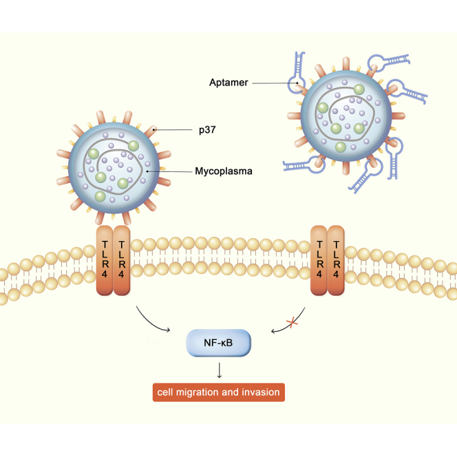
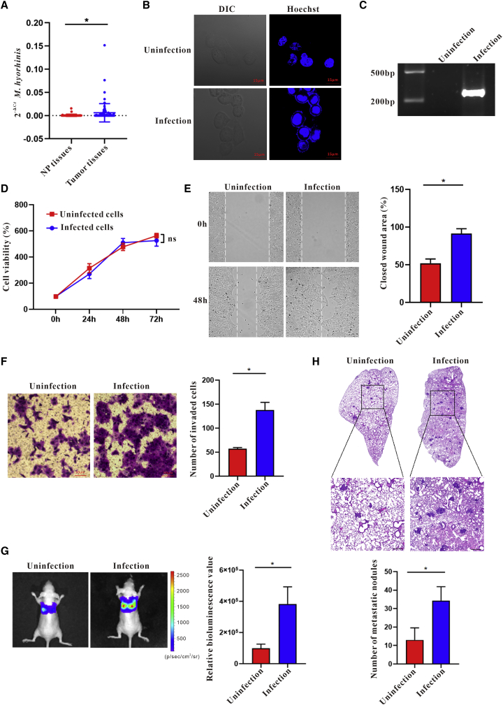
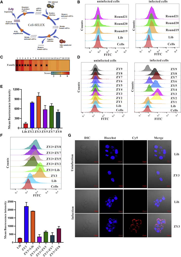
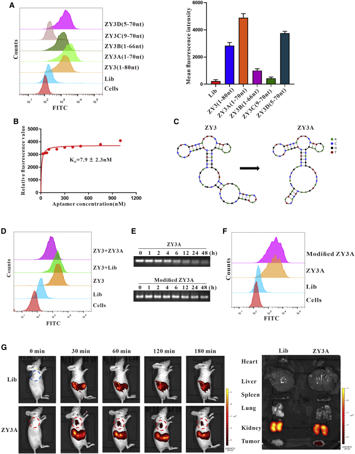
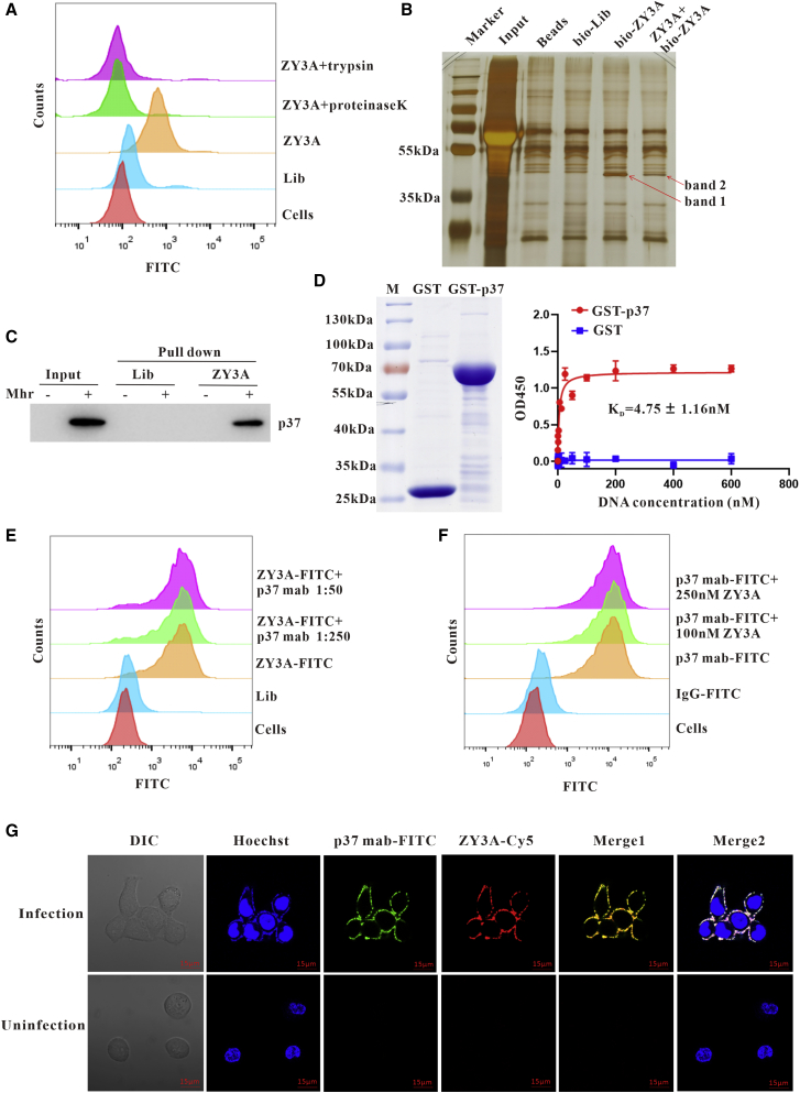
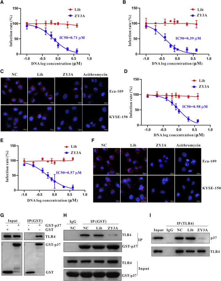
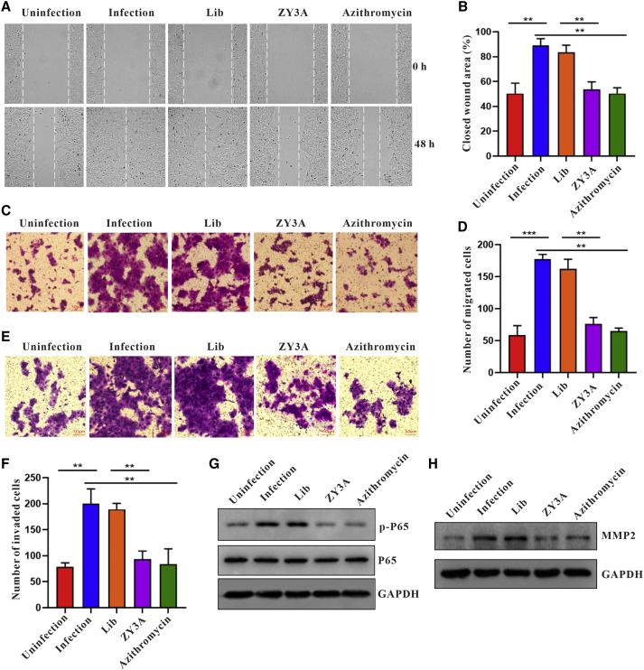
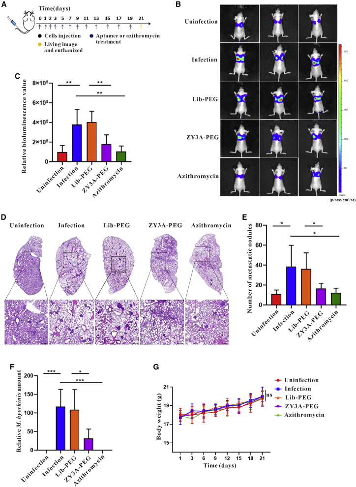
Esophageal cancer is one of the most frequent malignant tumors of the digestive tract, among which esophageal squamous cell carcinoma (ESCC) is the main pathological type worldwide. Previous studies have shown microbial infections in the upper digestive tract to be a potential risk factor in ESCC etiology. In this study, we identified that Mycoplasma hyorhinis infection promoted the malignancy of ESCC. In response, we generated a single-stranded DNA aptamer, ZY3A, against M. hyorhinis using the cell-SELEX strategy. The underlying recognition mechanism of ZY3A on M. hyorhinis involves its binding to M. hyorhinis-specific p37 protein. This tool allowed us to provide the first proof-of-concept evidence using a nucleic acid aptamer to control mycoplasma infection. More specifically, we found that ZY3A could neutralize M. hyorhinis infection on ESCC cells by blocking the interaction between p37 protein and its receptor TLR4 on the ESCC cell membrane. As a result, ZY3A inhibited the migration and invasion of M. hyorhinis-infected ESCC cells in vitro and metastasis in vivo. Taken together, these findings indicate that aptamer ZY3A is a potential candidate for development into a novel molecular tool for treatment of M. hyorhinis infection and a safe first-in-class M. hyorhinis-targeting antitumor agent.

Keywords: M. hyorhinis; esophageal squamous cell carcinoma; nucleic acid aptamer; p37 protein; tumor metastasis.


Conflict of interest statement

Declaration of interests The authors declare that they have no competing interests.

Figures

None
Graphical abstract
Figure 1
Figure 1
M. hyorhinis infection promotes the malignant behavior of ESCC cells (A) M. hyorhinis in ESCC tumor tissues and non-neoplastic esophageal tissues was measured by qPCR. Quantification of p37 levels relative to ATCB expression is shown. (B) Representative Hoechst staining images of Eca-109 cells before or after M. hyorhinis infection are shown. (C) PCR products of mycoplasma from infected or uninfected Eca-109 cells were analyzed by gel electrophoresis. (D) The proliferation ability of Eca-109 cells before or after M. hyorhinis infection was measured by CCK8 assay. (E) The migration ability of M. hyorhinis-infected or uninfected Eca-109 cells was detected by wound healing assay. The wound healing areas were calculated by ImageJ software. (F) The invasion ability of M. hyorhinis-infected or uninfected Eca-109 cells was detected by transwell invasion assay. (G) M. hyorhinis-infected or uninfected Eca-109/luc cells were i.v. injected into nude mice to evaluate metastasis ability in vivo. Representative images of living luciferase bioluminescence are shown. (H) H&E staining of mouse lung tissues were used to detect and count tumor metastasis nodules. Scale bars, 300 μm. ∗p < 0.05; ∗∗p < 0.01.
Figure 2
Figure 2
Screening and identification of aptamers targeting M. hyorhinis-infected ESCC cells by cell-SELEX (A) Schematic illustration of cell-SELEX based on M. hyorhinis-infected and uninfected Eca-109 cells. (B) The binding of enriched ssDNA pool (200 nM) to M. hyorhinis-infected or uninfected Eca-109 cells was analyzed by flow cytometry. (C) The sequencing results of round 21 were analyzed using FASTaptamer software. High-scoring families (asterisk marked) are shown. (D) The binding of selected aptamers (200 nM) to M. hyorhinis-infected or uninfected Eca-109 cells was analyzed by flow cytometry. (E) The binding of indicated aptamers (200 nM) to M. hyorhinis-infected Eca-109 cells was examined by flow cytometry. Mean fluorescence intensity was calculated by FlowJo software. (F) M. hyorhinis-infected Eca-109 cells were incubated with FITC-labeled ZY3 (200 nM) in the presence or absence of unlabeled library or indicated aptamers (400 nM). Competitive binding was analyzed by flow cytometry. (G) The binding of Cy5-labeled library or ZY3 (200 nM) to M. hyorhinis-infected or uninfected Eca-109 cells was examined by confocal microscopy. Representative images are shown.
Figure 3
Figure 3
Optimization and characterization of aptamer ZY3 (A) Flow cytometry was employed to determine the binding ability of different truncated versions of ZY3 (200 nM) to M. hyorhinis-infected Eca-109 cells. (B) Dissociation constant of ZY3A for M. hyorhinis-infected Eca-109 cells was determined by flow cytometry. (C) Secondary structures of ZY3 and ZY3A were predicted by NUPACK software. (D) M. hyorhinis-infected Eca-109 cells were incubated with FITC-labeled library or ZY3 (200 nM) in the presence or absence of unlabeled library or ZY3A (400 nM). Competitive binding was analyzed by flow cytometry. (E) Serum stability of ZY3A and modified ZY3A in cell medium with 10% FBS was examined by agarose gel electrophoresis. (F) Flow cytometry was employed to compare binding ability between ZY3A and modified ZY3A. (G) In vivo imaging and biodistribution analysis of aptamer in M. hyorhinis-infected Eca-109 cell-bearing nude mice after i.v. injection of Cy5-labeled ZY3A or library. The fluorescence of ZY3A in mice was collected by the IVIS Lumina II imaging system at the indicated time in vivo and ex vivo.
Figure 4
Figure 4
p37 was identified as the target of ZY3A (A) Flow cytometry was employed to determine the binding ability of ZY3A to M. hyorhinis-infected Eca-109 cells with or without pretreatment of proteinase K or trypsin. (B) Silver staining was used to analyze the proteins in SDS-PAGE, including input protein (Input), beads pulldown protein (Beads), biotin-labeled-library pulldown protein (bio-Lib), biotin-labeled-ZY3A pulldown protein (bio-ZY3A), and biotin-labeled-ZY3A pulldown protein after adding unlabeled ZY3A as a competitor (ZY3A + bio-ZY3A). (C) Membrane proteins from M. hyorhinis-infected or uninfected Eca-109 cells were incubated with beads conjugated with library or beads conjugated with ZY3A. Bound proteins were analyzed by western blotting using anti-p37 antibody. (D) Coomassie brilliant blue staining of GST-p37 or GST protein expressed by prokaryotic expression system is shown. Dissociation constant of ZY3A for GST-p37 or GST protein was determined by ELONA. (E) M. hyorhinis-infected Eca-109 cells were incubated with FITC-labeled ZY3A in the presence or absence of anti-p37 antibody with indicated dilution rate. Competitive binding was analyzed by flow cytometry. (F) M. hyorhinis-infected Eca-109 cells were incubated with FITC-labeled anti-p37 antibody in the presence or absence of unlabeled ZY3A with indicated concentrations. Competitive binding was analyzed by flow cytometry. (G) Confocal microscopy was used to analyze the colocalization of Hoechst stain, anti-p37 antibody, and ZY3A on Eca-109 cells.
Figure 5
Figure 5
Aptamer ZY3A can prevent and treat M. hyorhinis infection on ESCC cells by blocking the interaction between p37 and its cell membrane receptor TLR4 (A and B) After Eca-109 cells (A) or KYSE-150 cells (B) were incubated with different concentrations of library or ZY3A, cells were infected with M. hyorhinis. The infection rates were determined by p37 levels normalized to ACTB, and IC50 of ZY3A for preventing M. hyorhinis infection was calculated. (C) Immunofluorescence staining by anti-p37 antibody was employed to analyze the efficacy of ZY3A (2 μM) or azithromycin (5 μg/mL) in preventing M. hyorhinis infection on Eca-109 cells and KYSE-150 cells. (D and E) After Eca-109 cells (D) or KYSE-150 cells (E) were infected with M. hyorhinis, cells were treated with different concentrations of ZY3A. The infection rates were determined by p37 levels normalized to ACTB, and IC50 of ZY3A for treatment of M. hyorhinis infection was calculated. (F) Immunofluorescence assay by anti-p37 antibody staining was employed to evaluate the efficacy of ZY3A (2 μM) or azithromycin (5 μg/mL) in treating M. hyorhinis infection on Eca-109 cells and KYSE-150 cells. (G) GST pulldown assay was used to validate the interaction between GST-p37 and host receptor TLR4 from Eca-109 cell membrane lysates. (H) GST pulldown assay was employed to evaluate the interaction between GST-p37 and host receptor TLR4 from Eca-109 cells membrane lysates in the presence or absence of 2 μM ZY3A or library. (I) After M. hyorhinis-infected Eca-109 cells were treated with 2 μM ZY3A or library, the interaction between p37 and TLR4 in cell lysates was determined by co-immunoprecipitation assay.
Figure 6
Figure 6
Aptamer inhibits ESCC cell migration and invasion caused by M. hyorhinis in vitro (A) The wound healing assay was employed to detect the effects of M. hyorhinis infection on Eca-109 cell migration. M. hyorhinis-infected Eca-109 cells were treated with 2 μM library, 2 μM ZY3A, or 5 μg/mL azithromycin for 48 h. (B) The statistical analysis of (A), n = 3, t test. (C) The transwell migration assay was employed to evaluate the effects of M. hyorhinis infection on Eca-109 cell migration. M. hyorhinis-infected Eca-109 cells were treated with 2 μM library, 2 μM ZY3A, or 5 μg/mL azithromycin for 48 h. (D) The statistical analysis of (C), n = 3, t test. (E) The transwell invasion assay was employed to analyze the effects of M. hyorhinis infection on Eca-109 cell invasion. M. hyorhinis-infected Eca-109 cells were treated with 2 μM library, 2 μM ZY3A, or 5 μg/mL azithromycin for 48 h. (F) The statistical analysis of (E), n = 3, t test. (G and H) The effects of M. hyorhinis infection on NF-κB and MMP2 signal pathway in Eca-109 cell were detected by western blotting using indicating antibodies. M. hyorhinis-infected Eca-109 cells were treated with 2 μM library, 2 μM ZY3A, or 5 μg/mL azithromycin for 48 h, and the cell lysates were subjected to western blotting for analyzing NF-κB p65, p-p65 (G), or MMP2 (H) levels. ∗p < 0.05; ∗∗p < 0.01; ∗∗∗p < 0.001.
Figure 7
Figure 7
Aptamer inhibits M. hyorhinis-induced ESCC cell metastasis in vivo (A) Experimental timeline for cell injection and treatment of mice. Mice were injected with infected or uninfected Eca-109/luc cells, and then mice injected with infected cells were randomly grouped, followed by treatment with ZY3A-PEG, Lib-PEG, or azithromycin, respectively, while remaining mice were treated with DPBS as control. (B) Live imaging showing the distribution and intensity of metastatic tumor bioluminescence in mice through injection of D-luciferin after treatment. Representative pictures of living luciferase bioluminescence were shown. (C) The statistical analysis of (B), n = 5, t test. (D) H&E staining of lung tissues was used to detect and count the tumor metastasis nodules in lung of mice. Scale bars, 300 μm. Representative pictures were shown. (E) The statistical analysis of (D), n = 5, t test. (F) The DNA of mouse lung tissue was extracted and subjected to qPCR for analyzing the relative M. hyorhinis content. Data were analyzed using the 2-ΔΔCt method, the p37 levels were normalized to mouse ACTB. (G) Body weight of mice was measured to evaluate the effect of infection or therapeutic reagents. ∗p < 0.05; ∗∗p < 0.01; ∗∗∗p < 0.001.

Similar articles

Cited by

References

    1. Ajani J.A., D'Amico T.A., Bentrem D.J., Chao J., Corvera C., Das P., Denlinger C.S., Enzinger P.C., Fanta P., Farjah F., et al. Esophageal and esophagogastric junction cancers, version 2.2019. Natl. Comp. Canc. Netw. 2019;17:855–883. - PubMed
    1. Smyth E.C., Lagergren J., Fitzgerald R.C., Lordick F., Shah M.A., Lagergren P., Cunningham D. Oesophageal cancer. Nat. Rev. Dis. Prim. 2017;3:17048. - PMC - PubMed
    1. Domper Arnal M.J., Ferrández Arenas Á., Lanas Arbeloa Á. Esophageal cancer: risk factors, screening and endoscopic treatment in Western and Eastern countries. World J. Gastroenterol. 2015;21:7933–7943. - PMC - PubMed
    1. Huang F.L., Yu S.J. Esophageal cancer: risk factors, genetic association, and treatment. Asian J. Surg. 2018;41:210–215. - PubMed
    1. Napier K.J., Scheerer M., Misra S. Esophageal cancer: a Review of epidemiology, pathogenesis, staging workup and treatment modalities. World J. Gastrointest. Oncol. 2014;6:112–120. - PMC - PubMed

Publication types

MeSH terms