Review
. 2022 Jun:42:102542.
doi: 10.1016/j.nano.2022.102542. Epub 2022 Feb 19.

Glyco-nanotechnology: A biomedical perspective

Affiliations
Review

Glyco-nanotechnology: A biomedical perspective

Mausam Kalita et al. Nanomedicine. 2022 Jun.

Abstract

Glycans govern cellular signaling through glycan-protein and glycan-glycan crosstalk. Disruption in the crosstalk initiates 'rogue' signaling and pathology. Nanomaterials supply platforms for multivalent displays of glycans, mediate 'rogue' signal correction, and provide disease treatment modalities (therapeutics). The decorated glycans also target overexpressed lectins on unhealthy cells and direct metal nanoparticles such as gold, iron oxide, and quantum dots to the site of infection. The nanoparticles inform us about the state of the disease (diagnosis) through their distinct optical, magnetic, and electronic properties. Glyco-nanoparticles can sense disease biomarkers, report changes in protein-glycan interactions, and safeguard quality control (analysis). Here we review the current state of glyco-nanotechnology focusing on diagnosis, therapeutics, and analysis of human diseases. We highlight how glyco-nanotechnology could aid in improving diagnostic methods for the detection of disease biomarkers with magnetic resonance imaging (MRI) and fluorescence imaging (FLI), enhance therapeutics such as anti-adhesive treatment of cancer and vaccines against pneumonia, and advance analysis such as the rapid detection of pharmaceutical heparin contaminant and recombinant SARS-COV-2 spike protein. We illustrate these progressions and outline future potentials of glyco-nanotechnology in advancing human health.

Keywords: Analytical; Biomarkers; Cancer; Carbohydrates; Diagnostics; Glyco-magnetics; Glyconanotechnology; Infectious disease; Therapeutics.


Figures

Figure 1.
Figure 1.
Three main applications of glyco-nanotechnology: (1) diagnosis, (2) therapeutic, and (3) analysis.
Figure 2.
Figure 2.
“Self-assembly of the glyco-AuNP followed by formation of the supramolecular glycoprobe via glycoligand–receptor recognitions and use of the resulting glycoprobe to detect α- fetoprotein-L3 (AFP-L3) immobilized on a microplate from various biological samples” with permission from He X-P, Hu X-L, Jin H-Y, et al. Anal. Chem. 87(17), 9078–9083 (2015), copyright American Chemical Society, Washington, DC, 2015.
Figure 3.
Figure 3.
Fluorescence polarization-based assay to screen hemagglutinin blockers. Slow rotation of large QD-sialic acid (or QD-A2PC)@Bro-HA complex resulted in polarized emission with high polarization value. Addition of inihitior competed with QD-sialic acid for Bro-HA, formed inhibitor@Bro-HA complex, and freed the QD-sialic acid. The QD-sialic acid rotated faster leading to depolarized emission with low polarization value. This weighted average of the high and low polarization values gives fluorescence polarization (P).
Figure 4.
Figure 4.
Electrochemical assay for glycan measurement. The electrochemical assay starts with a self-assembled monolayer (SAM) of lectins on gold surface. Next, CdS-glycans and breast cancer’s T antigens compete for SAM of lectins. Finally, nitric acid dissolved CdS QDs to produce a solution containing Cd2+ for anodic stripping voltammetric detection of the cell surface T-antigen. Redrawn from Ref. [21].
Figure 5.
Figure 5.
Colorimetric assay for glycans sensing. The cholera toxin, AB5 hexameric protein (CTB) binds the pentasaccharide residue of the GM1 ganglioside in the small intestine. The Au-lactose mimics the pentasaccharide. The CTB mediates Au-lactose aggregation, broadens the surface plasmon absorption band (λ=534nm) and shifts the color of solution from red to deep purple (λ=580nm). Redrawn from Ref. [29].
Figure 6.
Figure 6.
HIV-1 drug development. Gold-low molecular weight mannoses (GNP-M2 or GNP-M3) compete with free glycans (M2 or M3) in presence of Cy5-cyanovirin-N (CV-N). The samples were shaken, centrifuged, and separated from nanoparticles pallets. Binding constants between GNP and CVNQ50C, KD1 and KD2 were calculated by using modified Cheng–Prusoff equation.
Figure 7.
Figure 7.
Biotin NeutrAvidin Adhesion Assay (BNAA). NetrAvidin coated plate was incubated with biotin-galactosylceramide (bGalCer). This follows incubation with horseradish peroxidase-recombinant gp120 (HRP-rgp120). Serially diluted galactose- and glucose-glyconanoparticles (GNPs) displaced HRP-rgp120. Finally, the plate was rinsed off. The efficacy of replacement and binding strength between GNPs and HRP-gp120 were determined via the chemiluminescence of HRP-gp120.
Figure 8.
Figure 8.
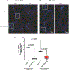
QD-antithombin probe pinpoints age-dependent 3-O-sulfated heparan sulfate. QD-AT staining of mouse aorta to image 3-O-sulfated heparan sulfate (3-OS-HS) in (A) control young aorta, heparitinases treated young aorta, and (B) control old aorta and heparitinases treated old aorta. (C) There is about 79-fold reduction in the fluorescence intensity of QD-AT staining (red) as aorta gets older from two weeks to six months (N = 3 mice, four different 20× cryosection images) (P < .0001), suggesting significant reduction in the anticoagulant HS structures. (Scale bar = 100 μm for 20×, 50 μm for 60×, red = QD-AT, blue = DAPI).
Figure 9.
Figure 9.
Heparin quality control. Oversulfated chondroitin sulfate (OSCS) contaminant in heparin was detected by gold-heparin-dye, a NSET nanoprobe. Heparitinase enzymes cleaved off dye labeled heparin polysaccharides into smaller oligosaccharide-dye, broke the NSET, and restored the dye fluorescence. The OSCS contaminant inhibited the heparitinase enzymes, resisted the heparin cleavage, and kept the fluorescence “off”. Serially diluted OSCS showed regular increase in the dye fluorescence.
Figure 10.
Figure 10.
Iron oxide-glycans image brain pathology. Iron oxide-sialyl LewisX targets activated E- and P-selectin in rat brains revealing brain pathology via MRI.
Figure 11.
Figure 11.
The Fe3O4/SiO2-sialic acid core-shell nanoparticle binds β-amyloid aggregates via GM1 ganglioside (sialic acid containing glycosphangolipid), reduces the T2* relaxation time, and detects 0.05 μM β-amyloid in in vitro. Redrawn from Ref. [14].
Figure 12.
Figure 12.
Overexpressed lectins on cancer cell surface mediate nanoparticle aggregation, reduce the relaxation time, and enhance the signal to noise ratio of T2 weighted images. Redrawn from Ref. [53].
Figure 13.
Figure 13.
AuNP stabilized with glycans and linked to tetraazacyclododecane triacetic acid (DO3A)—a chelator of MRI active gadolinium (III) detects glioma through MRI. Redrawn from Ref. [54].
Figure 14.
Figure 14.
Hyaluronic acid based nanoprobes. (A) HA nanoprobe binds LYVE-1 receptors on endothelium and reveals lymphatic vessel formation during cancer progression (Bhang SH, Won N, Lee T-J, et al. Hyaluronic acid-quantum dot conjugates for in vivo lymphatic vessel imaging. ACS Nano. 3(6), 1389–1398 (2009)). Inflammation upregulates CD44 expression on activated macrophages. HA nanoprobe identifies the CD44 and tracks activated macrophages (Kamat M, El-Boubbou K, Zhu DC, et al. Hyaluronic acid immobilized magnetic nanoparticles for active targeting and imaging of macrophages. Bioconjug. Chem. 21(11), 2128–2135 (2010)). (B) Gold-hyaluronic acid-dye probe assays hyaluronidase enzyme activity—a hallmark of rheumatoid arthritis and metastatic cancer. The fluorescence of the dye is ‘off’ before enzymatic cleavage due to nano-surface energy transfer (NSET) from the dye to gold nanoparticles. The hyaluronidase enzyme cleaves the nanoprobe, releases oligosaccharide-dye fragments, and switches ‘on’ the dye fluorescence by breaking the NSET phenomenon.
Figure 15.
Figure 15.
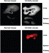
(A) Autoexposed fluorescent images of dissected livers (top), spleens (middle), and kidneys (bottom) 3 days after tail-vein injections of hyaluronic acid–quantum dot (HA-QDot) conjugates to normal and cirrhotic mice. (B) Fluorescent images of (A) at a detection wavelength of 790 nm.” With permission from Kim KS, Hur W, Park S-J, et al. Bioimaging for targeted delivery of hyaluronic acid derivatives to the livers in cirrhotic mice using quantum dots. ACS Nano. 4(6), 3005–3014 (2010)), copyright American Chemical Society, Washington, DC, 2010.
Figure 16.
Figure 16.
“Fluorescence microscope images of mouse ear tissues near the hyaluronic acid-quantum dot conjugate (HA-QD) injection site (A, B) and near the unconjugated QD injection site (C). The mouse ear tissues are vertically sectioned at 30 min after the subcutaneous injection of 200 nM HA-QD or unconjugated QD solution. The tissues are stained simultaneously by DAPI and fluorescent LYVE-1 antibodies. (A) Filter set is chosen to selectively show red fluorescence from QDs. (B, C) Fluorescence images are merged to overlay the red (QD), green (LYVE-1), and blue (DAPI) signals. The bright yellow co-localization spots of QD and LYVE-1 signals are indicated by arrow-heads in (B).” With permission from Bhang SH, Won N, Lee T-J, et al. Hyaluronic acid-quantum dot conjugates for in vivo lymphatic vessel imaging. ACS Nano. 3(6), 1389–1398 (2009), copyright American Chemical Society, Washington DC, 2009.
Figure 17.
Figure 17.
(A) N-acetylglucosamine (GlcNAc) labeled quantum dots (GlcNAc-QD) detect the localization of the GlcNAc binding proteins on the cell surface of a sperm. (B) “Confocal microscope imaging for staining of sperm with glycoquantum dots: (A) selective QDGLN labeling on the heads of sea-urchin sperm (scale bar = 20 mm), (B) close-up of QDGLN-labeled sea-urchin sperm, and C) close-up of QDMAN-labeled mouse sperm.” With permission of Robinson A, Fang J-M, Chou P–T, Liao K-W, Chu R-M, Lee S-J. Probing lectin and sperm with carbohydrate-modified quantum dots. Chembiochem. 6(10), 1899–1905 (2005), copyright Wiley & Sons, Hoboken, NJ.
Figure 18.
Figure 18.
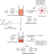
Glycosynapse disruption. Gold-lactose glyco-nanoparticle binds the GM3 glycosphingolipid on a cancer cell surface, sequesters the Gg3 lactosylceramide receptor on endothelial cells, and prevents adhesion and spreading of the cancer cells.
Figure 19.
Figure 19.
The gp120 glycoprotein on HIV-1 envelope binds high-mannose DC-SIGN receptor on dendritic cell and mediates viral entry. A glyco-nanoparticle mimetic consisting of mannoses (varied concentrations) arrests this interaction, isolates gp120 from DC-SIGN receptor, and inhibits the viral entry.
Figure 20.
Figure 20.
The B-subunit pentamer of Shiga toxin released by E. coli, S. dysentriae pathogens binds to Gb3 glycosphigolipid on an endothelial cell. Gold-trisaccharide nanoparticle mimics the Gb3 epitope and neutralizes the B-subunit. The glyco-nanoparticle inhibits internalization and cytosolic translocation of the Shiga toxin.
Figure 21.
Figure 21.
Anticancer glyco-nano vaccines. (A) Tetanus toxoid (TT) peptide-gold-silyl-Tn/LeY glyco-nanoparticle presents two cancer antigens: sialyl-Tn, a mucinassociated antigen expressed on epithelial cancer and LeY antigen for colon, liver, and prostate cancer. (B) C3d-peptide-gold-glycan vaccine exploits glycopeptides (consisting of Thomsen–Friedenreich (TF) disaccharide) of pancreatic adenocarcinoma and C3d adjuvant to elicit IgG and IgM antibody production.
Figure 22.
Figure 22.
Anti-pneumococcal vaccine. Gold nanoparticles decorated with tri-19F (trisaccharide fragment of serotype 19F), tetra-14 (tetrasaccharide fragment of serotype 14), and OVAp (T helper peptide) stimulate the immune system to generate IgG antibodies against tetra-14, while no IgG antibody was reported against tri-19F antigen.

References

    1. Kelly KL, Coronado E, Zhao LL, Schatz GC. The optical properties of metal nanoparticles: the influence of size, shape, and dielectric environment. J Phys Chem B 2003;107(3):668–77.
    1. Thakor AS, Jokerst JV, Ghanouni P, Campbell JL, Mittra E, Gambhir SS. Clinically approved nanoparticle imaging agents. J Nucl Med 2016;57(12):1833–7. - PMC - PubMed
    1. Ventola CL. Progress in nanomedicine: approved and investigational nanodrugs. P T 2017;42(12):742–55. - PMC - PubMed
    1. Bobo D, Robinson KJ, Islam J, Thurecht KJ, Corrie SR. Nanoparticle-based medicines: a review of FDA-approved materials and clinical trials to date. Pharm Res 2016;33(10):2373–87. - PubMed
    1. Abo-Zeid Y, Ismail NSM, McLean GR, Hamdy NM. A molecular-docking study repurposes FDA approved iron oxide nanoparticles to treat and control COVID-19 infection. Eur J Pharm Sci 2020;153105465. - PMC - PubMed

Publication types