Review
. 2022 Jun;126(11):1511-1528.
doi: 10.1038/s41416-022-01723-8. Epub 2022 Feb 21.

Future perspectives of uveal melanoma blood based biomarkers

Affiliations
Review

Future perspectives of uveal melanoma blood based biomarkers

Aaron B Beasley et al. Br J Cancer. 2022 Jun.

Abstract

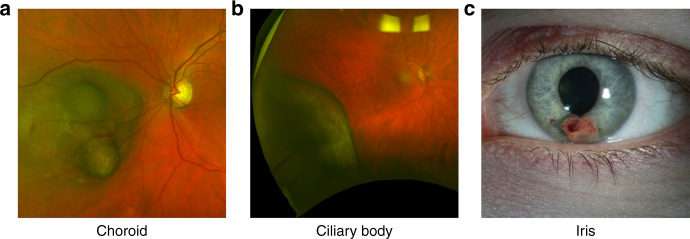
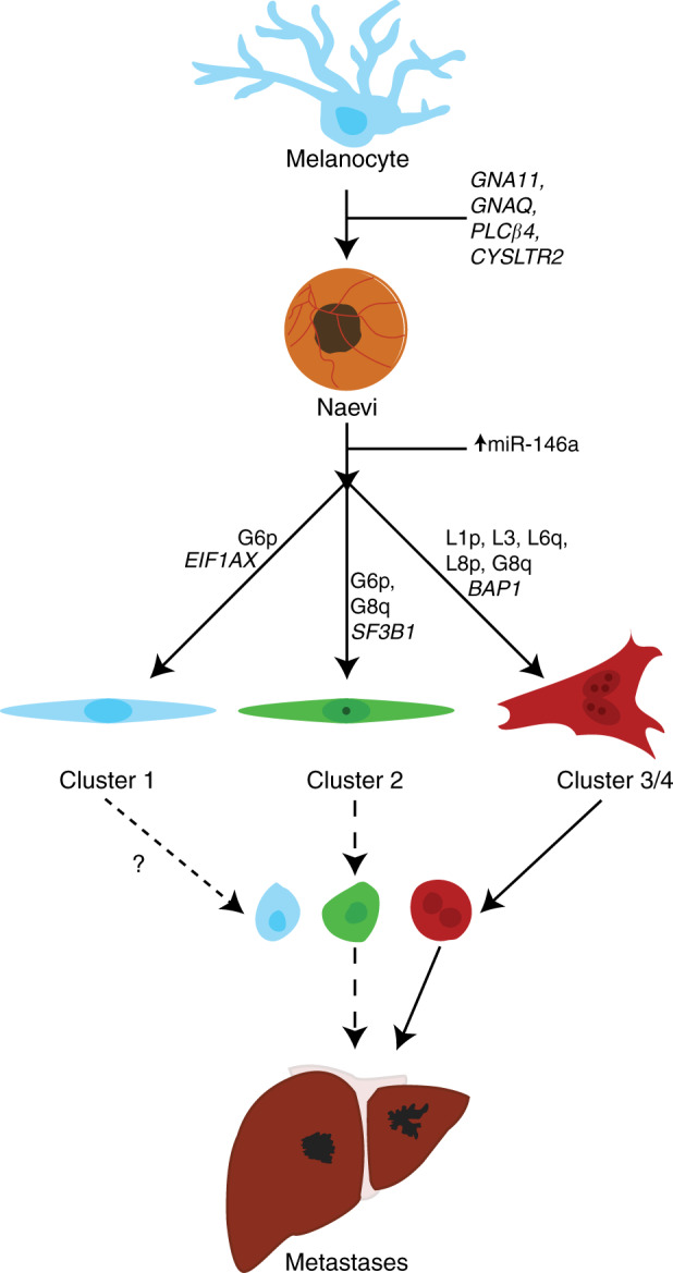
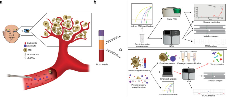
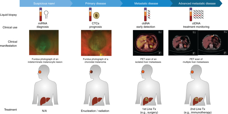
Uveal melanoma (UM) is the most common primary intraocular malignancy affecting adults. Despite successful local treatment of the primary tumour, metastatic disease develops in up to 50% of patients. Metastatic UM carries a particularly poor prognosis, with no effective therapeutic option available to date. Genetic studies of UM have demonstrated that cytogenetic features, including gene expression, somatic copy number alterations and specific gene mutations can allow more accurate assessment of metastatic risk. Pre-emptive therapies to avert metastasis are being tested in clinical trials in patients with high-risk UM. However, current prognostic methods require an intraocular tumour biopsy, which is a highly invasive procedure carrying a risk of vision-threatening complications and is limited by sampling variability. Recently, a new diagnostic concept known as "liquid biopsy" has emerged, heralding a substantial potential for minimally invasive genetic characterisation of tumours. Here, we examine the current evidence supporting the potential of blood circulating tumour cells (CTCs), circulating tumour DNA (ctDNA), microRNA (miRNA) and exosomes as biomarkers for UM. In particular, we discuss the potential of these biomarkers to aid clinical decision making throughout the management of UM patients.


Conflict of interest statement

The authors declare no competing interests.

Figures

Fig. 1
Fig. 1. Uveal Melanoma Locations.
Macroscopic images of uveal melanoma in the choroid (a), ciliary body (b), and iris (c).
Fig. 2
Fig. 2. Genetic pathways of uveal melanoma progression.
Non-prognostic mutations to GNA11, GNAQ, PLCβ4, and CYSLTR2 may drive the transformation from normal uveal melanocyte to early UM. Further genetic abnormalities dichotomise uveal melanoma into several prognostic groups. Patients with mutations to EIF1AX generally have the best prognosis and have no SCNA or G6p. Patients with mutations to SF3B1 have an intermediate prognosis and have G6p and/or abnormalities to chromosome 8. Lastly, patients with mutations to BAP1 have loss of chromosome 3 and gain of 8q, with larger gains in 8q equating to a shorter time to metastases. Each class of tumour may release CTCs at different rates and quantities, and may cause the formation of metastases.
Fig. 3
Fig. 3. Liquid biopsy.
a CTCs, ctDNA, and cmiRNA are minor components of the blood, mixed with the normal erythrocytes, leucocytes, and cell-free nucleic acids. A minimally invasive venous blood collection has the potential to provide genetic information of all tumours within the body. b Plasma derived circulating nucleic acids (DNA, RNA, miRNA) are generally extracted using commercially available kits. Following extraction, ctDNA or cmiRNA can be detected by real time-qPCR, digital PCR or NGS, to detect the presence of disease, to detect mutations or SCNAs for diagnostic, prognostic, or genetic changes in response to therapy. c Circulating tumour cells are isolated from the blood by antibody directed methods such as immunomagnetic beads, or through their physical properties such as size and deformability. Once isolated, CTCs can be indirectly quantified using RT-PCR, or directly quantified using immunocytochemistry. Fluorescence in-situ hybridisation (FISH) can be used to detect SCNAs or changes to mRNA or miRNA expression. Single cell sequencing of CTCs can provide mutational and/or chromosomal copy number information.
Fig. 4
Fig. 4. Suggested applications of liquid biopsy for the management of uveal melanoma.
Plasma cmiRNA profiling could be used to distinguish a suspicious naevus from a small UM lesion. Upon diagnosis of UM, analysis of CTCs can provide information on prognostically relevant SCNAs. After treatment of the primary tumour, regular monitoring of plasma ctDNA could serve as an early indicator metastatic disease. Plasma ctDNA can be also used for monitoring of response to treatment and disease progression in patients with metastatic disease.

References

    1. Dogrusoz M, Jager MJ, Damato B. Uveal melanoma treatment and prognostication. Asia Pac J Ophthalmol (Philos) 2017;6:186–96. - PubMed
    1. Damato B. Detection of uveal melanoma by optometrists in the United Kingdom. Ophthalmic Physiol Opt. 2001;21:268–71. doi: 10.1046/j.1475-1313.2001.00595.x. - DOI - PubMed
    1. Chattopadhyay C, Kim DW, Gombos DS, Oba J, Qin Y, Williams MD, et al. Uveal melanoma: From diagnosis to treatment and the science in between. Cancer. 2016;122:2299–312. doi: 10.1002/cncr.29727. - DOI - PMC - PubMed
    1. Ah-Fat FG, Damato BE. Delays in the diagnosis of uveal melanoma and effect on treatment. Eye (Lond) 1998;12:781–2. doi: 10.1038/eye.1998.202. - DOI - PubMed
    1. Shields CL, Furuta M, Thangappan A, Nagori S, Mashayekhi A, Lally DR, et al. Metastasis of uveal melanoma millimeter-by-millimeter in 8033 consecutive eyes. Arch Ophthalmol. 2009;127:989–98. doi: 10.1001/archophthalmol.2009.208. - DOI - PubMed

Publication types