. 2022 Jul;32(7):4537-4546.
doi: 10.1007/s00330-022-08568-y. Epub 2022 Feb 21.

Synthetic CT for the planning of MR-HIFU treatment of bone metastases in pelvic and femoral bones: a feasibility study

Affiliations

Synthetic CT for the planning of MR-HIFU treatment of bone metastases in pelvic and femoral bones: a feasibility study

Beatrice Lena et al. Eur Radiol. 2022 Jul.

Abstract

Objectives: Visualization of the bone distribution is an important prerequisite for MRI-guided high-intensity focused ultrasound (MRI-HIFU) treatment planning of bone metastases. In this context, we evaluated MRI-based synthetic CT (sCT) imaging for the visualization of cortical bone.

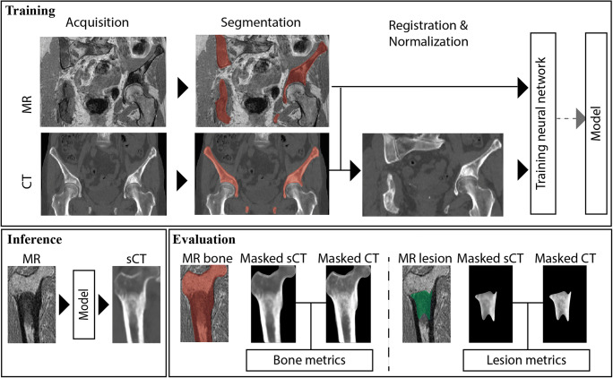
Methods: MR and CT images of nine patients with pelvic and femoral metastases were retrospectively analyzed in this study. The metastatic lesions were osteolytic, osteoblastic or mixed. sCT were generated from pre-treatment or treatment MR images using a UNet-like neural network. sCT was qualitatively and quantitatively compared to CT in the bone (pelvis or femur) containing the metastasis and in a region of interest placed on the metastasis itself, through mean absolute difference (MAD), mean difference (MD), Dice similarity coefficient (DSC), and root mean square surface distance (RMSD).

Results: The dataset consisted of 3 osteolytic, 4 osteoblastic and 2 mixed metastases. For most patients, the general morphology of the bone was well represented in the sCT images and osteolytic, osteoblastic and mixed lesions could be discriminated. Despite an average timespan between MR and CT acquisitions of 61 days, in bone, the average (± standard deviation) MAD was 116 ± 26 HU, MD - 14 ± 66 HU, DSC 0.85 ± 0.05, and RMSD 2.05 ± 0.48 mm and, in the lesion, MAD was 132 ± 62 HU, MD - 31 ± 106 HU, DSC 0.75 ± 0.2, and RMSD 2.73 ± 2.28 mm.

Conclusions: Synthetic CT images adequately depicted the cancellous and cortical bone distribution in the different lesion types, which shows its potential for MRI-HIFU treatment planning.

Key points: • Synthetic computed tomography was able to depict bone distribution in metastatic lesions. • Synthetic computed tomography images intrinsically aligned with treatment MR images may have the potential to facilitate MR-HIFU treatment planning of bone metastases, by combining visualization of soft tissues and cancellous and cortical bone.

Keywords: CT; High-intensity focused ultrasound ablation; MRI; Neoplasm metastasis; Synthetic CT.


Conflict of interest statement

The authors of this manuscript declare relationships with the following companies: PS and MvS are minority shareholders at MRIguidance BV.

Figures

Fig. 1
Fig. 1
Schematic description of the approach. The bone containing the lesion was segmented on MRI and CT and used to register the CT to the MRI. Patches of 24 × 24 × 24 voxels were then extracted from the MRI and registered CT to train a synthetic CT (sCT) generation model. Once trained, the model was used to create a sCT of the bone of interest from a patient not seen during the training. The sCT was then evaluated in the bone of interest and the metastatic lesion
Fig. 2
Fig. 2
MR, CT, synthetic CT (sCT), and sCT-to-CT difference (Δ) obtained for three patients presenting (a) osteoblastic, (b) osteolytic, and (c) mixed lesions. Red boxes on the MR images indicate the region that was zoomed in to compare CT and sCT images
Fig. 3
Fig. 3
Comparison of single slices out of the MR, CT, and sCT datasets of all patients, divided by their sCT-CT correspondence in the lesion. For most patients, lesions could be correctly identified on sCT images. For three patients, differences were observed between CT and sCT images, but it is hard to judge whether they are due to pathological changes (e.g., calcium-enriched bone visible in CT but not MRI in P8) or error in sCT reconstruction. For one patient, definite sCT reconstruction errors are visible with sclerotic regions not well depicted. Stars indicate different time spans between pretreatment CT and treatment MR: * < 10 days, ** 10–35 days, *** > 100 days
Fig. 4
Fig. 4
a Pretreatment CT images in the pelvis with conventional patient positioning and MR and intrinsically aligned synthetic CT (sCT) images with unconventional patient positioning for the HIFU treatment. Lesion is indicated by *. Since sCT scans are intrinsically registered with MR, they are able to provide bone cortex depiction with the patient in treatment position. b MR and a fused visualization of MR and sCT images depicting the cortical bone distribution, obtained for two patients with osteolytic lesions in the femur (left) and in the pelvic iliac crest (right). MR and sCT are inherently registered, allowing straightforward identification of soft tissues and bone. c MR images from transverse slices of two patients with lesions in the femur and pelvis and corresponding 3D bone renderings that provide an overview of the bone with the lesion. The bone renderings were obtained by thresholding the sCT at 150 Hounsfield units within the bone mask (created in MeVisLab v3.2, MeVis Medical Solutions AG). The red line identifies the same location in MR images and 3D bone renderings

References

    1. Najafi A, Sartoretti E, Binkert CA. Sacroiliac joint ablation using MR-HIFU. Cardiovasc Intervent Radiol. 2019;42:1363–1365. doi: 10.1007/s00270-019-02263-0. - DOI - PubMed
    1. Scipione R, Anzidei M, Bazzocchi A, et al. HIFU for bone metastases and other musculoskeletal applications. Semin Interv Radiol. 2018;35:261–267. doi: 10.1055/s-0038-1673363. - DOI - PMC - PubMed
    1. Napoli A, Bazzocchi A, Scipione R, et al. Noninvasive therapy for osteoid osteoma: a prospective developmental study with MR imaging-guided high-intensity focused ultrasound. Radiology. 2017;285:186–196. doi: 10.1148/radiol.2017162680. - DOI - PubMed
    1. Bing F, Vappou J, de Mathelin M, Gangi A. Targetability of osteoid osteomas and bone metastases by MR-guided high intensity focused ultrasound (MRgHIFU) Int J Hyperthermia. 2018;35:471–479. doi: 10.1080/02656736.2018.1508758. - DOI - PubMed
    1. Bitton RR, Rosenberg J, LeBlang S, et al. MRI-guided focused ultrasound of osseous metastasestreatment parameters associated with successful pain reduction. Invest Radiol. 2021;56:141–146. doi: 10.1097/rli.0000000000000721. - DOI - PubMed

MeSH terms