. 2022 Jun;66(4):483-494.
doi: 10.1111/1754-9485.13390. Epub 2022 Feb 22.

18 F-FDG PET/CT features of immune-related adverse events and pitfalls following immunotherapy

Affiliations

18 F-FDG PET/CT features of immune-related adverse events and pitfalls following immunotherapy

Martin H Cherk et al. J Med Imaging Radiat Oncol. 2022 Jun.

Abstract

18 F-FDG PET/CT scanning is routinely performed to stage and evaluate the treatment response in many malignancies. Immunotherapy is a rapidly growing treatment option for many cancers, and both clinicians and imaging specialists need to be familiar with 18 F-FDG PET/CT imaging characteristics unique to patients on this type of treatment. In particular, many immune-related adverse events (irAEs) can be detected on 18 F-FDG PET/CT and early accurate identification is critical to reduce treatment related morbidity and incorrect interpretation of malignant disease status. This pictorial essay reviews frequently encountered irAEs in clinical practice and their appearances on 18 F-FDG PET/CT along with a brief discussion on pseudoprogression and hyperprogression.

Keywords: 18F-FDG PET; nuclear imaging; nuclear medicine; oncologic imaging.


Figures

Fig. 1
Fig. 1
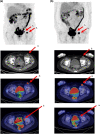
Reactive nodes. Fifty‐year‐old woman with metastatic melanoma treated with surgery and four cycles pembrolizumab. (a) PET MIP demonstrates mildly increased 18F‐FDG uptake in right axillary, right level IB and right level III cervical nodes. (b) Axial Fused PET/CT images of the same nodes. As the nodes are not in the drainage basin of the primary site of malignancy (lower limb) and are only mildly 18F‐FDG‐avid, they are more consistent with immunotherapy related nodal activation/reactive change rather than metastatic disease. [Colour figure can be viewed at wileyonlinelibrary.com]
Fig. 2
Fig. 2
Reactive marrow and spleen. Seventy‐three‐year‐old man with surgically resected metastatic melanoma treated with adjuvant pembrolizumab. (a) PET MIP demonstrates diffuse moderately increased 18F‐FDG uptake throughout the marrow of the axial and proximal appendicular skeleton in keeping with immune mediated marrow activation. (b) Axial Fused PET/CT images of the spleen demonstrate splenomegaly and diffuse mildly increased 18F‐FDG uptake throughout and subtle reversal of normal liver to spleen 18F‐FDG ratio. [Colour figure can be viewed at wileyonlinelibrary.com]
Fig. 3
Fig. 3
Thyroiditis. Sixty‐three‐year‐old woman with metastatic melanoma to nodes and subcutaneous tissues treated with pembrolizumab. (a) Baseline PET MIP demonstrating nodal and soft tissue metastases. (b) Post four cycles pembrolizumab PET MIP demonstrating complete resolution of metastatic disease but diffusely increased 18F‐FDG uptake in both lobes of thyroid gland in keeping with thyroiditis. (c) Axial Fused PET/CT images of the thyroid gland confirms increased 18F‐FDG uptake in both lobes. [Colour figure can be viewed at wileyonlinelibrary.com]
Fig. 4
Fig. 4
Synovitis/arthritis medium and large joints. Seventy‐four‐year‐old woman with metastatic melanoma treated with pembrolizumab. (a) PET MIP post six cycles pembrolizumab demonstrating diffuse mild to moderately increased 18F‐FDG uptake in the shoulders, knees, ankles and wrists consistent with immune mediated synovitis/reactive arthropathy. (b) Axial Fused PET/CT images of shoulders, knees and ankles demonstrating corresponding sites of increased 18F‐FDG uptake. [Colour figure can be viewed at wileyonlinelibrary.com]
Fig. 5
Fig. 5
Polymyalgia rheumatica and colitis. Fifty‐eight‐year‐old man with metastatic melanoma treated with combination ipilimumab and nivolumab followed by maintenance nivolumab. (a) Baseline PET MIP demonstrates widespread skeletal, liver and right submandibular nodal metastatic disease. (b) PET MIP and Axial fused PET/CT images post four cycles ipilimumab and nivolumab demonstrate severe relatively symmetrical synovitis/inflammation of the shoulders, sternoclavicular joints, hip joints and interspinous regions in a pattern typical for Polymyalgia Rheumatica. Colitis is also present in the descending colon. (c) PET MIP images following 100 mg weaning Prednisolone dose daily for 3 weeks and maintenance nivolumab demonstrates complete resolution of Synovitis/Polymyalgia Rheumatica and colitis and reduction in metastatic disease. [Colour figure can be viewed at wileyonlinelibrary.com]
Fig. 6
Fig. 6
Vasculitis and thyroiditis. Fifty‐two‐year‐old man with metastatic melanoma treated with combination ipilimumab and nivolumab. (a) Baseline PET MIP demonstrates widespread left pulmonary and pleural, left adrenal, small bowel and right thigh muscle metastases. (b) PET MIP Post three cycles ipilimumab and nivolumab demonstrates partial response at sites of metastatic disease, autoimmune thyroiditis (biochemically hypothyroid) and widespread severe medium and large vessel vasculitis of the upper and lower limbs. (c) PET MIP 3 weeks post 50 mg Prednisolone daily demonstrates reduction in medium and to a lesser extent large vessel vasculitis in the upper and lower limbs. (d) PET MIP 3 months post completion of ipilimumab and nivolumab demonstrates resolution of vasculitis and persistent low volume metastatic disease.
Fig. 7
Fig. 7
Myositis. Thirty‐nine‐year‐old woman with metastatic melanoma treated with combination ipilimumab and nivolumab followed by maintenance nivolumab. (a) Baseline PET MIP demonstrates extensive intra‐thoracic and to a lesser extent cervical and mesenteric nodal metastatic disease. (b) PET MIP and Axial fused PET/CT images post four cycles ipilimumab and nivolumab demonstrate diffuse mild to moderately increased 18F‐FDG uptake throughout skeletal muscles of the upper and lower arms and chest wall consistent with myositis. Marked reduction in metastatic disease also noted. (c) PET MIP images 4 months later on maintenance nivolumab demonstrates complete resolution of myositis and metastatic disease. [Colour figure can be viewed at wileyonlinelibrary.com]
Fig. 8
Fig. 8
Pneumonitis. (a) Seventy‐five‐year‐old man with metastatic melanoma on pembrolizumab. PET MIP and Axial fused PET/CT images demonstrate mildly 18F‐FDG‐avid ground glass change on CT in the left upper lobe typical of early immune mediated pneumonitis. (b) Eighty‐five‐year‐old man with metastatic melanoma on pembrolizumab. PET MIP, Axial Fused PET/CT and CT images demonstrate intensely 18F‐FDG ‐avid organizing pneumonia pattern left lower lobe, right upper and middle lobes consistent with advanced pneumonitis. This completely resolved subsequently with a 75 mg weaning dose of prednisolone over 3 weeks. [Colour figure can be viewed at wileyonlinelibrary.com]
Fig. 9
Fig. 9
Colitis. Seventy‐year‐old man with metastatic melanoma treated with four cycles pembrolizumab. (a) Baseline PET MIP demonstrates low volume metastatic disease right cervical nodes, subcutaneous tissues left upper neck and right upper back. (b) Post four cycles pembrolizumab PET MIP demonstrating complete resolution of metastatic disease but diffuse markedly increased 18F‐FDG uptake throughout the colon in keeping with Colitis. (c) Axial fused PET/CT images demonstrate corresponding diffuse intense 18F‐FDG uptake throughout the transverse colon. [Colour figure can be viewed at wileyonlinelibrary.com]
Fig. 10
Fig. 10
Pancreatitis. Sixty‐five‐year‐old man with metastatic melanoma treated with pembrolizumab. (a) Baseline PET MIP demonstrates complete metabolic tumour remission post four cycles pembrolizumab. Small focus intense 18F‐FDG uptake L2/3 spinous process in keeping with benign arthritic change. Further small focus intense 18F‐FDG uptake right pelvis in keeping with physiological urinary activity right ureter. (b) Post eight cycles pembrolizumab PET MIP demonstrates intense 18F‐FDG uptake in colon (colitis), small bowel (enteritis) and moderate uptake in pancreas (pancreatitis). (c) Axial Fused PET/CT images demonstrating intense 18F‐FDG uptake colon and small bowel loops and mild‐to‐moderate uptake in pancreas (arrow) in keeping with pancreatitis. [Colour figure can be viewed at wileyonlinelibrary.com]
Fig. 11
Fig. 11
Hypophysitis. Sixty‐one‐year‐old man with metastatic melanoma treated with combination ipilimumab and nivolumab. (a) Baseline Axial Fused PET/CT images of pituitary gland. (b) Post four cycles immunotherapy Axial Fused PET/CT images demonstrating focal intense 18F‐FDG uptake in the pituitary gland consistent with hypophysitis. [Colour figure can be viewed at wileyonlinelibrary.com]
Fig. 12
Fig. 12
Panniculitis/granulomatous disease. Sixty‐year‐old woman with metastatic melanoma treated with combination ipilimumab and nivolumab. (a) Baseline PET MIP demonstrates multiple liver metastases. (b) PET MIP and Axial fused PET/CT images post two cycles ipilimumab and nivolumab demonstrate relatively symmetrical intensely 18F‐FDG‐avid bilateral pulmonary hilar and mediastinal nodes in keeping with immune mediated granulomatous disease/sarcoidosis. Multiple intensely 18F‐FDG‐avid subcutaneous nodules bilateral elbow, gluteal, thigh and knee regions confirmed on biopsy as non‐necrotizing granulomata (panniculitis). Multiple new 18F‐FDG‐avid bony lesions in T10, T11, T12 and the left ilium in keeping with new metastatic disease or further bone granulomata. (c) PET MIP images following 75 mg weaning Prednisolone dose daily for 3 weeks demonstrating complete resolution of subcutaneous panniculitis and reduction of pulmonary hilar and mediastinal granulomatous disease. [Colour figure can be viewed at wileyonlinelibrary.com]
Fig. 13
Fig. 13
Melanoma pseudoprogression. Eighty‐six‐year‐old woman with metastatic melanoma treated with pembrolizumab. (a) Baseline PET MIP, Axial CT and fused PET/CT demonstrates metastatic left external iliac (Arrow 1) and left inguinal (Arrow 2) nodal metastases. (b) PET MIP, Axial CT, Axial fused PET/CT 12 weeks post pembrolizumab treatment demonstrates resolution of left external iliac node (Arrow 1) but significant increase in size and FDG‐avidity of left inguinal node (Arrow 2) suggestive of disease progression. Left inguinal node surgically excised 4 weeks later, with histopathology demonstrating fibrosis and chronic inflammatory change only with no evidence of melanoma (pseudoprogression). [Colour figure can be viewed at wileyonlinelibrary.com]

References

    1. Mellman I, Coukos G, Dranoff G. Cancer immunotherapy comes of age. Nature 2011; 480: 480–9. - PMC - PubMed
    1. Robert C, Long GV, Brady B et al. Nivolumab in previously untreated melanoma without BRAF mutation. N Engl J Med 2015; 372: 320–30. - PubMed
    1. Reck M, Rodriguez‐Abreu D, Robinson AG et al. Pembrolizumab versus chemotherapy for PD‐L1‐positive non‐small‐cell lung cancer. N Engl J Med 2016; 375: 1823–33. - PubMed
    1. Chan KK, Bass AR. Autoimmune complications of immunotherapy: pathophysiology and management. BMJ 2020; 369: m736. - PubMed
    1. Larsabal M, Marti A, Jacquemin C et al. Vitiligo‐like lesions occurring in patients receiving anti‐programmed cell death‐1 therapies are clinically and biologically distinct from vitiligo. J Am Acad Dermatol 2017; 76: 863–70. - PubMed

MeSH terms

Substances