. 2022 Feb 22;7(4):e155836.
doi: 10.1172/jci.insight.155836.

Renal tubular peroxisomes are dispensable for normal kidney function

Affiliations

Renal tubular peroxisomes are dispensable for normal kidney function

Camille Ansermet et al. JCI Insight. .

Abstract

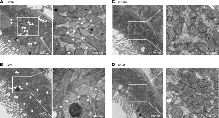
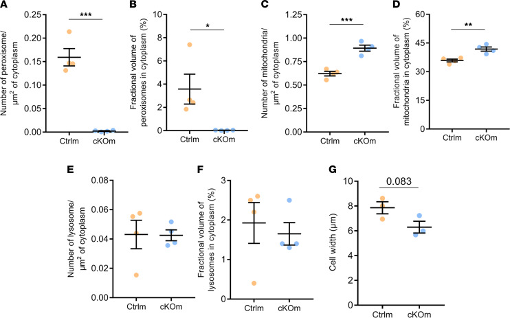
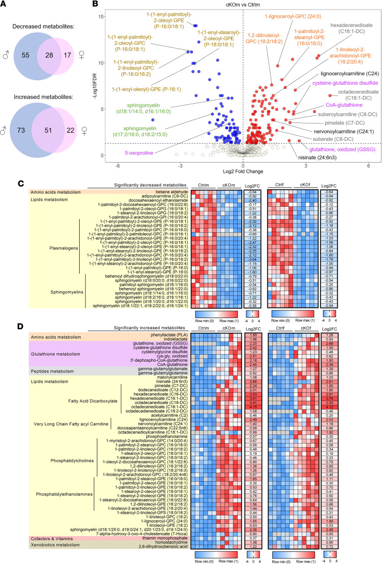
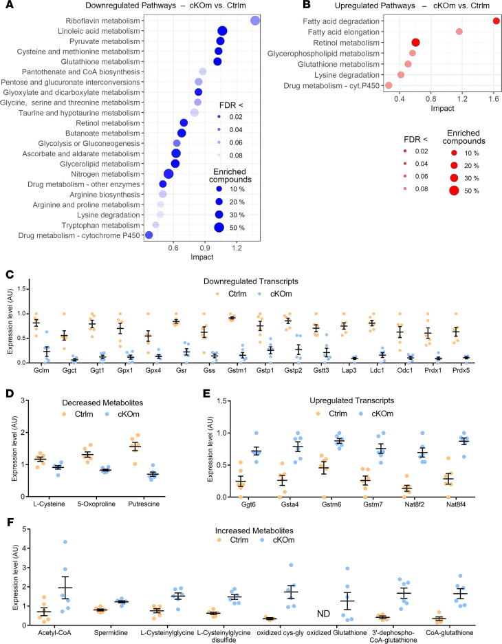
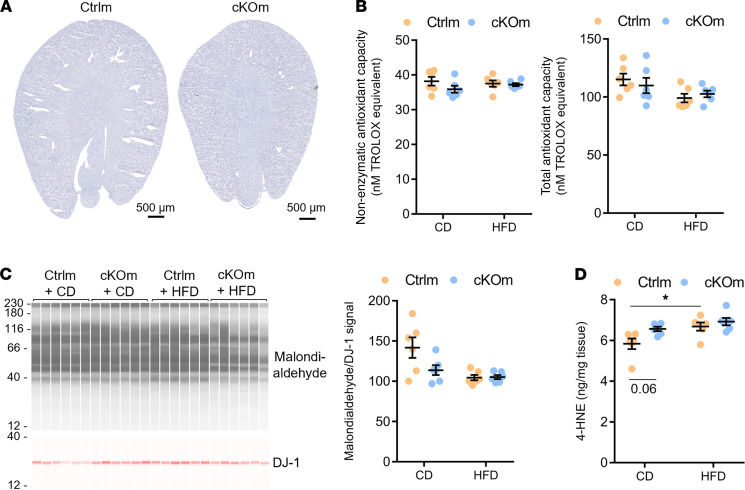
Peroxisomes are specialized cellular organelles involved in a variety of metabolic processes. In humans, mutations leading to complete loss of peroxisomes cause multiorgan failure (Zellweger's spectrum disorders, ZSD), including renal impairment. However, the (patho)physiological role of peroxisomes in the kidney remains unknown. We addressed the role of peroxisomes in renal function in mice with conditional ablation of peroxisomal biogenesis in the renal tubule (cKO mice). Functional analyses did not reveal any overt kidney phenotype in cKO mice. However, infant male cKO mice had lower body and kidney weights, and adult male cKO mice exhibited substantial reductions in kidney weight and kidney weight/body weight ratio. Stereological analysis showed an increase in mitochondria density in proximal tubule cells of cKO mice. Integrated transcriptome and metabolome analyses revealed profound reprogramming of a number of metabolic pathways, including metabolism of glutathione and biosynthesis/biotransformation of several major classes of lipids. Although this analysis suggested compensated oxidative stress, challenge with high-fat feeding did not induce significant renal impairments in cKO mice. We demonstrate that renal tubular peroxisomes are dispensable for normal renal function. Our data also suggest that renal impairments in patients with ZSD are of extrarenal origin.

Keywords: Genetic diseases; Nephrology.


Figures

Figure 1
Figure 1. Validation of the cKO model and basic characteristics of cKOm and cKOf mice.
(A) Relative Pex5 mRNA expression in kidneys of Ctrlm, cKOm, Ctrlf, and cKOf mice (n = 5–9). One hundred percent corresponds to the mean of Pex5 mRNA expression in kidneys of Ctrlm mice. (B) Western blot analysis of PEX5 protein expression in kidneys of cKOm and cKOf or of Ctrlf and cKOf mice. (C) Kidney weights of Ctrlm, cKOm, Ctrlf, and cKOf mice (n = 12–13). (D) Kidney weight/body weight ratio for Ctrlm, cKOm, Ctrlf, and cKOf mice (n = 5–6). Box and whiskers represent mean ± SEM; unpaired t test, ***P < 0.0001, *P < 0.05. The original full-length Western blot images for B are shown in Supplemental Figure 9.
Figure 2
Figure 2. Validation of the cKO model by electron microscopy.
(AD) Electron microscopy images of kidney proximal tubules in kidney cortex of Ctrlm (A), Ctrlf (B), cKOm (C), and cKOf (D) mice. The images are representative of 4 mice/genotype with 15 images analyzed/mice. White arrowheads indicate peroxisomes.
Figure 3
Figure 3. Stereological analysis of proximal tubule cells in kidneys of Ctrlm and cKOm mice.
(A) Number of peroxisomes/μm2 of cytoplasm; (B) fractional volume of peroxisomes in percentage of cytoplasm occupied by peroxisomes; (C) number of mitochondria/μm2 of cytoplasm; (D) fractional volume of mitochondria in percentage of cytoplasm occupied by mitochondria; (E) number of lysosomes/μm2 of cytoplasm; (F) fractional volume of lysosomes in percentage of cytoplasm occupied by lysosomes; (G) cell width. P = 0.083 (G). Stereology analysis was performed on n = 3–4 mice with 3 kidney cortex pieces per mouse, 15 micrographs per sample. Box and whiskers represent mean ± SEM; unpaired t test, ***P < 0.0001, **P < 0.001, *P < 0.05.
Figure 4
Figure 4. Transcriptional reprogramming in the kidneys of cKO mice.
(A and B) Volcano plot representing the relative transcriptional expression of all renal transcripts in cKOm versus Ctrlm (A) or cKOf versus Ctrlf (B). Transcripts depicted in blue are significantly downregulated while transcripts depicted in red are significantly upregulated. (C) Venn diagrams showing the number of transcripts significantly downregulated or upregulated in cKOm (in blue) or cKOf (in pink) mice versus Ctrl mice of the same sex. A significant transcript regulation is considered when the adjusted P value referred to as “FDR” is <0.05. (D) Enrichment analysis of a homemade gene set (based on the KEGG pathway mmu04146) targeting 100 transcripts related to peroxisomal functions, in cKOm versus Ctrlm mice. (E and F) Scatter plot of the top 25 most downregulated (E) or upregulated (F) metabolic pathways in cKOm versus Ctrlm mice, based on an untargeted GSEA using a database of 543 KEGG metabolic pathways. Pathways are sorted by their absolute normalized enrichment score. A significant pathway regulation can be considered when adjusted P value referred to as “q value” is <0.2.
Figure 5
Figure 5. Remodeling of the renal metabolome in cKO mice.
(A) Venn diagrams representing the number of detected renal metabolites showing a significantly decreased or increased abundance in cKOm (in blue) or cKOf (in pink) mice versus Ctrl mice of the same sex. (B) Volcano plot representing the relative abundance of all detected metabolites in kidneys of cKOm versus Ctrlm. Metabolites depicted with blue dots are significantly less abundant, and transcripts depicted with red dots are significantly more abundant in kidneys of cKOm mice as compared with Ctrlm mice. The names of some representative metabolites are depicted using colors shared for related metabolites. (C and D) Heatmaps of metabolites showing a significantly decreased (C) or increased (D) abundance in cKO mice of both sexes as compared with Ctrl mice. A significant difference of abundance is considered when adjusted P value from a 2-way ANOVA referred to as “FDR” is <0.05. Metabolites are identified by their biochemical name and sorted by related metabolisms and subclasses of metabolites. For each metabolite, the individual expression of 6 Ctrl and 6 cKO mice normalized between 0 and 1 and the log2-transformed mean fold change of expression (Log2FC) in cKO versus Ctrl mice are given, for both sexes. For calculation of the mean FC of expression, missing values (depicted in gray) have been replaced by the minimum value of both genotypes from the same sex.
Figure 6
Figure 6. Joint analysis of transcriptional and metabolic changes in cKOm mice.
(A and B) Scatter plot of significantly downregulated (A) or upregulated (B) metabolic pathways, based on a joint pathway analysis of both regulated transcripts and modulated metabolites in kidneys of cKOm versus Ctrlm mice. Pathways are sorted by their absolute impact, and a significant pathway regulation is considered when adjusted P value referred to as “FDR” is <0.1. The size of each dot depends on the percentage of all transcripts and metabolites (“compounds”) of the pathway that are significantly affected in cKOm mice. (C and D) Relative expression of glutathione-related transcripts (C) and metabolites (D) significantly less abundant (FDR < 0.05) in cKOm mice as compared with Ctrlm mice. (E and F) Relative expression of glutathione-related transcripts (E) and metabolites (F) significantly more abundant (FDR < 0.05) in cKOm mice as compared with Ctrlm mice. Individual values from 6 cKOm and 6 Ctrlm mice are depicted after transformation from raw individual data: values of metabolite abundance have been divided by the median value of both genotypes, while values of transcript expression from both genotypes have been normalized between 0 and 1. Box and whiskers represent the mean and the SEM, respectively. ND, not detected.
Figure 7
Figure 7. Analysis of the antioxidant capacity and level of lipid peroxidation in cKOm mice fed under HFD.
(A) Gross morphological structure and Oil Red O staining of neutral lipid depositions (in red) in the kidney of cKOm and Ctrlm adult mice fed under HFD for 4 weeks. The absence of the red color indicates the absence of lipid deposition. (B) Results of a Trolox assay performed on renal extracts showing nonenzymatic antioxidant capacity (left panel) and total antioxidant capacity (right panel) of Ctrlm and cKOm mice fed under control diet (CD) or HFD for 4 weeks. (C) Immunoblot performed on renal extracts targeting the product of lipid peroxidation malondialdehyde (left panel) and its quantification (right panel) in Ctrlm and cKOm mice fed under CD or HFD for 4 weeks. DJ-1 immunoblot is used as a loading control to normalize malondialdehyde abundance. (D) Amount of 4-HNE measured by competitive ELISA in kidney extracts from Ctrlm and cKOm mice fed under CD or HFD for 4 weeks. Box and whiskers represent mean ± SEM. Two-way ANOVA and post hoc Tukey’s multiple comparisons test, *P < 0.05.

References

    1. Rhodin JAG. Correlation of ultrastructural oganisation and function in normal and experimentally changed proximal tubule cells of the mouse kidney: an electron microscopic study. PhD Thesis. Karolinska Institutet; 1954.
    1. Goeckermann JA, Vigil EL. Peroxisome development in the metanephric kidney of mouse. J Histochem Cytochem. 1975;23(12):957–973. doi: 10.1177/23.12.440. - DOI - PubMed
    1. Reddy JK, et al. Microbody (peroxisome) proliferation in mouse kidney induced by methyl clofenapate. Virchows Arch B Cell Pathol. 1975;17(4):295–306. - PubMed
    1. Berendse K, et al. Zellweger spectrum disorders: clinical manifestations in patients surviving into adulthood. J Inherit Metab Dis. 2016;39(1):93–106. doi: 10.1007/s10545-015-9880-2. - DOI - PMC - PubMed
    1. Gilchrist KW, et al. Studies of malformation syndromes of man XIB: the cerebro-hepato-renal syndrome of Zellweger: comparative pathology. Eur J Pediatr. 1976;121(2):99–118. doi: 10.1007/BF00443065. - DOI - PubMed

Publication types