Review
. 2022 Apr;57(4):225-245.
doi: 10.1007/s00535-022-01857-9. Epub 2022 Feb 22.

Amendment of the Japanese consensus guidelines for autoimmune pancreatitis, 2020

Affiliations
Review

Amendment of the Japanese consensus guidelines for autoimmune pancreatitis, 2020

Kazuichi Okazaki et al. J Gastroenterol. 2022 Apr.

Abstract

In response to the latest knowledge and the amendment of the Japanese diagnostic criteria for autoimmune pancreatitis (AIP) in 2018, the Japanese consensus guidelines for managing AIP in 2013 were required to be revised. Three committees [the professional committee for developing clinical questions (CQs) and statements by Japanese specialists; the expert panelist committee for rating statements by the modified Delphi method; and the evaluating committee of moderators] were organized. Twenty specialists in AIP extracted the specific clinical statements from a total of 5218 articles (1963-2019) from a search in PubMed and the Cochrane Library. The professional committee made 14, 9, 5, and 11 CQs and statements for the current concept and diagnosis, extra-pancreatic lesions, differential diagnosis, and treatment, respectively. The expert panelists regarded the statements as valid after a two-round modified Delphi approach with individually rating these clinical statements, in which a clinical statement receiving a median score greater than 7 on a 9-point scale from the panel was regarded as valid. After evaluation by the moderators, the amendment of the Japanese consensus guidelines for AIP has been proposed in 2020.

Keywords: Autoimmune pancreatitis; Delphi method; Diagnosis; Guidelines; Treatment.


Figures

Fig. 1
Fig. 1
3D MRCP in AIP. 3D MRCP shows narrowing of the main pancreatic duct in the pancreatic body and tail (arrows)
Fig. 2
Fig. 2
Cholangiography and IDUS findings of IgG4-related sclerosing cholangitis. A Cholangiography reveals hilar stenosis. a. IDUS findings at a stenotic region showing entire circumferential and symmetrical wall thickening and homogeneous inner zone. b IDUS findings at a non-stenotic region showing similar wall thickening and homogeneous inner zone, with the smooth inner surface and outer margin observed in the stenotic region. B Differentiation by IDUS findings. In IgG4-SC, IDUS shows preservation of the three-layer structure, symmetrical thickening of the entire circumference of the wall, and homogeneous inner zone at affected stenotic regions. Even at non-stenotic regions, similar wall thickening and homogeneous inner zone can be seen. In PSC, IDUS displays asymmetrical wall thickening with irregular inner surface and interrupted outer margins, heterogeneous inner zone, destruction of the three-layer structure, and a diverticular-like pouch. In bile duct cancer, IDUS shows asymmetrical wall thickening with irregular inner surface and interrupted outer margin according to cancer invasion as well as heterogeneous inner zone. Unlike IgG4-SC, there is no wall thickening at non-stenotic regions
Fig. 3
Fig. 3
Enhanced CT images of AIP-related retroperitoneal fibrosis. A Enhanced CT (late phase) shows soft-tissue masses around both ureters (arrows). B Enhanced CT (arterial phase) reveals a soft-tissue mass anterior to the vertebra (arrow)
Fig. 4
Fig. 4
CT/MRI images of AIP-related kidney disease (AIP-KD). A Dynamic contrast-enhanced CT (arterial phase) shows multiple poorly enhanced nodules in both renal cortexes (arrows). B Diffusion-weighted MRI reveals multiple decreased diffusion areas in the right kidney (arrows)
Fig. 5
Fig. 5
Contrast-enhanced CT images of IgG4-related periaortitis/periarteritis. Contrast-enhanced CT (aortic phase) shows soft-tissue mass (arrow) around the right iliac artery
Fig. 6
Fig. 6
Contrast-enhanced CT images of focal AIP before and after corticosteroid therapy. A DCE-CT (pancreatic phase) shows a hypovascular lesion in the pancreatic head (arrow). B DCE-CT (pancreatic phase) after corticosteroid therapy reveals improvement of the pancreatic head lesion (arrow)
Fig. 7
Fig. 7
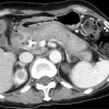
Contrast-enhanced CT images of AIP with diffuse pancreatic enlargement. DCE-CT (pancreatic phase) shows diffuse pancreatic enlargement, straightened pancreatic margin, and capsule-like rim
Fig. 8
Fig. 8
Contrast-enhanced CT images of AIP with focal pancreatic enlargement. a DCE-CT (pancreatic phase) shows a hypovascular lesion in the pancreatic head (arrows). b DCE-CT (delayed phase) depicts a pancreatic head lesion with homogeneous delayed enhancement (arrows)
Fig. 9
Fig. 9
Contrast-enhanced CT images of pancreatic head cancer. a DCE-CT (pancreatic phase) shows a hypovascular lesion in the pancreatic head (arrow). b DCE-CT (delayed phase) reveals a pancreatic head lesion with inhomogeneous delayed enhancement (poor enhancement in the central area; arrowhead)

References

    1. Yoshida K, Toki F, Takeuchi T, et al. Chronic pancreatitis caused by an autoimmune abnormality. Proposal of the concept of autoimmune pancreatitis. Dig Dis Sci. 1995;40:1561–1568. - PubMed
    1. Hamano H, Kawa S, Horiuchi A, et al. High serum IgG4 concentrations in patients with sclerosing pancreatitis. N Engl J Med. 2001;344:732–738. - PubMed
    1. Japan Pancreas Society. Clinical diagnostic criteria for autoimmune pancreaitis. (In Japanese) Suizo. 2002;17:585–7.
    1. Okazaki K, Kawa S, Kamisawa T, et al. Clinical diagnostic criteria of autoimmune pancreatitis: revised proposal. J Gastroenterol. 2006;41:626–631. - PMC - PubMed
    1. Members of the Autoimmune Pancreatitis Diagnostic Criteria Committee, the Research Committee of Intractable Diseases of the Pancreas supported by the Japanese Ministry of Health, Labor and Welfare, and Members of the Autoimmune Pancreatitis Diagnostic Criteria Committee, the Japan Pancreas Sociey. Clinical diagnostic criteria of autoimmune pancreatitis 2006. (In Japanese) Suizo. 2006;21:395–7.

Publication types

LinkOut - more resources