. 2022 May;477(5):1583-1595.
doi: 10.1007/s11010-022-04383-7. Epub 2022 Feb 22.

Bone morphogenetic protein receptor inhibitors suppress the growth of glioblastoma cells

Affiliations

Bone morphogenetic protein receptor inhibitors suppress the growth of glioblastoma cells

Joel Kaye et al. Mol Cell Biochem. 2022 May.

Abstract

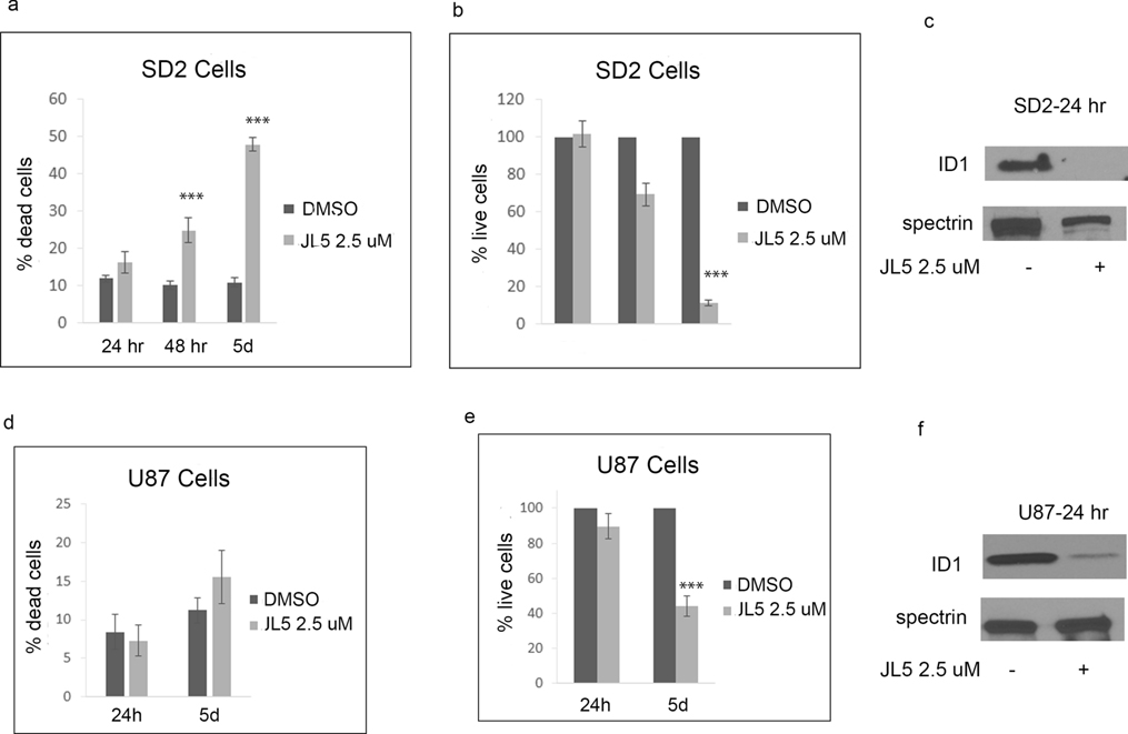
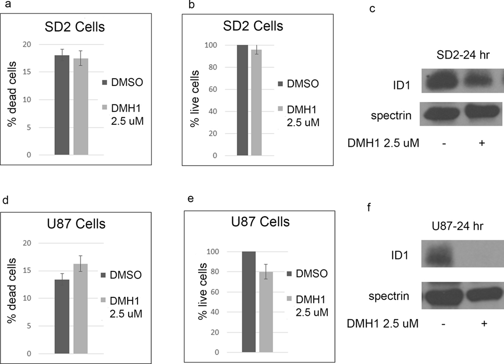
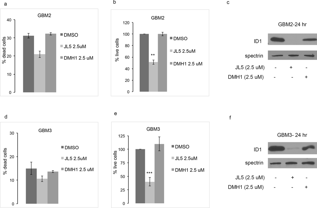
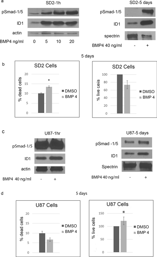
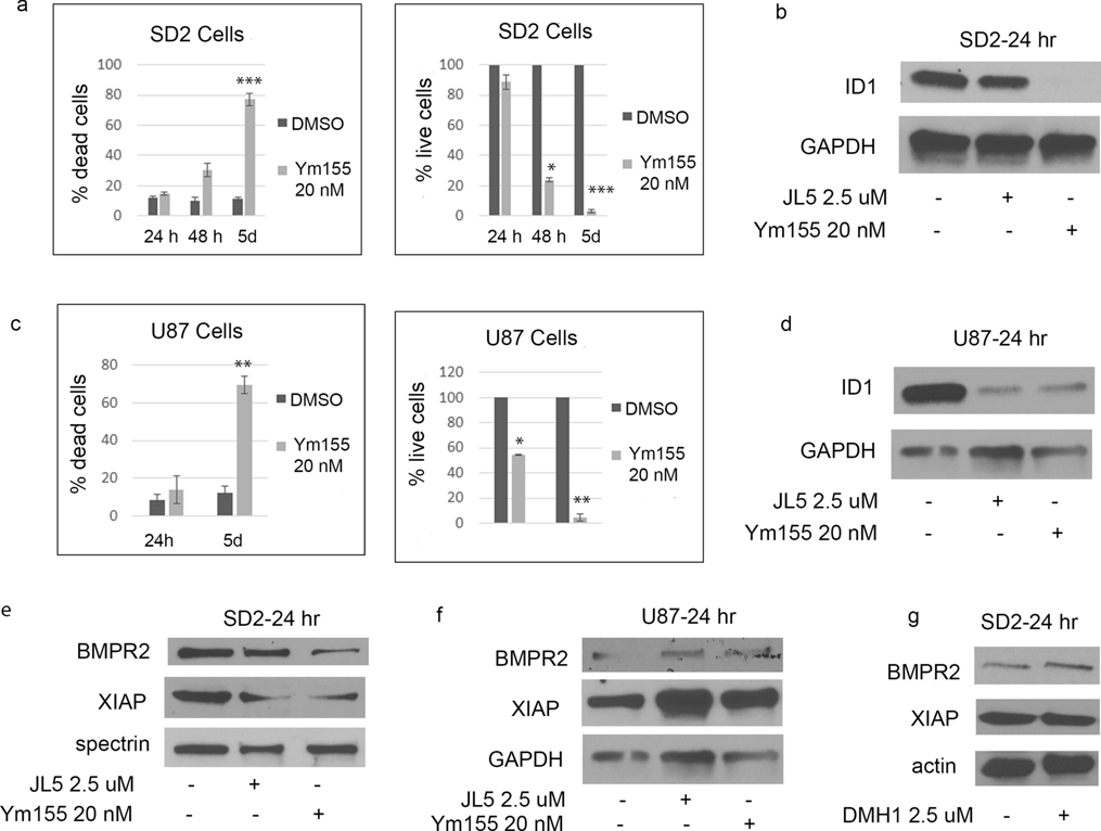
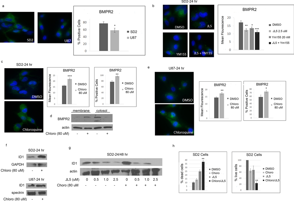
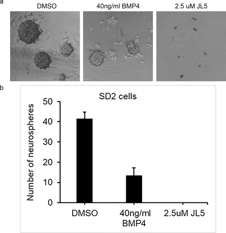
Glioblastomas (GBMs) are aggressive brain tumors that are resistant to chemotherapy and radiation. Bone morphogenetic protein (BMP) ligand BMP4 is being examined as a potential therapeutic for GBMs because it induces differentiation of cancer stem cells (CSCs) to an astrocyte phenotype. ID1 is reported to promote self-renewal and inhibit CSC differentiation. In most cancers, ID1 is transcriptionally upregulated by BMP4 promoting invasion and stemness. This conflicting data bring into question whether BMP signaling is growth suppressive or growth promoting in GBMs. We utilized BMP inhibitors DMH1, JL5, and Ym155 to examine the role of BMP signaling on the growth of GBMs. DMH1 targets BMP type 1 receptors whereas JL5 inhibits both the type 1 and type 2 BMP receptors. Ym155 does not bind the BMP receptors but rather inhibits BMP signaling by inducing the degradation of BMPR2. We show that JL5, DMH1, and Ym155 decreased the expression of ID1 in SD2 and U87 cells. JL5 and Ym155 also decreased the expression of BMPR2 and its downstream target inhibitor of apoptosis protein XIAP. JL5 treatment resulted in significant cell death and suppressed self-renewal to a greater extent than that induced by BMP4 ligand. The lysosome inhibitor chloroquine increases the localization of BMPR2 to the plasma membrane enhancing JL5-induced downregulation of ID1 and cell death in SD2 cells. We show that BMP signaling is growth promoting in GBMs. These studies suggest the need for development of BMP inhibitors and evaluation as potential therapeutic for GBMs.

Keywords: BMP; BMP inhibitors; BMPR2; Cancer stem cells; Glioblastoma; ID1.


Conflict of interest statement

Conflict of interests:

The authors declare that they have no conflict of interest.

Figures

Figure 1.
Figure 1.. JL5 suppresses growth of glioblastoma cell lines and down-regulates the expression of ID1.
(a,b,d,e) SD2 and U87 cells were treated with JL5 for 5 days and the percentage of dead cells and number of live cells determined. Data represents the mean of 4 independent experiments. (c,f) Western blot analysis of SD2 and U87 cells treated with JL5 demonstrating a decrease in ID1 expression. *** p <0.0005 compared to control.
Figure 2.
Figure 2.. Inhibitor of BMP type 1 receptor DMH1 decreases ID1 expression with little effect on cell survival
(a,b,d,e) SD2 and U87 cells were treated with DMH1 for 5 days and the percentage of dead cells and number of live cells determined. Data represents the mean of 4 independent experiments. (c,f) Western blot analysis of SD2 and U87 cells treated with DMH1 demonstrating a decrease in ID1 expression, which was more in U87 cells.
Figure 3.
Figure 3.
JL5 decrease cell growth and ID1 expression in primary glioblastomas GBM2 and GBM3. (a,b,d,e) GBM2 and GBM2 glioblastoma cell lines were treated with JL5 or DMH1 for 5 days and the percentage of dead and live cells determined. Data represents the mean of 4 experiments for GBM3 and 2 experiments for GBM2. (C,F) Western blot analysis of GBMs treated with JL5 or DMH1 for 24 hr. **p<0.005, *** p <0.0005 compared to control.
Figure 4.
Figure 4.. BMP4 activates ID1 expression and suppresses growth of SD2 cells.
(a) Western blot analysis of SD2 cells treated with BMP4. (b) SD2 cells were treated with BMP4 for 5 days and percentage of dead cells and number of live cells determined. Data represents the mean of 4 independent experiments. (c) Western blot analysis of U87 cells treated with BMP4. (d) U87 cells were treated with BMP4 for 5 days and percentage of dead cells and number of live cells determined. Data represents the mean of 4 independent experiments. * p <0.05, compared to control.
Figure 5.
Figure 5.. Ym155 inhibits BMP signaling. J5 and Ym155 decrease XIAP expression in SD2 cells.
(a,c) SD2 and U87 cells were treated with Ym155 and the percentage of dead cells and number of live cells determined. Data represents the mean of 4 independent experiments. (b,d) Western blot analysis of SD2 and U87 cells treated with Ym155 or JL5 demonstrating a decrease in ID1 expression. (e,f) Western blot analysis of cells treated with JL5 and Ym155 showing a decrease the expression of XIAP in SD2 but not the U87 cells. (g) Western blot analysis showing DMH1 does not decrease expression of XIAP. * p <0.05, ** p <0.005, *** p <.0005 compared to control.
Figure 6.
Figure 6.. Chloroquine increases BMPR2 expression and enhances cell death induced by JL5.
(a) Representative immunofluorescent images of BMPR2 in untreated cells. Graphs represent the percentage of cells expressing BMPR2. Approximately 80 cells were counted from each cell line. (b) Representative immunofluorescent images of BMPR2 following treatment with JL5 and Ym155. Graphs represent the mean fluorescence of approximately 20 cells from each treatment group. (c,e) Representative immunofluorescent images of BMPR2 following treatment with chloroquine. Graphs represent the mean fluorescence of approximately 30 cells from each treatment group and the percentages of cells that express BMPR2. (d) Western blot of membrane and cytosol fraction of SD2 cells treated with chloroquine for 24 hours. (f) Western blot showing chloroquine increases the expression of ID1. (g) Western blot analysis showing that chloroquine enhances JL5 induced down-regulation of ID1. (h) Cell counts of SD2 cells pre-treated with either DMSO or Chloroquine for 24 hours, then treated again with either DMSO or JL5 for 48 hours. The data represents the mean of 4 independent experiments. * p <0.05, ** p <0.005, *** p <0.0005 compared to control. (g) ** p < 0.005 chloroquine + JL5 in comparison to JL5 alone.
Figure 7.
Figure 7.. JL5 inhibits self-renew more than BMP4.
1000 SD2 cells were plated into 6 well plates and then treated with BMP4 or JL5 for 7 days. (a) Representative phase contrast images of sphere formation. (b) All the spheres in the 6 well plates were counted. Depicted is the mean number of spheres from 3 independent experiments.

References

    1. Blázquez-Medela AM, Jumabay M and Boström KI (2019) Beyond the bone: Bone morphogenetic protein signaling in adipose tissue. Obes Rev 20:648–658. doi: 10.1111/obr.12822 - DOI - PMC - PubMed
    1. Stupp R, Mason WP, van den Bent MJ, Weller M, Fisher B, Taphoorn MJ, Belanger K, Brandes AA, Marosi C, Bogdahn U, Curschmann J, Janzer RC, Ludwin SK, Gorlia T, Allgeier A, Lacombe D, Cairncross JG, Eisenhauer E and Mirimanoff RO (2005) Radiotherapy plus concomitant and adjuvant temozolomide for glioblastoma. N Engl J Med 352:987–96. doi: 10.1056/NEJMoa043330 - DOI - PubMed
    1. Chen J, Li Y, Yu TS, McKay RM, Burns DK, Kernie SG and Parada LF (2012) A restricted cell population propagates glioblastoma growth after chemotherapy. Nature 488:522–6. doi: 10.1038/nature11287 - DOI - PMC - PubMed
    1. Bao S, Wu Q, McLendon RE, Hao Y, Shi Q, Hjelmeland AB, Dewhirst MW, Bigner DD and Rich JN (2006) Glioma stem cells promote radioresistance by preferential activation of the DNA damage response. Nature 444:756–60. doi: 10.1038/nature05236 - DOI - PubMed
    1. Gross RE, Mehler MF, Mabie PC, Zang Z, Santschi L and Kessler JA (1996) Bone morphogenetic proteins promote astroglial lineage commitment by mammalian subventricular zone progenitor cells. Neuron 17:595–606. - PubMed

Substances

LinkOut - more resources