. 2022 Feb 22;18(2):e1010041.
doi: 10.1371/journal.pgen.1010041. eCollection 2022 Feb.

Histone demethylase IBM1-mediated meiocyte gene expression ensures meiotic chromosome synapsis and recombination

Affiliations

Histone demethylase IBM1-mediated meiocyte gene expression ensures meiotic chromosome synapsis and recombination

Chengpeng He et al. PLoS Genet. .

Abstract

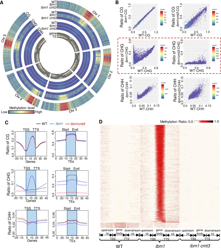
Histone methylation and demethylation play important roles in plant growth and development, but the involvement of histone demethylation during meiosis is poorly understood. Here we show that disruption of Arabidopsis thaliana INCREASE IN BONSAI METHYLATION 1 (IBM1) causes incomplete synapsis, chromosome entanglement and reduction of recombination during meiosis, leading to sterility. Interestingly, these ibm1 meiotic defects are rescued by mutations in either SUVH4/KYP or CMT3. Using transcriptomic analyses we show that mutation of IBM1 down-regulates thousands of genes expressed in meiocytes, and that expression of about 38% of these genes are restored to wild type levels in ibm1 cmt3 double mutants. Changes in the expression of 437 of these, including the ARABIDOPSIS MEI2-LIKE AML3-5 genes, are correlated with a significant reduction of gene body CHG methylation. Consistently, the aml3 aml4 aml5 triple have defects in synapsis and chromosome entanglement similar to ibm1. Genetic analysis shows that aml3 aml4 aml5 ibm1 quadruple mutants resembles the ibm1 single mutant. Strikingly, over expression of AML5 in ibm1 can partially rescue the ibm1 meiotic defects. Taken together, our results demonstrate that histone demethylase IBM1 is required for meiosis likely via coordinated regulation of meiocyte gene expression during meiosis.


Conflict of interest statement

The authors have declared that no competing interests exist.

Figures

Fig 1
Fig 1. IBM1 is required for male fertility.
(A) Plant images of wild type (WT), ibm1-4, ibm1-6 and ibm1-4/ibm1-6 whole plants. (B) Siliques from WT, ibm1-4, ibm1-6, and ibm1-4/ibm1-6. (C)—(E) Flowers (C), anthers after Alexander staining (D) and Toluidine blue stained tetrad-stage meiocytes (E) from WT (n = 112), ibm1-4 (n = 228), ibm1-6 (n = 332), and ibm1-4/ibm1-6 (n = 248) plants. Green arrows in (E) indicate micronuclei. (F) Quantification of viable pollen grains per anther from WT, ibm1-4, ibm1-6 and ibm1-4/ibm1-6 plants. ** represents p-value< 0.01 in two-tailed student t test. The data are shown as mean ± SD. (G) Histogram of different types of tetrads. ** represents p-value< 0.01 in two-tailed chi-square test. Scale bar: (A): 5 cm, (B): 1 mm, (C): 1 mm, (D): 50 μm, (E): 5 μm.
Fig 2
Fig 2. DAPI stained chromosome spreads from WT and ibm1 mutants.
Chromosome spreads showing meiotic chromosome morphologies from WT, ibm1-4 and ibm1-6 meiocytes. Yellow arrows on mutant pachytene chromosomes indicate the unsynapsed regions. Magenta triangles on metaphase I chromosomes show chromosome entanglements. Green arrows on anaphase I mutant chromosomes indicate chromosome fragments or lagging chromosomes. The magenta arrows on telophase II show chromosome fragments. Scale bar: 5 μm.
Fig 3
Fig 3. IBM1 is required for synapsis.
(A) Image showing dual signals of DAPI (blue) with chromosome 1 painting (red) at pachytene in WT, ibm1-4 and ibm1-6. (B) Immunofluorescence of ASY1 (red) in WT, ibm1-4 and ibm1-6 mutants at zygotene. (C) Localization of ZYP1 (green) and SYN1 (magenta) with DAPI (blue) at pachytene in WT, ibm1-4 and ibm1-6 meiocytes. (D) The merged ZYP1 (green) and SYN1 (magenta) signals from (C). The blue dotted-lined rectangles are magnified in (E) and show the regions with incomplete synapsis. Scale bar: 5 μm.
Fig 4
Fig 4. IBM1 is required for meiotic recombination.
(A) Images showing FISH with centromere probe of WT, ibm1-4, ibm1-6, spo11-1, ibm1-6 spo11-1, msh4, ibm1-6 msh4, mus81, ibm1-6 mus81 metaphase I chromosome spreads. (B) and (C) Zygotene and pachytene localization of γ-H2AX in WT, ibm1-4 and ibm1-6 meiocytes. (D) Quantification of γ-H2AX foci at zygotene and pachytene in WT, ibm1-4 and ibm1-6 mutants. (E) and (F) showing zygotene and pachytene localization of γ-H2AX and DMC1 in WT, ibm1-4 and ibm1-6 meiocytes. (G) Quantification of DMC1 at zygotene and pachytene in WT, ibm1-4 and ibm1-6 mutants. Box-plots show the median and interquartile ranges and the whiskers extend to the maximum and minimum of number of foci. ** p-value<0.01 with two-tailed student t test. Scale bar: 5 μm.
Fig 5
Fig 5. Reduction of COs observed in ibm1.
(A) 45S (green) and 5S (red) FISH of metaphase I from WT, ibm1-4, ibm1-6. (B) Quantification of the number of chiasmata in each WT, ibm1-4 and ibm1-6 meiocyte. The data are shown as mean ± SD. (C) Immunofluorescence analysis of HEI10 in WT, ibm1-4, ibm1-6 diakinesis chromosome spreads. (D) Quantification of the number of HEI10 foci in each WT, ibm1-4 and ibm1-6 meiocyte. The data are shown as mean ± SD. In image (B) and (D), ** represents p-value<0.01, *** represents p-value<0.001 with two-tailed student t test. Scale bar: 5 μm.
Fig 6
Fig 6. Immunofluorescence of IBM1 in the IBM1 trans-complemented plants at leptotene, zygotene, pachytene and diplotene.
Dual-immunofluorescence images with anti-FLAG antibody (magenta) and an antibody against the meiosis-specific cohesin protein SYN1 (green). An IBM1-FLAG tag fusion was expressed in ibm1-6 plants and IBM1 can be observed colocalizing with chromosomes (DAPI, blue) at leptotene, zygotene, pachytene but not diplotene. Scale bar: 5 μm.
Fig 7
Fig 7. suvh4/kyp and cmt3 suppress meiotic defects of in ibm1.
(A) DAPI stained pachytene (top row), diakinesis (middle row) and metaphase I (bottom row) chromosomes from WT, ibm1-4, ibm1-6, suvh4, cmt3, ibm1-4 cmt3, ibm1-4 suvh4, ibm1-6 cmt3 and ibm1-6 suvh4 meiocytes. Scale bar: 5 μm. (B) Quantification of viable pollen grains per anther from WT, ibm1-4, ibm1-6, suvh4, cmt3, ibm1-4 cmt3, ibm1-4 suvh4, ibm1-6 cmt3 and ibm1-6 suvh4 plants. *** represents p-value< 0.001 in two-tailed student t test. The data are shown as mean ± SD. (C) Image showing percentage of chromosome with (entanglement) or without (normal) entanglement in Metaphase I. *** represents p-value< 0.001 in two-tailed student chi-square test.
Fig 8
Fig 8. IBM1 is required for gene body CHG demethylation.
(A) The relative CG, CHG and CHH methylation levels along the 5 Arabidopsis chromosomes is shown as a heat map. Each type of DNA methylation was measured in WT (the outside lane), ibm1-6 (the middle lane) and ibm1-6 cmt3-7 (the inside lane). CHG methylation is elevated (redder colors) in ibm1, and reduced in ibm1 cmt3 (bluer colors). The distribution of genes and transposon elements (TEs) is shown in grey (inner rings). (B) Scatter diagram showing CG, CHG and CHH methylation ratios in WT, ibm1-6 and ibm1 cmt3. The red dotted line highlights CHG methylation. (C) The distribution of CG, CHG and CHH methylation level along genes, with gene bodies flanked by transcriptional start sites (TSS) and transcriptional termination sites (TTS), and TEs. (D) Heat map showing distribution of CHG methylation in genes with elevated CHG methylation within gene body in WT, ibm1-6 and ibm1 cmt3.
Fig 9
Fig 9. IBM1 specifically regulates many genes expressed in meiocytes.
(A) Venn diagram showing genes with significantly reduced or increased expression in ibm1-4 meiocytes and leaves. (B) Diagram showing Log2(fold change) in ibm1-4 meiocytes and leaves. Red rectangles indicate genes specifically downregulated in meiocytes. (C) Diagram showing Log2(fold change) in ibm1 meiocytes and ibm1 cmt3 meiocytes. Red rectangles indicate genes with restored expression in ibm1-6 cmt3-7 meiocytes. (D) Distribution of CG (top rows), CHG (middle rows) and CHH (bottom rows) methylation within gene body of AML3 (top row), AML4 (middle row) and AML5 (bottom row) in 4–7 stage anthers of WT and ibm1. (E) CHOP-PCR analysis of AML3-5 in 4–7 stage anthers of WT, ibm1-4, ibm1-6, cmt3, suvh4, ibm1-4 cmt3, ibm1-4 suvh4, ibm1-6 cmt3 and ibm1-6 suvh4. Genomic DNA was digested by MSP1, which recognizes the CCGG site. Methylation of the first C prevents cutting by MSP1. (F) RT-qPCR analysis of AML3-5 expression in meiocytes of WT, ibm1-4, ibm1-6 and ibm1-6 cmt3-7. * p-value<0.05, ** P-value<0.01 with two-tailed student t test. The error bars represent the SD of each group.
Fig 10
Fig 10. Mutation of AML family reduces fertility and causes similar meiotic defects with ibm1.
(A) Alexander staining of anthers in WT, ibm1-6, aml1 aml4, aml3 aml4 aml5 and aml3 aml4 aml5 ibm1-6. (B) Chromosome spreads showing meiotic chromosome morphologies of WT, ibm1-6, aml1 aml4, aml3 aml4 aml5 and aml3 aml4 aml5 ibm1-6 meiocytes. Yellow arrows on mutant pachytene chromosomes indicate the unsynapsed regions. (C) Quantification of viable pollen grains per anther of WT, ibm1-6, aml1 aml4, aml3 aml4 aml5 and aml3 aml4 aml5 ibm1-6. *** represents p-value< 0.001 in two-tailed student t test. The data are shown as mean ± SD. Scale bars: A: 50 μm, B: 5 μm.

Similar articles

Cited by

References

    1. Speijer D, Lukes J, Elias M (2015) Sex is a ubiquitous, ancient, and inherent attribute of eukaryotic life. Proceedings of the National Academy of Sciences of the United States of America 112: 8827–8834. doi: 10.1073/pnas.1501725112 - DOI - PMC - PubMed
    1. Yang HX, Lu PL, Wang YX, Ma H (2011) The transcriptome landscape of Arabidopsis male meiocytes from high-throughput sequencing: the complexity and evolution of the meiotic process. Plant Journal 65: 503–516. doi: 10.1111/j.1365-313X.2010.04439.x - DOI - PubMed
    1. Dukowic-Schulze S, Sundararajan A, Mudge J, Ramaraj T, Farmer AD, et al.. (2014) The transcriptome landscape of early maize meiosis. Bmc Plant Biology 14. doi: 10.1186/1471-2229-14-118 - DOI - PMC - PubMed
    1. Mercier R, Mezard C, Jenczewski E, Macaisne N, Grelon M (2015) The molecular biology of meiosis in plants. Annu Rev Plant Biol 66: 297–327. doi: 10.1146/annurev-arplant-050213-035923 - DOI - PubMed
    1. Wang YX, Copenhaver GP (2018) Meiotic Recombination: Mixing It Up in Plants. Annual Review of Plant Biology, Vol 69 69: 577–609. doi: 10.1146/annurev-arplant-042817-040431 - DOI - PubMed

Publication types