. 2022 May 1;71(5):1149-1165.
doi: 10.2337/db21-0830.

Endothelial Phospholipase Cγ2 Improves Outcomes of Diabetic Ischemic Limb Rescue Following VEGF Therapy

Affiliations

Endothelial Phospholipase Cγ2 Improves Outcomes of Diabetic Ischemic Limb Rescue Following VEGF Therapy

Yashika Rustagi et al. Diabetes. .

Abstract

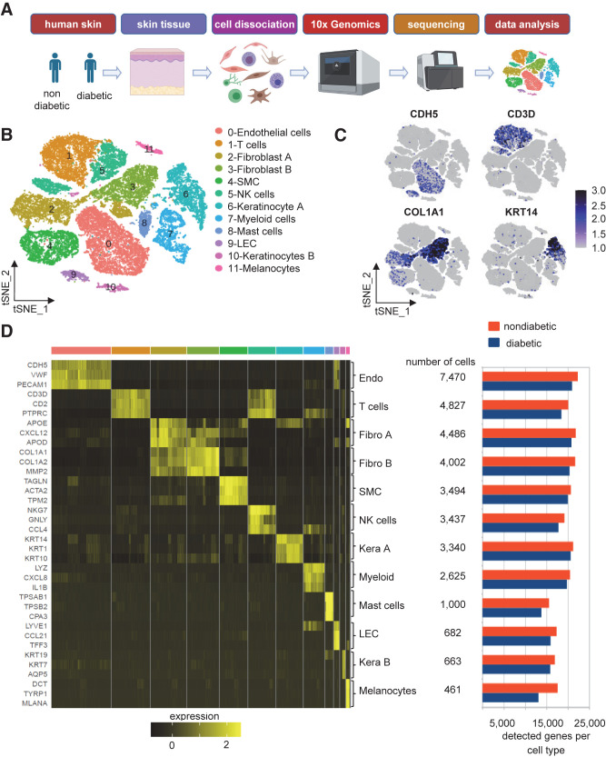
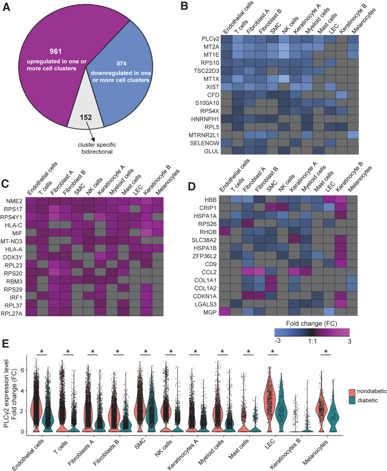
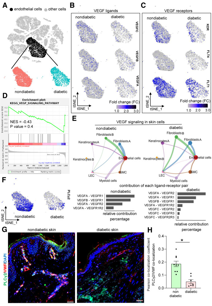
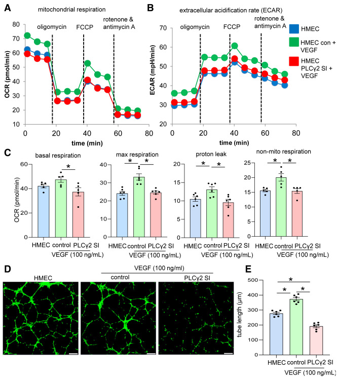
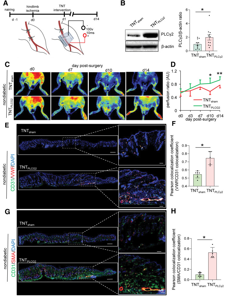
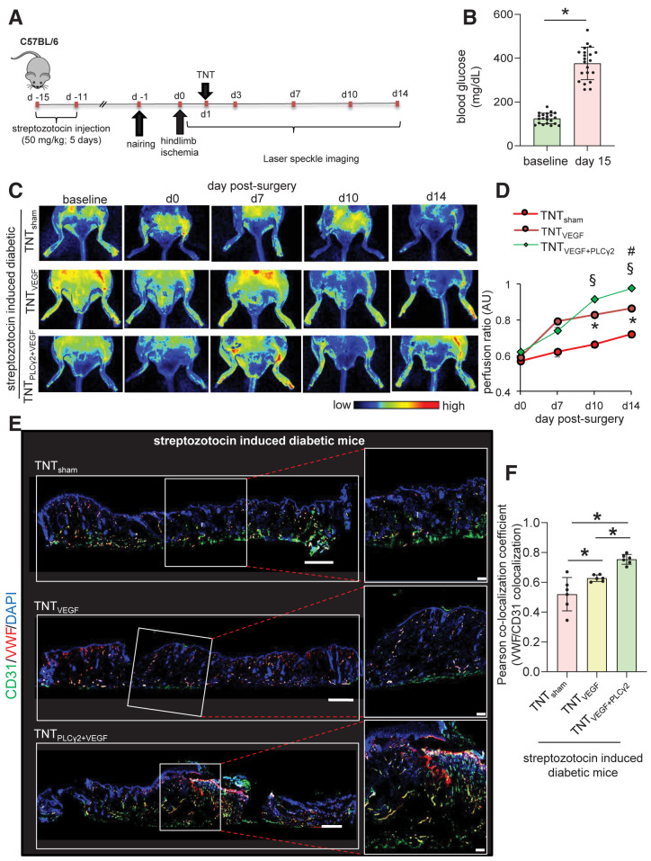
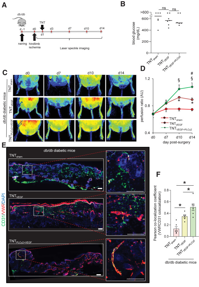
Therapeutic vascular endothelial growth factor (VEGF) replenishment has met with limited success for the management of critical limb-threatening ischemia. To improve outcomes of VEGF therapy, we applied single-cell RNA sequencing (scRNA-seq) technology to study the endothelial cells of the human diabetic skin. Single-cell suspensions were generated from the human skin followed by cDNA preparation using the Chromium Next GEM Single-cell 3' Kit v3.1. Using appropriate quality control measures, 36,487 cells were chosen for downstream analysis. scRNA-seq studies identified that although VEGF signaling was not significantly altered in diabetic versus nondiabetic skin, phospholipase Cγ2 (PLCγ2) was downregulated. The significance of PLCγ2 in VEGF-mediated increase in endothelial cell metabolism and function was assessed in cultured human microvascular endothelial cells. In these cells, VEGF enhanced mitochondrial function, as indicated by elevation in oxygen consumption rate and extracellular acidification rate. The VEGF-dependent increase in cell metabolism was blunted in response to PLCγ2 inhibition. Follow-up rescue studies therefore focused on understanding the significance of VEGF therapy in presence or absence of endothelial PLCγ2 in type 1 (streptozotocin-injected) and type 2 (db/db) diabetic ischemic tissue. Nonviral topical tissue nanotransfection technology (TNT) delivery of CDH5 promoter-driven PLCγ2 open reading frame promoted the rescue of hindlimb ischemia in diabetic mice. Improvement of blood flow was also associated with higher abundance of VWF+/CD31+ and VWF+/SMA+ immunohistochemical staining. TNT-based gene delivery was not associated with tissue edema, a commonly noted complication associated with proangiogenic gene therapies. Taken together, our study demonstrates that TNT-mediated delivery of endothelial PLCγ2, as part of combination gene therapy, is effective in diabetic ischemic limb rescue.


Figures

Figure 1
Figure 1
Identification of 12 distinct clusters within the skin tissue in individuals with diabetes and without diabetes. A: The overall design of the study. Schematic diagram was created with BioRender.com. B: tSNE projection of the filtered data (36,487 cells). Each cell is represented as a dot. C: Expression level of selected markers: endothelial cells, CDH5 (further elaborated in Supplementary Fig. 4A); T cells, CD3D; fibroblasts, COL1A1; and keratinocytes, KRT14. D: Heat map showing relative expression of the top three markers for each cluster (left) and the total number of detected cells and genes in each cluster (right).
Figure 2
Figure 2
Diabetes-related DEGs in distinct cell types of skin. A: Pie chart showing the number of the upregulated, downregulated, or bidirectional genes across all identified clusters. Heat map showing a subset of genes that has shared downregulation (B) or shared upregulation (C) (FDR-adjusted P < 0.05 and logFC ±0.25) in diabetic skin. D: Heat map of logFC showing a subset of diabetic-related genes (FDR-adjusted P < 0.05 and logFC ±0.25) that has bidirectional genes (down- or upregulated) in different clusters of the skin. Heat maps were created using Genesis software. E: Violin plot representing expression level of PLCγ2 in each cellular cluster in diabetic and nondiabetic skin. Each cell is represented as a dot. PLCγ2 was downregulated in 11 cellular clusters in diabetic skin (not significantly different in only keratinocyte B cluster) (*FDR-adjusted P < 0.05).
Figure 3
Figure 3
Identification and extraction of endothelial cell clusters within the skin samples of individuals with diabetes and without diabetes. A: Identification and extraction of the endothelial cell cluster in human skin by visualizing the expression level of CDH5, VWF, and PECAM1 gene expression enrichment (Supplementary Fig. 4A). B: tSNE plots showing nonsignificant difference of VEGF ligands between diabetic and nondiabetic skin endothelial cells (VEGFC, FDR-adjusted P value: 1; VEGFB, adjusted P value: 1; VEGFA, not expressed in >20% of the cells in either group). C: tSNE plots showing nonsignificant difference of VEGF receptors between diabetic and nondiabetic skin endothelial cells (KDR, FDR-adjusted P value: 1; FLT1, FDR-adjusted P value: 0.04, logFC < ±0.25; FLT4, not expressed in >20% of the cells in either group). D: GSEA showing that the VEGF signaling was not significantly altered between diabetic and nondiabetic skin endothelial cells (normalized enrichment score [NES] −0.43; P = 0.4). E: Signaling interaction analysis of VEGF pathway across different cell types in diabetic and nondiabetic skin. E, top: VEGF signaling network in nondiabetic (left) and diabetic (right) skin. E, bottom: Ligands and receptors contributed in VEGF signaling in nondiabetic (left) and diabetic (right) skin cells. F: tSNE plots showing PLCγ2 expression level between nondiabetic (left) and diabetic (right) skin endothelial cells (logFC: −0.52; FDR-adjusted P value: 5.98E-100). Immunohistochemical analysis of VWF+/PLCγ2+ colocalization in human nondiabetic and diabetic skin (G) and its colocalization coefficient (H). Scale bars, 50 µm. n = 9 and 7. *P < 0.05 (Student t test). Data are represented as the mean ± SEM.
Figure 4
Figure 4
VEGF-mediated increase in endothelial cell functionality is PLCγ2 dependent. OCR (A) and ECAR (B) calculation of HMECs in normal conditions and after VEGF exposure (100 ng/mL) and cotreatment with either control (con) siRNA or PLCγ2 siRNA (SI) using the Seahorse XF 96-well plate reader. OCR was determined during sequential treatments with oligomycin (ATP synthase inhibitor), carbonyl cyanide-4 (trifluoromethoxy) phenylhydrazone (FCCP), a protonophore that lowers the mitochondrial membrane potential to create conditions for maximal oxidative respiration, and antimycin A plus rotenone to inhibit the electron transport chain. C: Basal respiration, maximal respiration, proton leak, and nonmitochondrial (non-mito) respiration measurement in HMECs in normal conditions and supplemented with VEGF (100 ng/mL) and cotreatment with control (con) siRNA or PLCγ2 siRNA (SI). Matrigel tube formation images (D) and tube length (E) analysis in HMECs in normal conditions and after cotreatment with control (con) siRNA or PLCγ2 siRNA. Scale bars, 200 µm. n = 6. *P < 0.05 (one-way ANOVA, followed by Tukey HSD post hoc test).
Figure 5
Figure 5
TNT-mediated overexpression of PLCγ2 improves perfusion in nondiabetic ischemic limbs. A: Schematic diagram showing TNT-assisted delivery of PLCγ2 ORF in murine ischemic limbs. Schematic diagram was created with BioRender.com. B: Western blot analysis of TNT-mediated PLCγ2 overexpression in murine skin. Data represent 10 different sites from 3 mice. *P < 0.05 (Student t test). Data are represented as the mean ± SEM. PeriMed laser speckle–assisted perfusion images (C) and their analysis (D) in ischemic limbs on which TNT procedure was done with sham or PLCγ2 ORF in nondiabetic conditions. Perfusion was calculated based on the ratio of the ischemic versus normal/contralateral limb. n = 6 to 7. *P < 0.05 (Student t test); **P < 0.005. E: Immunohistochemical analysis of CD31+/VWF+. G: CD31+/SMA+ colocalization in ischemic limbs of mice treated with sham or PLCγ2 ORF at day 14 postsurgery and their colocalization coefficient (F and H) (n = 5–7). *P < 0.05, Student t test. Data are represented as the mean ± SEM. AU, arbitrary unit.
Figure 6
Figure 6
Endothelial cell–targeted delivery of PLCG2 augments VEGF therapy rescue of STZ-induced diabetic ischemic limb. A: Schematic diagram showing TNT-mediated delivery of VEGF ORFs or/and endothelial PLCγ2 ORFs in ischemic hindlimb of STZ-induced diabetic mice. B: Blood glucose level in mice used in this study before and after injection of STZ (50 mg/kg). PeriMed laser speckle–assisted perfusion images (C) and their analysis (D) in ischemic limbs on which TNT procedure was done with sham, VEGF only, or VEGF plus CDH5 promoter–driven (endothelial) PLCγ2 cocktail. Perfusion was calculated based on the ratio of the ischemic vs. normal/contralateral limb. n = 6 to 7. *P < 0.05 TNTVEGF vs. TNTsham; §P < 0.05 TNTVEGF+PLCγ2 vs. TNTsham; #P < 0.05 TNTVEGF+PLCγ2 vs. TNTVEGF (one-way ANOVA, followed by Tukey HSD post hoc test). Immunohistochemical analysis of CD31+/VWF+ colocalization in diabetic ischemic limbs of mice treated with above-mentioned cocktails at day 14 postsurgery (E) and the colocalization coefficient (F). n = 6 to 7. *P < 0.05 (one-way ANOVA, followed by Tukey HSD post hoc test). AU, arbitrary unit.
Figure 7
Figure 7
Endothelial cell–targeted delivery of PLCG2 augments VEGF therapy rescue of the T2DM (db/db) diabetic ischemic limb. A: Schematic diagram showing TNT-mediated delivery of VEGF ORFs or/and endothelial PLCγ2 ORFs in the ischemic hindlimb of db/db diabetic mice. B: Blood glucose level in db/db mice used in this study. TNTsham (n = 6; 4 male and 2 female), TNTVEGF (n = 7; 4 male and 3 female), and TNTVEGF+PLCG2 (n = 6; 3 male and 3 female). PeriMed laser speckle–assisted perfusion images (C) and their analysis (D) in ischemic limbs on which TNT procedure was done with sham, VEGF only, or VEGF plus CDH5 promoter–driven (endothelial) PLCγ2 cocktail. Perfusion was calculated based on the ratio of the ischemic vs. normal/contralateral limb. n = 6 to 7. *P < 0.05 TNTVEGF vs. TNTsham; §P < 0.05 TNTVEGF+PLCγ2 vs. TNTsham; #P < 0.05 TNTVEGF+PLCγ2 vs. TNTVEGF (one-way ANOVA, followed by Tukey HSD post hoc test). Immunohistochemical analysis of CD31+/VWF+ colocalization in diabetic ischemic limbs of db/db mice treated with above-mentioned cocktails at day 14 postsurgery (E) and the colocalization coefficient (F). n = 6. *P < 0.05 (one-way ANOVA, followed by Tukey HSD post hoc test). AU, arbitrary unit.

References

    1. Spreen MI, Gremmels H, Teraa M, et al.; PADI and JUVENTAS Study Groups . Diabetes is associated with decreased limb survival in patients with critical limb ischemia: pooled data from two randomized controlled trials. Diabetes Care 2016;39:2058–2064 - PubMed
    1. Wang SK, Murphy MP. Getting a leg up on cell therapy for critical limb ischemia. Circ Res 2017;120:1227–1228 - PMC - PubMed
    1. Lüscher TF, Creager MA, Beckman JA, Cosentino F. Diabetes and vascular disease: pathophysiology, clinical consequences, and medical therapy: part II. Circulation 2003;108:1655–1661 - PubMed
    1. Abbott RD, Brand FN, Kannel WB. Epidemiology of some peripheral arterial findings in diabetic men and women: experiences from the Framingham Study. Am J Med 1990;88:376–381 - PubMed
    1. Jude EB, Oyibo SO, Chalmers N, Boulton AJ. Peripheral arterial disease in diabetic and nondiabetic patients: a comparison of severity and outcome. Diabetes Care 2001;24:1433–1437 - PubMed

Publication types

MeSH terms

Substances