. 2022 Apr;130(7):994-1010.
doi: 10.1161/CIRCRESAHA.121.319718. Epub 2022 Feb 23.

Effects of Atrial Fibrillation on the Human Ventricle

Affiliations

Effects of Atrial Fibrillation on the Human Ventricle

Steffen Pabel et al. Circ Res. 2022 Apr.

Abstract

Rationale: Atrial fibrillation (AF) and heart failure often coexist, but their interaction is poorly understood. Clinical data indicate that the arrhythmic component of AF may contribute to left ventricular (LV) dysfunction.

Objective: This study investigates the effects and molecular mechanisms of AF on the human LV.

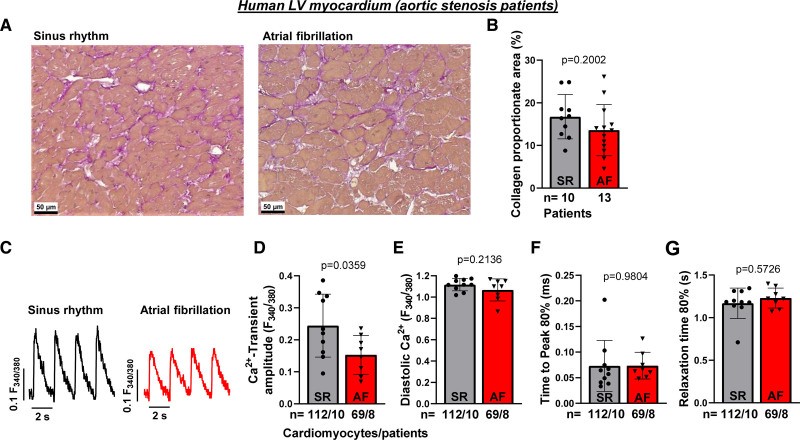
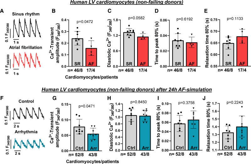
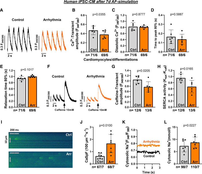
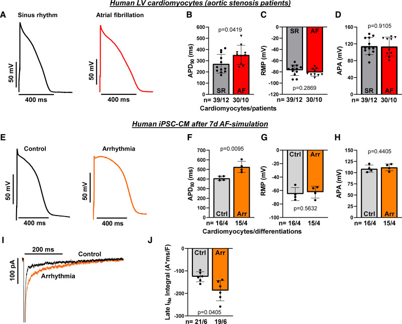
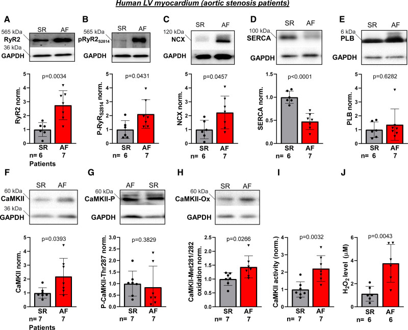
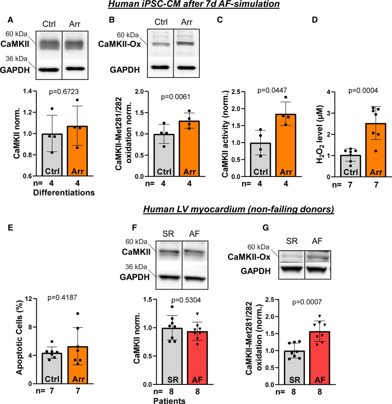
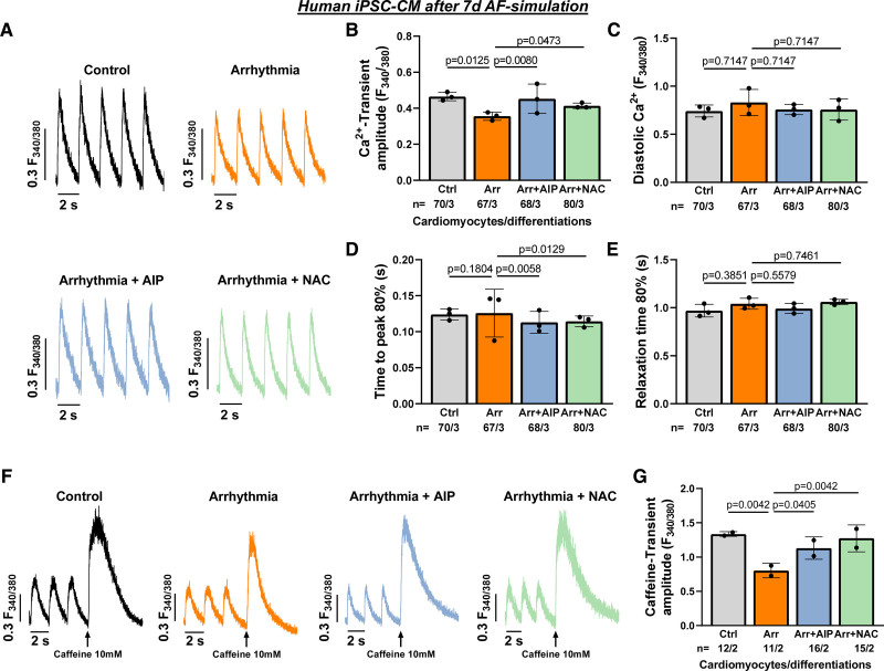
Methods and results: Ventricular myocardium from patients with aortic stenosis and preserved LV function with sinus rhythm or rate-controlled AF was studied. LV myocardium from patients with sinus rhythm and patients with AF showed no differences in fibrosis. In functional studies, systolic Ca2+ transient amplitude of LV cardiomyocytes was reduced in patients with AF, while diastolic Ca2+ levels and Ca2+ transient kinetics were not statistically different. These results were confirmed in LV cardiomyocytes from nonfailing donors with sinus rhythm or AF. Moreover, normofrequent AF was simulated in vitro using arrhythmic or rhythmic pacing (both at 60 bpm). After 24 hours of AF-simulation, human LV cardiomyocytes from nonfailing donors showed an impaired Ca2+ transient amplitude. For a standardized investigation of AF-simulation, human iPSC-cardiomyocytes were tested. Seven days of AF-simulation caused reduced systolic Ca2+ transient amplitude and sarcoplasmic reticulum Ca2+ load likely because of an increased diastolic sarcoplasmic reticulum Ca2+ leak. Moreover, cytosolic Na+ concentration was elevated and action potential duration was prolonged after AF-simulation. We detected an increased late Na+ current as a potential trigger for the detrimentally altered Ca2+/Na+-interplay. Mechanistically, reactive oxygen species were higher in the LV of patients with AF. CaMKII (Ca2+/calmodulin-dependent protein kinase IIδc) was found to be more oxidized at Met281/282 in the LV of patients with AF leading to an increased CaMKII activity and consequent increased RyR2 phosphorylation. CaMKII inhibition and ROS scavenging ameliorated impaired systolic Ca2+ handling after AF-simulation.

Conclusions: AF causes distinct functional and molecular remodeling of the human LV. This translational study provides the first mechanistic characterization and the potential negative impact of AF in the absence of tachycardia on the human ventricle.

Keywords: atrial fibrillation; calcium-calmodulin-dependent protein kinase type 2; excitation contraction coupling; heart failure; oxidative stress.


Figures

Figure 1.
Figure 1.
Fibrosis and Ca2+ handling in the atrial fibrillation (AF) ventricle. Human left ventricular (LV) myocardium was studied from patients with aortic stenosis (AS) with preserved LV function with sinus rhythm (SR) or AF (clinical characteristics are given in Table 1). A, Representative stainings (Elastica van Gieson) and (B) mean values for collagen proportionate area in ventricular SR (n=10 patients) and AF myocardium (n=13). C, Representative original recordings of stimulated Ca2+ transients (epifluorescence microscopy, Fura-2) of human LV cardiomyocytes from patients with SR or AF. D, Mean values of Ca2+ transient amplitude of human LV cardiomyocytes from patients with SR (n=112 cardiomyocytes/10 patients) compared with AF (n=69/8) and (E) diastolic Ca2+ levels, (F) time to peak 80%, (G) relaxation time 80%. Data are presented as scatter plot with mean±SD. Each data point represents the mean value of measurements from one patient. P were computed using Student t test.
Figure 2.
Figure 2.
Effects of atrial fibrillation (AF) in human left ventricular (LV) myocardium from nonfailing donors. A, Original Ca2+ transients from LV cardiomyocytes from nonfailing donors (clinical characteristics are provided in Table 2) with sinus rhythm (SR) or with AF (epifluorescence microscopy, Fura-2). B, Mean values of human LV cardiomyocytes from patients with SR (n=46 cardiomyocytes/8 patients) compared with AF (n=17/4) for Ca2+ transient amplitude, (C) diastolic Ca2+ level, (D) time to peak 80%, (E) relaxation time 80%. F, Original Ca2+ transients of human LV cardiomyocytes from nonfailing donors (epifluorescence microscopy, Fura-2) after 24-h in vitro AF-simulation (arrhythmic pacing: Arr; 60 bpm, 40% beat-to-beat variability) vs rhythmic pacing (control: Ctrl; 60 bpm). G, Mean values of human LV cardiomyocytes after AF-simulation (n=43/8) or rhythmic pacing (n=52/8) for Ca2+ transient amplitude, (H) diastolic Ca2+ level, (I) time to peak 80%, (J) relaxation time 80%. Data are presented as scatter plot with mean±SD. Each data point represents the mean value of measurements from one patient. P were calculated using Student t test.
Figure 3.
Figure 3.
Atrial fibrillation (AF)-simulation in induced pluripotent stem cell cardiomyocytes (iPSC-CMs). Human iPSC-CMs treated either with AF-simulation (arrhythmic pacing: Arr; 60 bpm, 40% beat-to-beat-variability) or rhythmic pacing (control [Ctrl]; 60 bpm) chronically for 7 d. A, Representative recordings of stimulated Ca2+ transients (epifluorescence microscopy, Fura-2) and (B) mean values for Ca2+ transient amplitude, (C) diastolic Ca2+ levels, (D) time to peak 80%, (E) relaxation time 80% of human iPSC-CM upon chronic AF-simulation (n=69 cardiomyocytes/6 differentiations/4 donors) or rhythmic pacing (n=71/6/4). F, Original recordings of caffeine-induced Ca2+ transients (10 mmol/l caffeine, epifluorescence microscopy, Fura-2), (G) mean caffeine-transient amplitude indicating the sarcoplasmic reticulum Ca2+ load and (H) SERCA2a activity (Ksys-Kcaff) of iPSC-CM after chronic AF-simulation (n=13/6/4) vs control (n=12/6/4). I, Representative confocal line scans (Fluo-4) showing diastolic sarcoplasmic reticulum Ca2+ sparks and (J) mean Ca2+ spark frequency (CaSpF) after chronic AF-simulation (n=68/7/4) vs control (n=67/7/4). K, Original recordings of cytosolic Na+ levels (epifluorescence microscopy, SBFI) and (L) mean values of cytosolic Na+ concentration of human iPSC-CM after chronic AF-simulation (n=110/7/4) compared with control (98/7/4). Data are provided as scatter plot with mean±SD. Each data point is calculated as mean value per differentiation. P were calculated using Student t test (A–C, E–L) or Mann-Whitney U test (D).
Figure 4.
Figure 4.
Electrophysiological alterations in response to atrial fibrillation (AF). A, Original stimulated action potential recordings (whole-cell current-clamp) of human left ventricular (LV) cardiomyocytes from patients with aortic stenosis (AS) with preserved LV function with either sinus rhythm (SR) or AF. B, Effects of AF (n=30 cardiomyocytes/10 patients) compared with SR (n=39/12) on action potential (AP) duration at 90% repolarization (APD90), C, Resting membrane potential (RMP) and (D) action potential amplitude (APA) in isolated human LV cardiomyocytes. E, Representative stimulated action potential recordings (whole-cell current-clamp) of iPSC-CM after chronic (7 d) AF-simulation (arrhythmic pacing: Arr; 60 bpm, 40% beat-to-beat-variability) compared with rhythmic pacing (control: ctrl; 60 bpm). F, Mean values for APD80, (G) RMP and (H) APA after chronic AF-simulation (n=15 cardiomyocytes/4 differentiations/4 donors) compared with control (n=16/4/4). I, Original traces of late Na+ current (INaL, whole-cell voltage-clamp) in iPSC-CM after chronic AF-simulation compared with control. J, Mean data of INaL (integral 100–500 ms) after AF-simulation (n=19/6/2) compared with control (n=21/6/2). Data are presented as scatter plot with mean±SD. Each data point is calculated as mean value per patient or differentiation. P were calculated using Student t test.
Figure 5.
Figure 5.
Molecular remodeling in the atrial fibrillation (AF) ventricle. Original representative Western Blots of human left ventricular (LV) myocardium from aortic stenosis patients with preserved LV function with sinus rhythm (SR, n=6-7) or AF (n=7) and expression levels (normalized to SR) for (A) ryanodine receptor type 2 (RyR2), (B) RyR2 phosphorylation at Ser2814 (normalized to total RyR2 expression), (C) NCX (Na+/Ca2+ exchanger), (D) SERCA (sarcoplasmic reticulum Ca2+ ATPase 2a), and (E) PLB (phospholamban). Representative Western Blots for (F) CaMKII (Ca2+/calmodulin-dependent protein kinase IIδc), (G) CaMKII phosphorylation at Thr287 (CaMKII-P), and (H) CaMKII oxidation at Met281/282 (CaMKII-ox). GAPDH was used as loading control. I, CaMKII activity (CycLex CaMKII activity ELISA kit) and (J) H2O2 levels (colorimetric peroxidase assay) in LV myocardium from patients with SR or AF (n=6–7 each). Data are provided as scatter plot with mean±SD. Groups were statistically analysed using Student t test or Mann-Whitney U test (for E and G).
Figure 6.
Figure 6.
Molecular remodeling in response to atrial fibrillation (AF) in induced pluripotent stem cell cardiomyocytes (iPSC-CM) and in non-failing myocardium. Original representative Western Blots of human iPSC-CMs after chronic (7 d) AF-simulation (Arr, n=4 differentiations/2 donors) or rhythmic pacing (control [Ctrl]; 60 bpm, n=4 differentiations/2 donors) and expression levels (normalized to SR) for (A) CaMKII (Ca2+/calmodulin-dependent protein kinase IIδc) and (B) CaMKII oxidation at Met281/282 (CaMKII-ox). GAPDH was used as loading control. C, CaMKII activity (CycLex CaMKII activity ELISA kit, n=4 differentiations/2 donors each), (D) H2O2 levels (colorimetric peroxidase assay, n=7 differentiations/3 donors each) and (E) percentage of apoptotic cells (n=7 differentiations/3 donors each) in iPSC-CM after chronic (7 d) AF-simulation compared with control. F, Representative Western Blots for CaMKII and (G) CaMKII oxidation at Met281/282 (CaMKII-ox) in LV myocardium from nonfailing donors with SR or AF (n=6–7 donors each). GAPDH was used as loading control. All Blots were on the same respective gels but on different positions. Thus, membrane was cut (black middle line). Data are given as scatter plot with mean±SD. P were calculated using Student t test.
Figure 7.
Figure 7.
Effects of CaMKII and oxidative stress on Ca2+ handling after atrial fibrillation (AF)-simulation. Human induced pluripotent stem cell cardiomyocytes (iPSC-CM) after chronic (7 d) AF-simulation (arrhythmic pacing: Arr; 60 bpm, 40% beat-to-beat-variability) or rhythmic pacing (control [Ctrl]; 60 bpm) and effects of CaMKII inhibition (AIP [autocamtide-2-related inhibitory peptide], 1 μmol/L) or oxidative stress reduction (NAC [N-acetylcysteine], 200 μmol/L). A, Representative recordings of stimulated Ca2+ transients (epifluorescence microscopy, Fura-2) and (B) mean values for Ca2+ transient amplitude, (C) diastolic Ca2+ levels, (D) time to peak 80%, (E) relaxation time 80% of human iPSC-CM upon chronic AF-simulation (n=67 cardiomyocytes/3 differentiations/2 donors), rhythmic pacing (n=70/3/2), AF-simulation + AIP (n=68/3/2) or AF-simulation+NAC (n=80/3/2). F, Original recordings of caffeine-induced Ca2+ transients (10 mmol/L caffeine, epifluorescence microscopy, Fura-2) and (G) mean caffeine-transient amplitude indicating the sarcoplasmic reticulum Ca2+ load of iPSC-CM after chronic AF-simulation (n=11/2/2), rhythmic pacing (n=12/2/2), AF-simulation+AIP (n=16/2/2) or AF-simulation+NAC (n=15/2/2). Data are provided as scatter plot with mean±SD. Each data point is calculated as mean value of all cardiomyocytes per differentiation. P were calculated using nested 1-way ANOVA and corrected for multiple comparisons using Holm-Sidak test.

Comment in

References

    1. Benjamin EJ, Wolf PA, D’Agostino RB, Silbershatz H, Kannel WB, Levy D. Impact of atrial fibrillation on the risk of death: the Framingham Heart Study. Circulation. 1998;98:946–952. doi: 10.1161/01.cir.98.10.946 - PubMed
    1. Sossalla S, Vollmann D. Arrhythmia-induced cardiomyopathy. Dtsch Arztebl Int. 2018;115:335–341. doi: 10.3238/arztebl.2018.0335 - PMC - PubMed
    1. Anter E, Jessup M, Callans DJ. Atrial fibrillation and heart failure: treatment considerations for a dual epidemic. Circulation. 2009;119:2516–2525. doi: 10.1161/CIRCULATIONAHA.108.821306 - PubMed
    1. Wang TJ, Larson MG, Levy D, Vasan RS, Leip EP, Wolf PA, D’Agostino RB, Murabito JM, Kannel WB, Benjamin EJ. Temporal relations of atrial fibrillation and congestive heart failure and their joint influence on mortality: the Framingham Heart Study. Circulation. 2003;107:2920–2925. doi: 10.1161/01.CIR.0000072767.89944.6E - PubMed
    1. Marrouche NF, Brachmann J, Andresen D, Siebels J, Boersma L, Jordaens L, Merkely B, Pokushalov E, Sanders P, Proff J, et al. ; CASTLE-AF Investigators. Catheter ablation for atrial fibrillation with heart failure. N Engl J Med. 2018;378:417–427. doi: 10.1056/NEJMoa1707855 - PubMed

Publication types

MeSH terms

Substances