. 2022 Feb 22;22(1):95.
doi: 10.1186/s12935-022-02507-z.

A novel qualitative signature based on lncRNA pairs for prognosis prediction in hepatocellular carcinoma

Affiliations

A novel qualitative signature based on lncRNA pairs for prognosis prediction in hepatocellular carcinoma

Xiaoyun Bu et al. Cancer Cell Int. .

Abstract

Background: Prognostic assessment is imperative for clinical management of patients with hepatocellular carcinoma (HCC). Most reported prognostic signatures are based on risk scores summarized from quantitative expression level of candidate genes, which are vulnerable against experimental batch effects and impractical for clinical application. We aimed to develop a robust qualitative signature to assess individual survival risk for HCC patients.

Methods: Long non-coding RNA (lncRNA) pairs correlated with overall survival (OS) were identified and an optimal combination of lncRNA pairs based on the majority voting rule was selected as a classification signature to predict the overall survival risk in the cancer genome atlas (TCGA). Then, the signature was further validated in two external datasets. Besides, biomolecular characteristics, immune infiltration status, and chemotherapeutics efficacy of different risk groups were further compared. Finally, we performed key lncRNA screening and validated it in vitro.

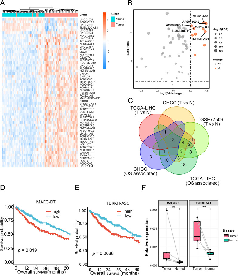
Results: A signature consisting of 50 lncRNA pairs (50-LPS) was identified in TCGA and successfully validated in external datasets. Patients in the high-risk group, when at least 25 of the 50-LPS voted for high risk, had significantly worse OS than the low-risk group. Multivariate Cox, receiver operating characteristic (ROC) curve and decision curve analyses (DCA) demonstrated that the 50-LPS was an independent prognostic factor and more powerful than other available clinical factors in OS prediction. Comparison analyses indicated that different risk groups had distinct biomolecular characteristics, immune infiltration status, and chemotherapeutics efficacy. TDRKH-AS1 was confirmed as a key lncRNA and associated with cell growth of HCC.

Conclusions: The 50-LPS could not only predict the prognosis of HCC patients robustly and individually, but also provide theoretical basis for therapy. Besides, TDRKH-AS1 was identified as a key lncRNA in the proliferation of HCC. The 50-LPS might guide personalized therapy for HCC patients in clinical practice.

Keywords: Hepatocellular carcinoma; LncRNA pairs; Precision medicine; Risk stratification.


Conflict of interest statement

The authors declare no conflict of interest.

Figures

Fig. 1
Fig. 1
Overview of the study workflow
Fig. 2
Fig. 2
The overall survival outcome of the two risk subgroups stratified by the 50-LPS in the training cohort (TCGA-LIHC). The Kaplan–Meier curves of overall survival for the entire training cohort (A) and patients in TNM I + II (B) and TNM III + IV (C). The time-dependent ROC analysis of the 50-LPS in the training cohort (D). Univariate and multivariate Cox regression analyses of OS in the training cohort (E). DCA of prognostic factors identified from univariate cox in training cohort (F)
Fig. 3
Fig. 3
Performance of the 50-LPS in two external cohorts. The Kaplan–Meier curves of overall survival for the entire CHCC cohort (A) and TNM I + II (B) and TNM III + IV (C) patients. The Kaplan–Meier curves of overall survival for the entire LIRI cohort (D) and TNM I + II (E) and TNM III + IV (F) patients. The time-dependent ROC analysis of the 50-LPS in the CHCC cohort (G) and LIRI cohort (J). Univariate and multivariate Cox regression analyses of OS in the CHCC cohort (H) and LIRI cohort (K). DCA of prognostic factors identified from univariate cox in CHCC cohort (I) and LIRI cohort (L)
Fig. 4
Fig. 4
Biological features and immune cell infiltration of the two risk subgroups stratified by the 50-LPS. Pathway enrichment analysis by GSEA (A). The difference of immune cell infiltrated in the tumor microenvironment between the two groups (B). *p < 0.05, **p < 0.01, ***p < 0.001, ****p < 0.0001
Fig. 5
Fig. 5
Clinical, molecular and genomic characteristics of the two risk subgroups stratified by the 50-LPS. Clinical characteristics of HCC subgroups and association with previous HCC molecular subtypes (A). The difference of frequencies in gene mutation between the two groups (B). *p < 0.05, **p < 0.01, ***p < 0.001
Fig. 6
Fig. 6
Correlations of the HCC subgroups with the response to different chemotherapeutics. Subclass mapping correlation analysis between HCC subgroups and samples with different sensitivities to TACE (A), and several anticancer drugs (B). TACE-NR, TACE non-responder; TACE-R, TACE responder. ** p<0.01,*** p<0.001,**** p<0.0001
Fig. 7
Fig. 7
Hub lncRNA screening identified TDRKH-AS1 as a key lncRNA in HCC. Expression heatmap of the 55 lncRNAs included in the 50-LPS in the 50 paired tumor and normal liver tissues of TCGA-LIHC cohort (A). Volcano plot of the differential test and the top 6 differentially expressed lncRNAs with log2FC > 1 and FDR < 0.05 were marked in orange (B). Venn diagram identified two lncRNAs (TDRKH-AS1 and MAFG-DT) that were differentially expressed in TCGA-LIHC, CHCC and GSE77509, as well as associated with overall survival in TCGA-LIHC and CHCC cohort (C). The Kaplan–Meier curves of overall survival for TDRKH-AS1 and MAFG-DT in TCGA-LIHC (D, E). Real-time PCR validated the expression difference of TDRKH-AS1 and MAFG-DT in five paired HCC and normal liver tissues (F). *p < 0.05, **p < 0.01, ***p < 0.001. T, tumor; N, normal
Fig. 8
Fig. 8
Validation of the biological function of TDRKH-AS1 in HCC. TDRKH-AS1 was successfully knocked down in Huh7 and MHCC97H cells (A). CCK8 assay in Huh7 (B) and MHCC97H (C) after knockdown of TDRKH-AS1. Colony formation assay in Huh7 and MHCC97H after knockdown of TDRKH-AS1 (D, E). Apoptosis assay in Huh7 and MHCC97H after knockdown of TDRKH-AS1 (FH). Cleaved caspase3 and phospho-Akt (Ser473) were detected in Huh7 and MHCC97H by western blotting after knockdown of TDRKH-AS1 (I). *p < 0.05, **p < 0.01, ***p < 0.001

References

    1. Sung H, Ferlay J, Siegel RL, Laversanne M, Soerjomataram I, Jemal A, Bray F. Global cancer statistics 2020: GLOBOCAN estimates of incidence and mortality worldwide for 36 cancers in 185 countries. CA Cancer J Clin. 2021;71(3):209–249. doi: 10.3322/caac.21660. - DOI - PubMed
    1. Forner A, Reig M, Bruix J. Hepatocellular carcinoma. Lancet. 2018;391(10127):1301–1314. doi: 10.1016/S0140-6736(18)30010-2. - DOI - PubMed
    1. Villanueva A. Hepatocellular carcinoma. N Engl J Med. 2019;380(15):1450–1462. doi: 10.1056/NEJMra1713263. - DOI - PubMed
    1. Subramaniam S, Kelley RK, Venook AP. A review of hepatocellular carcinoma (HCC) staging systems. Chin Clin Oncol. 2013;2(4):33. - PubMed
    1. Toyoda H, Kumada T, Tada T, Sone Y, Kaneoka Y, Maeda A. Tumor markers for hepatocellular carcinoma: simple and significant predictors of outcome in patients with HCC. Liver Cancer. 2015;4(2):126–136. doi: 10.1159/000367735. - DOI - PMC - PubMed

LinkOut - more resources