A novel apolipoprotein E mutation, ApoE Ganzhou (Arg43Cys), in a Chinese son and his father with lipoprotein glomerulopathy: two case reports
- PMID: 35193676
- PMCID: PMC8864814
- DOI: 10.1186/s13256-022-03302-0
A novel apolipoprotein E mutation, ApoE Ganzhou (Arg43Cys), in a Chinese son and his father with lipoprotein glomerulopathy: two case reports
Abstract
Background: Lipoprotein glomerulopathy is a rare and newly recognized glomerular disease that can lead to kidney failure. Its pathological features include the presence of lipoprotein embolus in the loop cavity of glomerular capillaries. It is believed that apolipoprotein E gene mutation is the initiator of the disease. Since the discovery of lipoprotein glomerulopathy, 16 different apolipoprotein E mutations have been reported worldwide, but most of these cases are sporadic. Here we report two cases of lipoprotein glomerulopathy, a Chinese son and his father, with a novel apolipoprotein E mutation, ApoE Ganzhou (Arg43Cys).
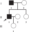
Case presentation: Case 1, a 33-year-old Chinese man, was hospitalized on 3 March 2014 owing to edema and weakness of facial and lower limbs for 1 month. Laboratory data showed urine protein 3+, hematuria 2+, serum creatinine 203 μmol/L, uric acid 670 μmol/L, total cholesterol 12.91 mmol/L, triglyceride 5.61 mmol/L, high-density lipoprotein 1.3 mmol/L, low-density lipoprotein 7.24 mmol/L, apolipoprotein B 2.48 g/L, and lipid protein (a) 571 mg/L. Renal tissue examined by immunofluorescence and electron microscopy indicated lipoprotein glomerulopathy. Case 2, 55-year-old father of case 1, was hospitalized on 12 January 2016 owing to edema of his lower extremities for 6 months. Laboratory data showed urine protein 2+, hematuria 2+, serum creatinine 95 μmol/L, uric acid 440 μmol/L, total cholesterol 4.97 mmol/L, triglyceride 1.91 mmol/L, high-density lipoprotein 1.18 mmol/L, low-density lipoprotein 3.12 mmol/L, apolipoprotein B 2.48 g/L, and lipid protein (a) 196 mg/L. Renal tissue examined by immunofluorescence and electron microscopy indicated lipoprotein glomerulopathy. Apolipoprotein E mutation test showed that they had the same gene mutation, a novel type of apolipoprotein E mutation. Based on their clinical presentation and examination findings, they were diagnosed with lipoprotein glomerulopathy. Case 1 was treated with prednisone and dual plasma replacement, followed by simvastatin, nifedipine, triptolide, and angiotensin II receptor blocker drug therapy. After 1 month, the edema symptoms of the patient were alleviated, and urinary protein, serum creatinine, and uric acid were quantitatively reduced. Case 2 was treated with Tripterygium wilfordii and angiotensin II receptor blocker drugs for 3 weeks, and his edema symptoms were alleviated, and urinary protein, serum creatinine, and uric acid were quantitatively reduced.
Conclusions: The apolipoprotein E mutation in the two cases we reported was a familial aggregation phenomenon, and the mutation is a novel type, which we named ApoE Ganzhou (Arg43Cys). The location of the gene mutation is close to the most common mutation type of lipoprotein glomerulopathy, ApoE Kyoto (Arg25Cys), so we speculate that its pathogenic role might be the similar to that of ApoE Kyoto (Arg25Cys).
Keywords: ApoE Ganzhou; Apolipoprotein E; Case report; Lipoprotein glomerulopathy.
© 2022. The Author(s).
Conflict of interest statement
The authors declare that they have no competing interests.
Figures


Similar articles
-
Lipoprotein glomerulopathy associated with the Osaka/Kurashiki APOE variant: two cases identified in Latin America.Diagn Pathol. 2021 Jul 26;16(1):65. doi: 10.1186/s13000-021-01119-x. Diagn Pathol. 2021. PMID: 34311745 Free PMC article.
-
Normolipidemic lipoprotein glomerulopathy with IgA nephropathy - ApoE Kyoto mutation: a case report.Diagn Pathol. 2025 Apr 5;20(1):36. doi: 10.1186/s13000-025-01636-z. Diagn Pathol. 2025. PMID: 40186270 Free PMC article.
-
Lipoprotein glomerulopathy resulting from compound heterogeneous mutations of APOE gene: A case report.Medicine (Baltimore). 2022 Feb 4;101(5):e28718. doi: 10.1097/MD.0000000000028718. Medicine (Baltimore). 2022. PMID: 35119017 Free PMC article.
-
Case Report: A Pediatric Case of Lipoprotein Glomerulopathy in China and Literature Review.Front Pediatr. 2021 Aug 27;9:684814. doi: 10.3389/fped.2021.684814. eCollection 2021. Front Pediatr. 2021. PMID: 34513758 Free PMC article. Review.
-
Lipoprotein glomerulopathy in China.Clin Exp Nephrol. 2014 Apr;18(2):218-9. doi: 10.1007/s10157-013-0873-x. Epub 2013 Oct 29. Clin Exp Nephrol. 2014. PMID: 24165683 Review.
Cited by
-
The first case of lipoprotein glomerulopathy complicated with collagen type III glomerulopathy and literature review.J Nephrol. 2023 Apr;36(3):663-667. doi: 10.1007/s40620-022-01491-x. Epub 2022 Nov 12. J Nephrol. 2023. PMID: 36370330 Free PMC article. Review.
-
Impact of Apolipoprotein E Variants: A Review of Naturally Occurring Variants and Clinical Features.J Atheroscler Thromb. 2025 Mar 1;32(3):281-303. doi: 10.5551/jat.65393. Epub 2025 Jan 8. J Atheroscler Thromb. 2025. PMID: 39779225 Free PMC article. Review.
-
A boy and his mother with lipoprotein glomerulopathy: Two case reports and literature review.Medicine (Baltimore). 2025 Feb 21;104(8):e41628. doi: 10.1097/MD.0000000000041628. Medicine (Baltimore). 2025. PMID: 39993083 Free PMC article. Review.
References
Publication types
MeSH terms
Substances
Supplementary concepts
Grants and funding
LinkOut - more resources
Full Text Sources
Medical
Miscellaneous

