

Proteome analysis of the Gram-positive fish pathogen Renibacterium salmoninarum reveals putative role of membrane vesicles in virulence
- PMID: 35194033
- PMCID: PMC8863785
- DOI: 10.1038/s41598-022-06130-w
Proteome analysis of the Gram-positive fish pathogen Renibacterium salmoninarum reveals putative role of membrane vesicles in virulence
Abstract
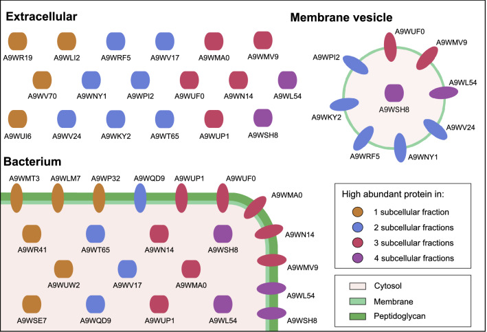
Bacterial kidney disease (BKD) is a chronic bacterial disease affecting both wild and farmed salmonids. The causative agent for BKD is the Gram-positive fish pathogen Renibacterium salmoninarum. As treatment and prevention of BKD have proven to be difficult, it is important to know and identify the key bacterial proteins that interact with the host. We used subcellular fractionation to report semi-quantitative data for the cytosolic, membrane, extracellular, and membrane vesicle (MV) proteome of R. salmoninarum. These data can aid as a backbone for more targeted experiments regarding the development of new drugs for the treatment of BKD. Further analysis was focused on the MV proteome, where both major immunosuppressive proteins P57/Msa and P22 and proteins involved in bacterial adhesion were found in high abundance. Interestingly, the P22 protein was relatively enriched only in the extracellular and MV fraction, implicating that MVs may play a role in host-pathogen interaction. Compared to the other subcellular fractions, the MVs were also relatively enriched in lipoproteins and all four cell wall hydrolases belonging to the New Lipoprotein C/Protein of 60 kDa (NlpC/P60) family were detected, suggesting an involvement in the formation of the MVs.
© 2022. The Author(s).
Conflict of interest statement
The authors declare no competing interests.
Figures

References
-
- Bandin I, Ellis A, Barja J, Secombes C. Interaction between rainbow trout macrophages and Renibacterium salmoninarum in vitro. Fish Shellfish Immunol. 1993;3:25–33.
-
- Gutenberger S, Duimstra J, Rohovec J, Fryer J. Intracellular survival of Renibacterium salmoninarum in trout mononuclear phagocytes. Dis. Aquat. Organ. 1997;28:93–106.
-
- Evelyn T, Ketcheson J, Prosperi-Porta L. Further evidence for the presence of Renibacterium salmoninarum in salmonid eggs and for the failure of povidone-iodine to reduce the intra-ovum infection rate in water-hardened eggs. J. Fish Dis. 1984;7:173–182.
-
- Balfry S, Albright L, Evelyn T. Horizontal transfer of Renibacterium salmoninarum among farmed salmonids via the fecal-oral route. Dis. Aquat. Organ. 1996;25:63–69.
-
- Wiens, G. Bacterial kidney disease (Renibacterium salmoninarum). In Fish diseases and disorders. Volume 3: viral, bacterial and fungal infections (eds. Woo, P. T. K. & Bruno, D. W.) 338–374 (CABI, 2011). 10.1079/9781845935542.0000
Publication types
MeSH terms
Substances
Supplementary concepts
LinkOut - more resources
Full Text Sources
Molecular Biology Databases
Research Materials
