

Choroidal vascularity index of patients with coronary artery disease
- PMID: 35194148
- PMCID: PMC8864027
- DOI: 10.1038/s41598-022-07120-8
Choroidal vascularity index of patients with coronary artery disease
Abstract
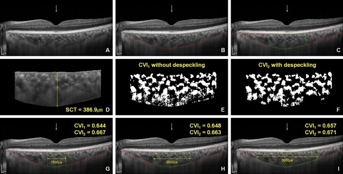
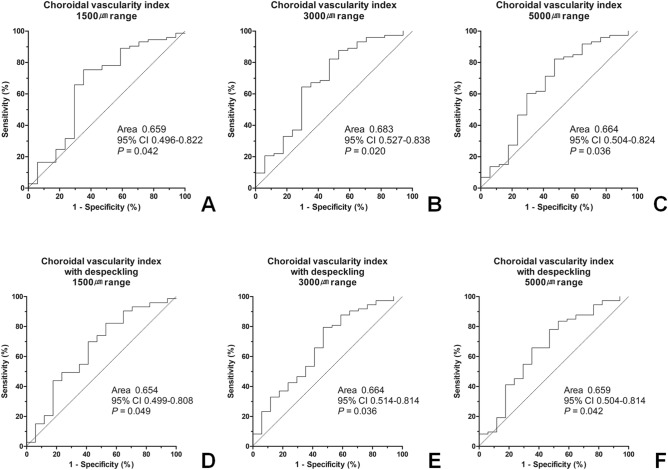
We investigated the changes in subfoveal choroidal thickness and choroidal vascularity index (CVI) and their relationship with the severity of coronary artery stenosis in patients with cardiovascular risk factors and symptoms suggestive of coronary artery disease (CAD). Ninety patients who underwent coronary angiography (CAG) for evaluation of their coronary artery status and cardiac symptoms were included. Forty-two patients showed no evidence of CAD; 31 patients had one to two vessel disease; and 17 had a triple vessel disease. There were no significant differences in the subfoveal choroidal thickness among the three groups; however, the CVI in the triple vessel disease group was lower than those in the other groups. The CVI values were good predictors of the presence of triple-vessel disease (p = 0.020). Multivariate logistic regression analysis results revealed that male sex (odds ratio 5.4, p = 0.049), hypertension (odds ratio 4.9, p = 0.017), and CVI (%, odds ratio 0.8, p = 0.016) were significant factors associated with the presence of triple vessel disease. Although CVI may not be a sensitive marker for detecting early changes in the coronary artery, it may be helpful in indicating severe CAD.
© 2022. The Author(s).
Conflict of interest statement
The authors declare no competing interests.
Figures
References
-
- 2021 Heart Disease and Stroke Statistics Update Fact Sheet.
-
- Knuuti J, Ballo H, Juarez-Orozco LE, et al. The performance of non-invasive tests to rule-in and rule-out significant coronary artery stenosis in patients with stable angina: a meta-analysis focused on post-test disease probability. Eur. Heart J. 2018;39(35):3322–3330. doi: 10.1093/eurheartj/ehy267. - DOI - PubMed
Publication types
MeSH terms
LinkOut - more resources
Full Text Sources
Medical
Miscellaneous
