. 2022 Apr;41(15):2139-2151.
doi: 10.1038/s41388-022-02235-8. Epub 2022 Feb 22.

Induction of APOBEC3-mediated genomic damage in urothelium implicates BK polyomavirus (BKPyV) as a hit-and-run driver for bladder cancer

Affiliations

Induction of APOBEC3-mediated genomic damage in urothelium implicates BK polyomavirus (BKPyV) as a hit-and-run driver for bladder cancer

Simon C Baker et al. Oncogene. 2022 Apr.

Abstract

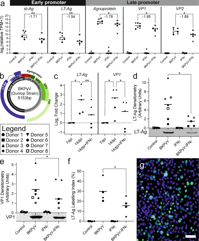
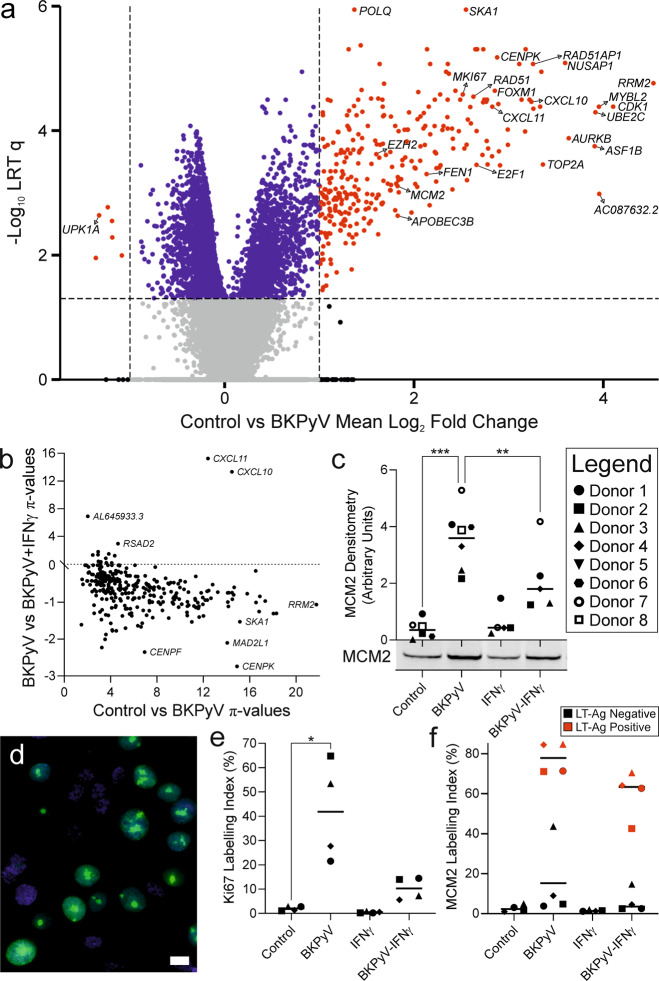
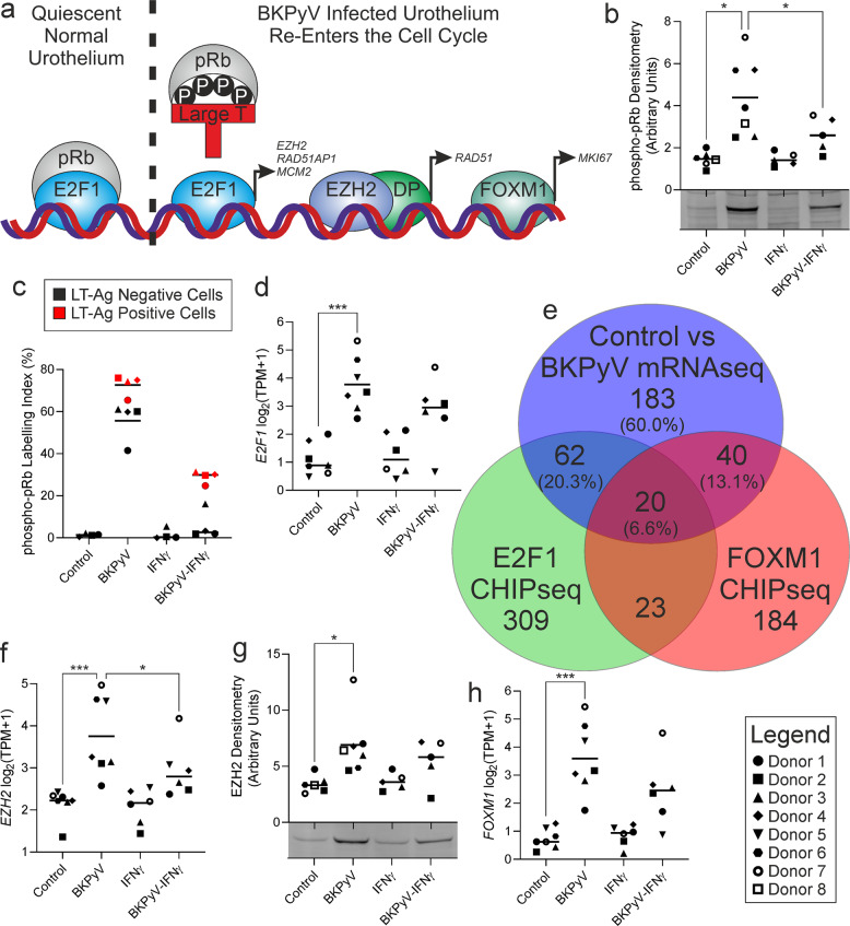
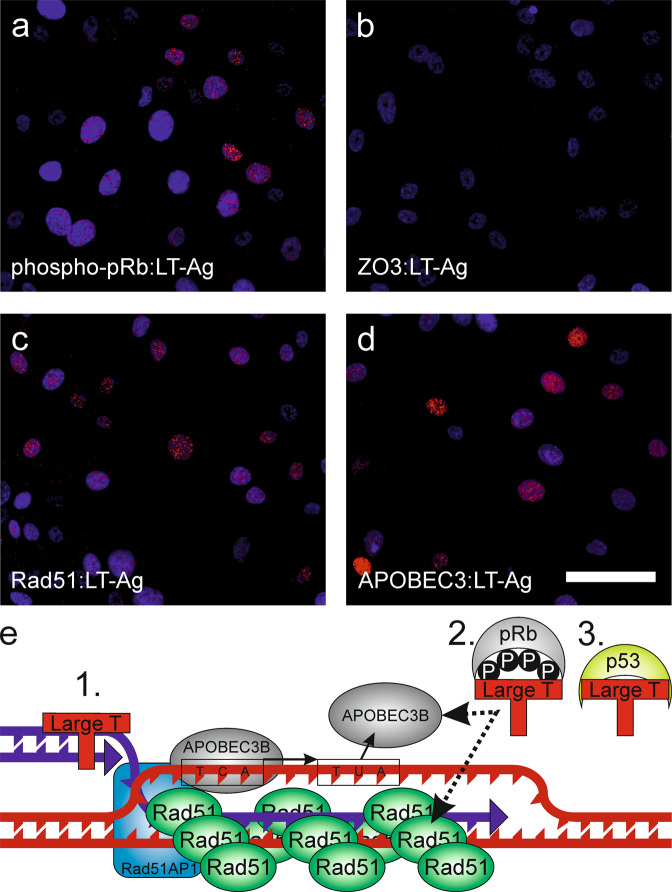
Limited understanding of bladder cancer aetiopathology hampers progress in reducing incidence. Mutational signatures show the anti-viral apolipoprotein B mRNA editing enzyme catalytic polypeptide (APOBEC) enzymes are responsible for the preponderance of mutations in bladder tumour genomes, but no causative viral agent has been identified. BK polyomavirus (BKPyV) is a common childhood infection that remains latent in the adult kidney, where reactivation leads to viruria. This study provides missing mechanistic evidence linking reactivated BKPyV-infection to bladder cancer risk. We used a mitotically-quiescent, functionally-differentiated model of normal human urothelium to examine BKPyV-infection. BKPyV-infection led to significantly elevated APOBEC3A and APOBEC3B protein, increased deaminase activity and greater numbers of apurinic/apyrimidinic sites in the host urothelial genome. BKPyV Large T antigen (LT-Ag) stimulated re-entry from G0 into the cell cycle through inhibition of retinoblastoma protein and activation of EZH2, E2F1 and FOXM1, with cells arresting in G2. The single-stranded DNA displacement loops formed in urothelial cells during BKPyV-infection interacted with LT-Ag to provide a substrate for APOBEC3-activity. Addition of interferon gamma (IFNγ) to infected urothelium suppressed expression of the viral genome. These results support reactivated BKPyV infections in adults as a risk factor for bladder cancer in immune-insufficient populations.


Conflict of interest statement

The authors declare no competing interests.

Figures

Fig. 1
Fig. 1. Schematic model of BKPyV hit-and-run carcinogenesis hypothesis.
Immune-insufficiency leads to reactivation of latent BKPyV, sloughing of actively-infected renal “decoy” cells and BKPyV viruria. BKPyV infects the G0-arrested urothelium but remains episomal. In infected urothelial cells, BKPyV LT-Ag inhibits host retinoblastoma (pRb) and disables p53, releasing urothelial cells from G0 into the cell cycle for arrest at the G2/M checkpoint. BKPyV stimulates APOBEC3 enzyme activity and causes host genome damage that inactivates tumour suppressors. The immune system clears the virus but initiated cells persist and over a period of years expand to form a tumour.
Fig. 2
Fig. 2. Expression of viral genes and proteins during BKPyV infection of normal human urothelium.
a mRNAseq analysis of BKPyV gene expression at 14 days post infection (dpi) showed late promoter genes including Agnoprotein were the most expressed viral genes, whilst early promoter genes such as LT-Ag were less transcriptionally-active. All viral gene expression was significantly suppressed by the indirect actions of IFNγ on the urothelium; however, the efficacy was widely variable between donors. Statistically-significant differences are indicated by stars with the mean log2 fold change in gene expression reported beneath (n = 6 or 7 independent donors). b BKPyV genome map showing the non-coding control region (NCCR) which regulates both the early and late genes that are expressed in opposing orientations. c RT-qPCR analysis of BKPyV LT-Ag and VP1 transcript abundance in normal human urothelial (NHU) cell cultures. Data is displayed as log2 fold-change normalised to abundance at 7 dpi. LT-Ag and VP1 abundance increased significantly between 7 and 14 dpi. The addition of IFNγ led to a significant reduction in LT-Ag and VP1 abundance at 14 dpi. d 14 dpi western blot densitometry for large T antigen (LT-Ag) showing significant reduction by IFNγ (mean log2 fold change −1.77), with exemplar blot image from a representative patient shown below the x-axis. Truncated LT-Ag (truncT-Ag) was also expressed; densitometry analysis of truncT-Ag and whole blots for all donor lines probed with LT-Ag can be found in Supplementary Fig. 1 and the β-actin loading controls in Supplementary Fig. 2. e 14 dpi western blot densitometry for viral capsid protein 1 (VP1) showing significant (mean log2 fold change −0.64) reduction by IFNγ, with exemplar blot image from a representative patient shown below x-axis. Full VP1 blots for all donor lines are shown as Supplementary Fig. 3 and the β-actin loading controls in Supplementary Fig. 2 (n = 5 independent donors). f LT-Ag indirect immunofluorescence labelling index found a mean of 29.8% of urothelial cells expressed detectable protein. n > 2900 cells per condition per donor. g Exemplar LT-Ag indirect immunofluorescence image from Donor 3 BKPyV-infected urothelial cells (all conditions shown in Supplementary Fig. 4). Scale bar denotes 100 μm. Significance was assessed in panels a by LRT test and cf by paired t-test.
Fig. 3
Fig. 3. mRNAseq analysis of differentiated human urothelium post BKPyV-infection.
a Volcano plot highlighting the significant induction of cell cycle and DNA-damage genes 14 dpi with BKPyV compared with controls (n = 7 independent donors). b The 305 genes significantly induced by BKPyV-infection are plotted to show their induction (x-axis) against the effect of post-infection addition of IFNγ (y-axis). Addition of IFNγ suppressed expression of the vast majority of BKPyV-induced genes (t = −17.57; p = 1.97 × 10−47). The chemokines CXCL10 and CXCL11 were notable exceptions, where the addition of IFNγ further increased expression. c Western blot densitometry for the DNA replication licensing factor “MCM2” showing significant mean 31-fold induction (p < 0.001) at 14 dpi with BKPyV and significant inhibition by IFNγ (p < 0.01); a representative blot image from a single patient shown is shown below the x-axis. Whole MCM2 blots for all donors are shown in Supplementary Fig. 6 and the β-actin loading controls in Supplementary Fig. 2. d Indirect immunofluorescence labelling of Ki67 (green) in BKPyV-infected urothelial tissues shows nearly all Ki67-positive cells have few large nucleolar granules, characteristic of the G2 cell cycle stage. A single image of BKPyV-infected cells is shown here and larger images of all conditions in a representative donor are shown in Supplementary Fig. 7. DNA was stained with Hoechst 33258 (blue). White scale bar denotes 10 μm. e Ki67 labelling indices for quiescent, G0-arrested control cultures were low (mean = 1.99% ± 0.94). BKPyV infection led to a significant mean 27-fold (p = 0.0319) increase in Ki67 labelling index (mean = 41.8% ± 20.62). f BKPyV infection also led to a significant increase in MCM2 labelling index (p = 0.0281; Supplementary Fig. 9). When cells from infected cultures were separated into LT-Ag positive and negative populations, there was a mean 5.6-fold ( ± 4.0) increase in MCM2 labelling of LT-Ag negative cells from BKPyV-infected cultures, implying a field effect. The red colouration for LT-Ag positive cells applies only to panel f.
Fig. 4
Fig. 4. BKPyV stimulates urothelial cell cycle re-entry by inactivating phosphorylation of retinoblastoma protein.
a Schematic summary of proposed BKPyV cell cycle regulation. pRb Retinoblastoma protein, DP Dimerization partners, P Phosphorylation. b The RB1 transcript that encodes pRB showed no changes in response to infection or IFNγ (Supplementary Fig. 11a); however, western blotting of pRb phosphorylated at serine 807/811 showed a significant infection-associated increase (mean 3.0-fold) that was suppressed by IFNγ. A representative blot from a single donor is shown here, with full blots for all donors in Supplementary Fig. 11b and the β-actin loading controls in Supplementary Fig. 2. c BKPyV infection also led to a significant mean 88-fold increase in phosphorylated-pRb labelling index (p < 0.01; representative images are shown in Supplementary Fig. 12). When cells from infected cultures were split into LT-Ag positive and negative populations, there was a mean 63.1-fold ( ± 57.1) increase in phosphorylated-pRb labelling of LT-Ag negative cells from BKPyV-infected cultures from all donors compared to non-infected controls, supporting the proposed field effect. The red colouration for LT-Ag positive cells applies only to panel c. d mRNAseq showed significant induction of the pRb-regulated E2F1 transcription factor, which was induced more prominently than other members of the E2F family (Supplementary Fig. 13). e Comparison of genes significantly 2-fold induced by BKPyV-infection with those reported to possess proximal E2F1 [21] and FOXM1 [23] ChIP-seq peaks, suggested 40% of the BKyV-induced transcriptome may be activated by these transcription factors. E2F1 ChIPseq peaks [21] and BKPyV-induced genes had a significant overlap of 82 genes (exact hypergeometric probability p < 4.95 × 10−106; representation factor = 38.6). There was a 60-gene overlap (exact hypergeometric probability p < 1.30 × 10−80; representation factor = 43.8) between genes up-regulated upon BKPyV-infection and those associated with FOXM1 binding [23]. Gene lists provided as Supplementary Fig. 14. Increased E2F and FOXM1-activity was further supported by GSEA (Supplementary Fig. 10d–f and 10h). f and g mRNAseq and western blotting respectively show a BKPyV-mediated increase in expression of the DREAM complex member EZH2. EZH2 joins the polycomb repressive complex 2 (PRC2) dimerization partners to drive transcription as shown by GSEA (Supplementary Fig. 10g). A representative blot from a single donor is shown here, with full EZH2 blots shown in Supplementary Fig. 15 and the β-actin loading controls in Supplementary Fig. 2. h mRNAseq shows significant induction of FOXM1 transcription by BKPyV-infection.
Fig. 5
Fig. 5. Homologous recombination was induced by BKPyV-infection of human urothelium.
a Western blotting for p53 showed significant protein stabilisation (no increase in transcription was observed) during BKPyV infection. Stabilisation of p53 in the nucleus was confirmed by indirect immunofluorescence. A representative blot from a single donor is shown here, with full p53 blots shown in Supplementary Fig. 16 and the β-actin loading controls in Supplementary Fig. 2. b There was no increased expression of verified p53 target genes [51]. c RAD51 and d RAD51AP1 transcripts were significantly induced by BKPyV-infection. e Western blotting confirmed significant Rad51 protein induction (mean 2.7-fold) and phosphorylation was observed as a slight increase in molecular weight when Rad51 was induced by BKPyV. A slight increase in Rad51 molecular size during infection indicated possible activating-phosphorylation of the induced Rad51 by the Chk1 kinase [24], whose transcription was also significantly induced. A representative blot from a single donor is shown here, with full Rad51 blots shown in Supplementary Fig. 17 and the β-actin loading controls in Supplementary Fig. 2. f Analysis of indirect immunofluorescence for Rad51 found nuclear speckles were significantly increased in BKPyV infection (from mean 0.05% positive cells in controls to 11.13% in BKPyV-infected; images in Supplementary Fig. 17). In some cells, indirect immunofluorescence revealed LT-Ag and Rad51 protein co-localisation to large granular deposits in the nuclei of BKPyV-infected urothelial cells. Scale bar denotes 10 μm, immunofluorescence performed on n = 3 independent donors with representative images shown.
Fig. 6
Fig. 6. APOBEC3 expression and activity in normal human urothelium.
mRNAseq analysis of APOBEC3A and APOBEC3B transcripts found only APOBEC3B was significantly (mean log2 fold change 1.78 ± 1.31; q = 0.0023) induced by BKPyV-infection (a and b, respectively). mRNAseq expression data for the whole APOBEC enzyme family is provided as Supplementary Fig. 18. c Western blotting for APOBEC3A found a significant (p = 0.0133) 2.04-fold induction by BKPyV infection (whole blots for all donors shown in Supplementary Fig. 20); note “A3A”; full-length (“Met1”) and truncated protein from the internal Met13 start site were analysed together as both possess catalytic activity [29]. d APOBEC3B (“A3B”) and APOBEC3G (“A3G”) bands appear close together on the blots and densitometry shown here is for APOBEC3B only (as APOBEC3G expression was not altered; Supplementary Fig. 20). APOBEC3B protein was significantly (mean 1.93-fold) induced by BKPyV infection (p = 0.0025). Western blot densitometry and TPM for APOBEC3B were significantly correlated (Pearson Rho = 0.77; p = 0.0001). Densitometry analysis suggested that during BKPyV infection, the ratio of APOBEC3A to APOBEC3B protein was 1.95:1 ( ± 1.18; n = 7). c and d Representative APOBEC blot images from a single donor are shown here, with full blots for all donors shown in Supplementary Fig. 20 and the β-actin loading controls in Supplementary Fig. 2. e Variant transcript frequency (VTF) for APOBEC-mediated C > U editing of DDOST RNA at cytosine 558 quantified by mRNAseq was increased slightly, but not significantly, in 6/7 donors by BKPyV infection. Donors 5 and 7 appeared to show an IFNγ-mediated induction of APOBEC3A-function. Across all mRNAseq samples (n = 26) DDOST C558U variant transcript frequency (VTF) was significantly correlated with APOBEC3A TPM (Pearson Rho = 0.867; p < 0.0001) but not APOBEC3B TPM (Pearson Rho = 0.368; p = 0.0642; Supplementary Fig. 21). f Deaminase assays for urothelial cell lysates against a linear RTCA probe, preferentially targeted by APOBEC3B, showed significant induction of activity following BKPyV infection (mean log2 fold change 1.66 ± 1.05; p = 0.0313; gels for all donors shown in Supplementary Fig. 22). Deaminase assays were also performed against a YTCA hairpin probe, in the presence and absence of RNA, to evaluate APOBEC3A activity (Supplementary Fig. 24). g mRNAseq expression data for the uracil-DNA glycosylase gene UNG found it was significantly induced by BKPyV-infection (q = 0.0213). h An apurinic/apyrimidinic (AP) sites assay was performed on genomic DNA from five independent donors analysing 2–3 independent cultures per donor. T74D breast cancer cells were included as a positive control known to generate apurinic/apyrimidinic sites in their genomes [50]. The legend in the top right identifies the donor by specific point shapes in all dot plot panels.
Fig. 7
Fig. 7. Proximity ligation assays of human urothelium during BKPyV-infection.
Proximity ligation assays indicated that nuclear complexes formed involving large T antigen (LT-Ag) and a phospho-Retinoblastoma (phospho-pRb), b Zonula occludins 3 (ZO3) is an intercellular tight junction protein and included here as a non-nuclear-localized negative control, c Rad51 and d APOBEC3 proteins. Scale bar denotes 50 μm, images from one representative donor during BKPyV-infection are shown here with images from three donors, including non-infected controls, provided as Supplementary Figs. 26–28. e Schematic of hypothetical model of urothelial DNA-damage at displacement loops at the G2/M checkpoint during BKPyV infection. LT-Ag interactions might derive from (1.) its DNA-binding helicase activity or (2.) LT-Ag protein dimerization with another involved protein, such as pRb. (3.) LT-Ag inhibition of p53 is likely important for this process as p53 would normally prevent Rad51 oligomerisation by direct binding [52]. The evidence from this study is most robust for BKPyV-induced APOBEC3B-activity but APOBEC3A protein was consistently more abundant during infection and its role requires further validation.

References

    1. Alexandrov LB, Kim J, Haradhvala NJ, Huang MN, Tian Ng AW, Wu Y, et al. The repertoire of mutational signatures in human cancer. Nature. 2020;578:94–101. doi: 10.1038/s41586-020-1943-3. - DOI - PMC - PubMed
    1. Baker SC, Mason AS, Southgate J. Procarcinogen activation and mutational signatures model the initiation of carcinogenesis in human urothelial tissues in vitro. Eur Urol. 2020;78:143–7. doi: 10.1016/j.eururo.2020.03.049. - DOI - PMC - PubMed
    1. Robertson AG, Kim J, Al-Ahmadie H, Bellmunt J, Guo G, Cherniack AD, et al. Comprehensive molecular characterization of muscle-invasive bladder cancer. Cell. 2017;171:540–56.e25. - PMC - PubMed
    1. Kean JM, Rao S, Wang M, Garcea RL. Seroepidemiology of human polyomaviruses. PLoS Pathog. 2009;5:e1000363. doi: 10.1371/journal.ppat.1000363. - DOI - PMC - PubMed
    1. Chesters PM, Heritage J, McCance DJ. Persistence of DNA sequences of BK virus and JC virus in normal human tissues and in diseased tissues. J Infect Dis. 1983;147:676–84. doi: 10.1093/infdis/147.4.676. - DOI - PubMed