. 2022;56(1):72-89.
doi: 10.1134/S0026893322010022. Epub 2022 Feb 12.

Genetic Engineering Systems to Study Human Viral Pathogens from the Coronaviridae Family

Affiliations

Genetic Engineering Systems to Study Human Viral Pathogens from the Coronaviridae Family

S O Galkin et al. Mol Biol. 2022.

Abstract

The COVID-19 pandemic caused by the previously unknown SARS-CoV-2 Betacoronavirus made it extremely important to develop simple and safe cellular systems which allow manipulation of the viral genome and high-throughput screening of its potential inhibitors. In this review, we made an attempt at summarizing the currently existing data on genetic engineering systems used to study not only SARS-CoV-2, but also other viruses from the Coronaviridae family. In addition, the review covers the basic knowledge about the structure and the life cycle of coronaviruses.

Keywords: COVID-19; SARS-CoV-2; pseudoviruses; replicons.


Conflict of interest statement

COMPLIANCE WITH ETHICAL STANDARDSConflict of interest. The authors declare no conflict of interest. Statement on the welfare of humans or animals. This article does not contain any studies involving humans or animals performed by any of the authors.

Figures

Fig. 1.
Fig. 1.
Schematic representation of the Coronaviridae family virion structure. The helical symmetry nucleocapsid made up by the nucleocapsid (N) protein and one RNA genome molecule is surrounded by the supercapsid of cellular origin with embedded virus S, M, and E proteins. The hemagglutinin esterase (HE) protein is included in the virions of the human coronaviruses HCoV-OC43 and HCoV-HKU1, but is not found in HCoV-229E, HCoV-NL63, MERS-CoV, SARS-CoV-1, and SARS-CoV-2. The coronavirus virion contains about 300 S protein molecules, 2000 М protein molecules, 1000 N protein molecules, and 100 E protein molecules [11].
Fig. 2.
Fig. 2.
Genome structure of coronaviruses circulating in human populations. In the figure, encoded virus proteins are indicated in capital letters to the right, virus-specific ORF nos. are indicated with numbers and small letters. UTR—untranslated region. Numbers to the left indicate encoded NSP nos.
Fig. 3.
Fig. 3.
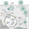
Schematic representation of Coronaviridae family viruses. Infection starts with the virus particle binding with its receptor on the host cell surface which leads to virus and cell membrane fusion (early entry pathway), or virion encapsulation into endosomes and further virus and cell membrane fusion (late entry pathway). Viral RNA in complex with N protein enters the cytoplasm. ORF1a/b first open reading frame translation results in the pp1a and pp1ab polyproteins which further undergo autoproteolysis to obtain NSP2s. NSPs form the RdRp complex which carries out viral RNA replication and transcription. This process takes place in the double-membrane-bound organelles which are formed from the rough endoplasmic reticulum in the presence of pp1a and pp1ab components. Replication/transcription results in full-size and subgenomic viral RNAs. Structural and accessory proteins are translated from subgenomic RNAs. Structural proteins are assembled into the intermediate structure in the endoplasmic reticulum–Golgi intermediate compartment (ERGIC) which then form new virions together with the N protein/genomic viral RNA complex. Newly assembled virions are accumulated in the intracellular vesicles, and then leave the infected cell by exocytosis [modified from 54].
Fig. 4.
Fig. 4.
Schematic representation of coronavirus subgenomic RNA synthesis strategies. (a) Internal initiation model. IBS—internal RdRp binding site. (b) Premature transcription termination model. Termination may occur both during (+)-chain and (–)-chain RNA synthesis. PTS—premature termination site for RdRp. (c) Discontinuous transcription model. Transcription may be interrupted both during (+)-chain and (–)-chain RNA synthesis [58]. LS–leader sequence; TRS—transcription regulatory sequence; L-TRS—leader TRS, and B-TRS—TRS in the coding region.
Fig. 5.
Fig. 5.
DI-RNA system functioning principle. In coronavirus-infected cells, DI-RNAs may be included in the virion (1), and/or replicated (2), and/or express reporter proteins (3) in the presence of viral proteins translated from genomic coronavirus RNA.
Fig. 6.
Fig. 6.
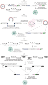
Schematic representation of full-size virus cDNA production using artificial bacterial chromosomes (a), TAR cloning technique (b), in vitro ligation technique (c), or recombinant poxviruses (d). CMV—cytomegalovirus promoter; pA—poly(A) tail; Rz—HDV ribozyme, and MCS—polylinker.
Fig. 7.
Fig. 7.
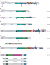
Key structural elements of coronavirus replicons. (a) SARS-CoV-2 schematic genome structure (provided as a reference structure). (b‒h) Structure of several different replicons [–118]; regulatory elements (5'UTR, 3'UTR, TRS) are always encoded in a replicon, pp1a/ab components are absent only in minimal replicons (h) [118]. Replicon coding part (body) may be put under the control of either T7, or a cellular promoter. In the first case, it is possible to both carry out in vitro transcription using the obtained RNA for cell transfection and transfect cells with the DNA construct with the simultaneous T7 RNA polymerase expression [–118]. Encoded proteins are indicated with letters across the diagram. TRS are indicated with red lines. GFP—green fluorescent protein; LUC—luciferase; BSD—blasticidin-resistance protein; NEO—neomycin-resistance protein, and IRES—internal ribosome entry site.

Similar articles

Cited by

References

    1. Yin Y., Wunderink R.G. MERS, SARS and other coronaviruses as causes of pneumonia. Respirology. 2018;23:130–137. - PMC - PubMed
    1. Wu F., Zhao S., Yu B., Chen Y.-M., Wang W., Song Z.-G., Hu Y., Tao Z.-W., Tian J.-H., Pei Y.-Y., Yuan M.-L., Zhang Y.-L., Dai F.-H., Liu Y., Wang Q.-M. A new coronavirus associated with human respiratory disease in China. Nature. 2020;579:265–269. - PMC - PubMed
    1. Rothe C., Schunk M., Sothmann P., Bretzel G., Guggemos W., Seilmaier M., Drosten C., Vollmar P., Zwirglmaier K., Zange S., Wölfel R., Hoelscher M. Transmission of 2019-nCoV infection from an asymptomatic contact in Germany. N. Engl. J. Med. 2020;382:970–971. - PMC - PubMed
    1. Perez-Saez J., Lauer S.A., Kaiser L., Regard S., Delaporte E., Guessous I., Stringhini S., Azman A.S., Serocov-POP Study Group Serology-informed estimates of SARS-CoV-2 infection fatality risk in Geneva, Switzerland. Lancet Infect. Dis. 2021;21:e69–e70. - PMC - PubMed
    1. Rebello C. J., Kirwan J.P., Greenway F.L. Obesity, the most common comorbidity in SARS-CoV-2: is leptin the link? Int. J. Obes. (Lond.) 2020;44:1810–1817. - PMC - PubMed

LinkOut - more resources