Review
. 2022 Oct 14;43(39):3971-3980.
doi: 10.1093/eurheartj/ehac061.

Heart-brain interactions in cardiac and brain diseases: why sex matters

Affiliations
Review

Heart-brain interactions in cardiac and brain diseases: why sex matters

Alexia Rossi et al. Eur Heart J. .

Abstract

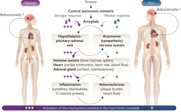
Cardiovascular disease and brain disorders, such as depression and cognitive dysfunction, are highly prevalent conditions and are among the leading causes limiting patient's quality of life. A growing body of evidence has shown an intimate crosstalk between the heart and the brain, resulting from a complex network of several physiological and neurohumoral circuits. From a pathophysiological perspective, both organs share common risk factors, such as hypertension, diabetes, smoking or dyslipidaemia, and are similarly affected by systemic inflammation, atherosclerosis, and dysfunction of the neuroendocrine system. In addition, there is an increasing awareness that physiological interactions between the two organs play important roles in potentiating disease and that sex- and gender-related differences modify those interactions between the heart and the brain over the entire lifespan. The present review summarizes contemporary evidence of the effect of sex on heart-brain interactions and how these influence pathogenesis, clinical manifestation, and treatment responses of specific heart and brain diseases.

Keywords: Brain; Dementia; Depression; Gender; Heart; Heart failure; Ischaemic heart disease; Sex; Stroke; Takotsubo syndrome.


Conflict of interest statement

none declared.

Figures

Graphical Abstract
Graphical Abstract
Mechanisms involved in the heart–brain crosstalk. Simplified representation of sex differences seen in the main mechanisms and neurohumoral circuits involved in heart–brain interactions. The intensity of activation is represented by a colour code scale, with red indicating the maximal activation. In brief, specific triggers (e.g. stress, acute myocardial infarction) induce the activation of the amygdala via the central autonomic system. Efferent projections increase the activation of the sympathetic nervous system and initiate neurohormonal output through the hypothalamic–pituitary–adrenal axis leading to catecholamine release, myelopoiesis activation, and release of pro-inflammatory cytokines with deleterious effect on the heart. This pro-inflammatory state initiates and promotes atherosclerosis. Current evidence on the pathophysiology of the specific heart and brain disease discussed in this review has shown that the activation of all these mechanisms is more pronounced in women as compared with men. The bidirectionality of heart–brain interactions is still under investigation.
Figure 1
Figure 1
Imaging modalities used to investigate the mechanisms involved in the heart–brain crosstalk. (A) Functional MR illustrates activated regions of the brain (A1). 123I-mIBG-SPECT shows a perfusion defect involving the infero-lateral wall of the left ventricle (A2). 11C-mHED-PET demonstrates reduced tracer uptake in the lateral wall of the left ventricle (A3). The findings shown in A2 and A3 are indicative of cardiac sympathetic denervation and, indirectly, of increased sympathetic tone. The uptake scales used for image visualization are reported on the right. (B) 18F-FDG-PET images show an increased 18F-FDG uptake at the level of the right amygdala (B1 – white arrow), myocardium (B2), and bone marrow of the spine (B3). The SUV scale used for image visualization is reported on the right. (C) Straight multiple curve reconstructions from CCTA show a mixed plaque with positive remodelling of mid RCA (C1), calcified plaque of mid LAD (C2), and spotty calcification of the mid LCx (C3). A cross-section at the level of the corresponding plaque is also shown for each vessel (red box). (D) SPECT images acquired during stress (D1) show a reversible myocardial perfusion defect of the left ventricular inferior wall which is not present at rest (D2). Hypoperfusion is detected as a relative decrease of the uptake of the inferior wall (50–62%) as compared to the myocardial territory with the highest tracer uptake. PET images acquired during stress indicate a low MBF (mL/g/min) in the myocardial territory supplied by the LAD (D3). In the LAD territory MBF did not increase during stress (D3) as compared to rest (D4). The uptake and MBF scales used for image visualization and MBF quantification are reported on the right. CCTA: coronary computed tomography angiography; 11C-mHED: 11C-meta-hydroxyephedrine; 18F-FDG: 18F-fluorodeoxyglucose; fMRI: functional magnetic resonance imaging; 123I-mIBG: 123I-metaiodobenzylguanidine; LAD: left anterior descending coronary artery; LCx: left circumflex coronary artery; MBF: myocardial blood flow; MR: magnetic resonance; 13N-NH3: 13N-ammonia; PET: positron emission tomography; RCA: right coronary artery; SPECT: single-positron emission computed tomography; SUV: standard uptake value; 99Tc-MIBI: 99Technetium-methoxyisobutyl isonitrile.123I-mIBG-SPECT image was provided through the courtesy of Dr Renata Chequer and Prof. François Rouzet from the Nuclear Medicine department of the Bichat Hospital—Assistance Publique Hôpitaux de Paris.

References

    1. Tahsili-Fahadan P, Geocadin RG. Heart-brain axis: effects of neurologic injury on cardiovascular function. Circ Res 2017;120:559–572. - PubMed
    1. Fiechter M, Roggo A, Burger IA, Bengs S, Treyer V, Becker A, et al. Association between resting amygdalar activity and abnormal cardiac function in women and men: a retrospective cohort study. Eur Heart J Cardiovasc Imaging 2019;20:625–632. - PubMed
    1. Ishai A, Osborne MT, Tung B, Wang Y, Hammad B, Patrich T, et al. Amygdalar metabolic activity independently associates with progression of visceral adiposity. J Clin Endocrinol Metab 2019;104:1029–1038. - PMC - PubMed
    1. Osborne MT, Ishai A, Hammad B, Tung B, Wang Y, Baruch A, et al. Amygdalar activity predicts future incident diabetes independently of adiposity. Psychoneuroendocrinology 2019;100:32–40. - PMC - PubMed
    1. Radfar A, Abohashem S, Osborne MT, Wang Y, Dar T, Hassan MZO, et al. Stress-associated neurobiological activity associates with the risk for and timing of subsequent Takotsubo syndrome. Eur Heart J 2021;42:1898–1908. - PMC - PubMed

Publication types