. 2022 Feb 23;14(633):eabm1375.
doi: 10.1126/scitranslmed.abm1375. Epub 2022 Feb 23.

Hematopoietic cell transplantation donor-derived memory-like NK cells functionally persist after transfer into patients with leukemia

Affiliations

Hematopoietic cell transplantation donor-derived memory-like NK cells functionally persist after transfer into patients with leukemia

Melissa M Berrien-Elliott et al. Sci Transl Med. .

Abstract

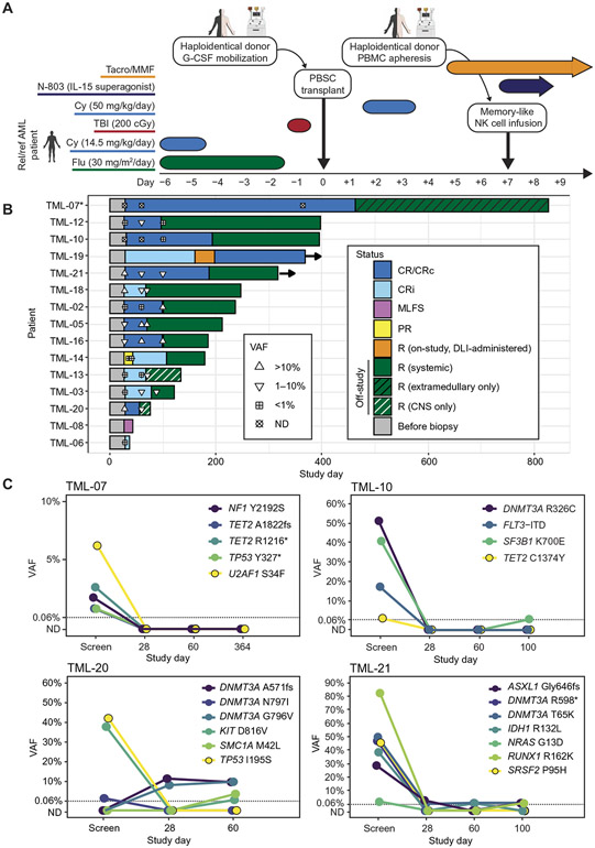
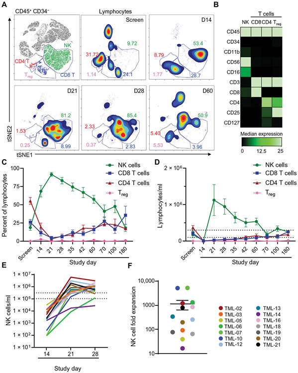
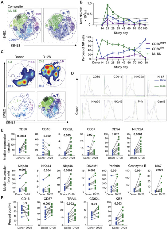
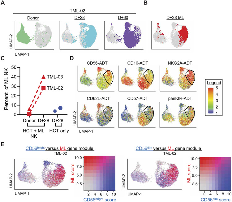
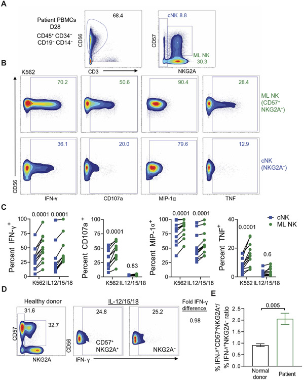
Natural killer (NK) cells are innate lymphoid cells that eliminate cancer cells, produce cytokines, and are being investigated as a nascent cellular immunotherapy. Impaired NK cell function, expansion, and persistence remain key challenges for optimal clinical translation. One promising strategy to overcome these challenges is cytokine-induced memory-like (ML) differentiation, whereby NK cells acquire enhanced antitumor function after stimulation with interleukin-12 (IL-12), IL-15, and IL-18. Here, reduced-intensity conditioning (RIC) for HLA-haploidentical hematopoietic cell transplantation (HCT) was augmented with same-donor ML NK cells on day +7 and 3 weeks of N-803 (IL-15 superagonist) to treat patients with relapsed/refractory acute myeloid leukemia (AML) in a clinical trial (NCT02782546). In 15 patients, donor ML NK cells were well tolerated, and 87% of patients achieved a composite complete response at day +28, which corresponded with clearing high-risk mutations, including TP53 variants. NK cells were the major blood lymphocytes for 2 months after HCT with 1104-fold expansion (over 1 to 2 weeks). Phenotypic and transcriptional analyses identified donor ML NK cells as distinct from conventional NK cells and showed that ML NK cells persisted for over 2 months. ML NK cells expressed CD16, CD57, and high granzyme B and perforin, along with a unique transcription factor profile. ML NK cells differentiated in patients had enhanced ex vivo function compared to conventional NK cells from both patients and healthy donors. Overall, same-donor ML NK cell therapy with 3 weeks of N-803 support safely augmented RIC haplo-HCT for AML.


Figures

Fig. 1.
Fig. 1.. RIC haplo-HCT was augmented by ML NK cells and N-803.
(A) Study schema. Patients with rel/ref AML underwent RIC with fludarabine (Flu), cyclophosphamide (Cy), and total body irradiation (TBI) and then received a donor-derived G-CSF–mobilized peripheral blood stem cell (PBSC) graft on day 0, posttransplant cyclophosphamide on day +3/+4, and donor-derived memory-like (ML) NK cells (produced using nonmobilized PBMCs) on day +7 with N-803 (IL-15 superagonist) support. GvHD prophylaxis consisted of tacrolimus (Tacro) and mycophenolate mofetil (MMF). (B) Swimmer’s plot of patient outcomes. Disease status is indicated by bar color: complete response (CR), cytogenetic complete response (CRc; marked by *), morphologic leukemia-free state (MLFS), partial response (PR), and relapse (R). DLI indicates donor lymphocyte infusion. CNS indicates central nervous system. Patients alive at the last follow-up are indicated by right arrows. Symbols indicate the highest variant allele fraction (VAF) at a given time point of mutations initially identified at the time of screening. ND indicates not detected. (C) Mutation VAF trends over time for patients TML-07, TML-10, TML-20, and TML-21. Dotted lines at VAF = 0.06% represent the limit of detection upper bound across all variants. Variants detected at VAF 0.06% or lower are plotted on the dotted line; variants not detected are plotted below on the y axis at ND.
Fig. 2.
Fig. 2.. NK cells are the most abundant lymphocyte after NK cell administration.
Patient PB was obtained and assessed by mass cytometry at the indicated time points. (A) FlowSOM identified the major lymphocyte populations. Representative overlay and density plots are shown for samples isolated from TML-05, with each lymphocyte population indicated. Gray indicates nonlymphocyte/myeloid populations. Inset numbers depict frequency of cells within the indicated gate on lymphocytes (T, B, and NK cells). (B) Markers used for FlowSOM clustering for each population shown in (A). Treg, regulatory T cell. (C) Summary data from (A) are shown for 14 patients. (D) Total lymphocyte concentrations per milliliter of PB are shown for 14 patients. (E) Total NK cells in PB are shown for each patient. (F) Summary data are shown depicting maximum fold expansion from days +7 to +21 or +28. Each dot represents a patient. The dashed lines in (D) and (E) indicate average NK cell counts for healthy donors. Data are presented as means ± SEM.
Fig. 3.
Fig. 3.. ML NK cells persist in patient PB after transfer in the RIC-HCT setting.
The multidimensional phenotype of FlowSOM-gated ML NK cells was assessed using viSNE. (A) Representative viSNE plots for patient TML-05 are shown, demonstrating NK cell subset (CD56bright, CD56dim, and ML NK) binning. (B) Summary percent (bottom) and total numbers (top) of NK cell subsets are shown for 14 patients. (C) Representative density plots of donor and patient NK cells at days +28 and +60 are shown, and inset numbers represent frequency of cells within the gate (top). A second FlowSOM was used to identify NK cell subsets (see figs. S5 and S6), and distinct subsets within viSNE were identified (bottom). (D) Overlay histograms are shown of donor (blue) and day +28 (green) PB bulk NK cells for the expression of the indicated marker. (E and F) Summary data from (D) depicting median expression (E) or percent positive (F) are shown for 12 donor-patient pairs. Data are presented in arcsinh transformed scale and are depicted as means ± SEM and were compared using paired t or Wilcoxon test, as appropriate. P values are indicated above each plot. TRAIL, TNF-related apoptosis-inducing ligand.
Fig. 4.
Fig. 4.. CITE-seq identifies ML NK cells persisting for 2 months in patients.
(A) UMAP visualization of TML-02 with cells highlighted by color at each time point: Donor (sea green), +D28 (turquoise), +D60 (purple). (B) ML NK cells identified by classifier are highlighted in red on the UMAP visualization of TML-02; see also fig. S9. (C) Proportions of ML NK in total NK cells at baseline/donor, day +28, and in patients receiving haplo-HCT transplant without ML NK cells are shown. (D) Expression of key ML antibody-derived tags (ADTs) on TML-02 are shown; min.cutoff = “q02,” max.cutoff = “q98.” (E) CD56bright and CD56dim as annotated (blue), and ML (red) NK cell gene module scoring was overlaid on TML-02 UMAP visualization. Cells are ordered on the UMAP by binned expression.
Fig. 5.
Fig. 5.. ML NK cells are transcriptionally distinct from cNK cells.
(A to C) Select differentially expressed genes conserved across both patients in ML NK cells (indicated within the gate) compared to all other clusters were visualized on TML-02 UMAP. Expression of activation, effector function, and NK receptors (A); adhesion and co-receptors (B); and transcription factors (C) are shown. Data were analyzed using a Wilcoxon rank sum test. n = 2 independent experiments. Cells are ordered on UMAPs according to normalized binned expression; see figs. S10 and S11. Adjusted P value or q value of <0.05 for all.
Fig. 6.
Fig. 6.. ML NK cells differentiate in patients.
(A) A monocle trajectory plot is shown for samples from TML-02, depicting three cell states and their proportions over time. (B) ADT expression of CD56, CD16, and CD57 was overlaid on the TML-02 trajectory. (C) ML NK cells are abundant in cell state 3 (red). (D) Select significantly changed genes across time in state 3 are shown. Points are colored by time, and the line depicts expression as a function of pseudotime. Differential gene test in Monocle2 using a regression model was used to determine genes changing as a function of time. Adjusted P value or q value of <0.05 for all.
Fig. 7.
Fig. 7.. In vivo differentiated ML NK cells demonstrate enhanced functionality ex vivo.
Freshly isolated patient PBMCs (day +28) were stimulated with K562 or IL-12/IL-15/IL-18 for 6 hours and characterized by mass cytometry. (A) A representative gating strategy shows identification of in vivo differentiated ML NK cells (NKG2A+CD57+) and cNK cells (NKG2A). (B) Representative plots show ML NK (green) and cNK (blue) responses to K562. Inset numbers represent the frequency of cells within the gate. (C) Summary data from (B) are shown, with each patient depicted. (D)Patient and healthy donor PBMCs were stimulated with IL-12/IL-15/IL-18, and IFN-γ responses were compared. Representative flow plots comparing relative IFN-γ responses in NKG2A+CD57+ and NKG2A from healthy donors are shown. The ratio of IFN-γ+ cells in each subset was quantified (right) as % IFN-γ+NKG2A+CD57+/% IFN-γ+NKG2A. (E) Summary data from (D) are shown. Data in (E) are from 12 healthy donors and 12 patients. Each dot represents an individual patient. Data were compared using a (C) two-way ANOVA or (E) Mann-Whitney test.

References

    1. Estey EH, Acute myeloid leukemia: 2019 update on risk-stratification and management. Am. J. Hematol 93, 1267–1291 (2018). - PubMed
    1. Araki D, Wood BL, Othus M, Radich JP, Halpern AB, Zhou Y, Mielcarek M, Estey EH, Appelbaum FR, Walter RB, Allogeneic hematopoietic cell transplantation for acute myeloid leukemia: Time to move toward a minimal residual disease-based definition of complete remission? J. Clin. Oncol 34, 329–336 (2016). - PMC - PubMed
    1. Scott BL, Pasquini MC, Logan BR, Wu J, Devine SM, Porter DL, Maziarz RT, Warlick ED, Fernandez HF, Alyea EP, Hamadani M, Bashey A, Giralt S, Geller NL, Leifer E, Le-Rademacher J, Mendizabal AM, Horowitz MM, Deeg HJ, Horwitz ME, Myeloablative versus reduced-intensity hematopoietic cell transplantation for acute myeloid leukemia and myelodysplastic syndromes. J. Clin. Oncol 35, 1154–1161 (2017). - PMC - PubMed
    1. McCurdy SR, Luznik L, How we perform haploidentical stem cell transplantation with posttransplant cyclophosphamide. Blood 2019, 513–521 (2019). - PMC - PubMed
    1. Jaiswal SR, Zaman S, Chakrabarti A, Sen S, Mukherjee S, Bhargava S, Ray K, O’Donnell PV, Chakrabarti S, Donnell PVO, Chakrabarti S, O’Donnell PV, Chakrabarti S, Improved outcome of refractory/relapsed acute myeloid leukemia after post-transplantation cyclophosphamide-based haploidentical transplantation with myeloablative conditioning and early prophylactic granulocyte colony-stimulating factor–mobilized donor lymphocyte infusions. Biol. Blood Marrow Transplant 22, 1867–1873 (2016). - PubMed

Publication types

Associated data