. 2022 Feb 25;8(8):eabk0052.
doi: 10.1126/sciadv.abk0052. Epub 2022 Feb 23.

Activation of PDGFRA signaling contributes to filamin C-related arrhythmogenic cardiomyopathy

Affiliations

Activation of PDGFRA signaling contributes to filamin C-related arrhythmogenic cardiomyopathy

Suet Nee Chen et al. Sci Adv. .

Abstract

FLNC truncating mutations (FLNCtv) are prevalent causes of inherited dilated cardiomyopathy (DCM), with a high risk of developing arrhythmogenic cardiomyopathy. We investigated the molecular mechanisms of mutant FLNC in the pathogenesis of arrhythmogenic DCM (a-DCM) using patient-specific induced pluripotent stem cell-derived cardiomyocytes (iPSC-CMs). We demonstrated that iPSC-CMs from two patients with different FLNCtv mutations displayed arrhythmias and impaired contraction. FLNC ablation induced a similar phenotype, suggesting that FLNCtv are loss-of-function mutations. Coimmunoprecipitation and proteomic analysis identified β-catenin (CTNNB1) as a downstream target. FLNC deficiency induced nuclear translocation of CTNNB1 and subsequently activated the platelet-derived growth factor receptor alpha (PDGFRA) pathway, which were also observed in human hearts with a-DCM and FLNCtv. Treatment with the PDGFRA inhibitor, crenolanib, improved contractile function of patient iPSC-CMs. Collectively, our findings suggest that PDGFRA signaling is implicated in the pathogenesis, and inhibition of this pathway is a potential therapeutic strategy in FLNC-related cardiomyopathies.


Figures
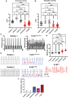
Fig. 1.
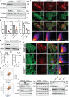
Fig. 1.. FLNCG1891Vfs61X and FLNCE2189X iPSC-CMs exhibited impaired contractility and arrhythmia.
(A) Contraction and (B) relaxation velocities of both patient-derived iPSC-CMs were lower than those of healthy controls (n = 20 to 400 replicates from two different batches). (C) Representative contraction traces of healthy control iPSC-CMs and FLNCG1891Vfs61X iPSC-CMs suggest spontaneous arrhythmia in the patient cells (red arrow). (D) Beating rate variations were higher in both patient-derived iPSC-CM lines (n = 20 to 400 replicates from two different batches). (E) Representative spontaneous action potential recordings from healthy and arrhythmic iPSC-CMs. Healthy iPSC-CMs do not manifest arrhythmias but mutants FLNC and isogenic FLNCKO−/− iPSC-CMs display delayed afterdepolarizations indicated by the red arrows. (F) Percentage of proarrhythmic cells were higher in patient-derived and FLNCKO−/− iPSC-CMs. KO, knockout; ns, not significant. *P < 0.05, **P < 0.01, ***P <0.001, and ****P < 0.0001.
Fig. 2.
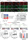
Fig. 2.. FLNCG1891Vfs61X and FLNCE2189X are loss-of-function mutations leading to haploinsufficiency in FLNC-related cardiomyopathy.
(A) Expression levels of FLNC in patient-derived iPSC-CMs were significantly decreased as detected by immunoblot. (B) Quantitative representation of the immunoblots in (A). (C) FLNC mRNA expression levels were significantly decreased in patient-derived iPSC-CMs. (D) Detection of FLNC (red) by immunofluorescence staining in control and patients’ iPSC-CMs, α-actinin (green), a cardiac myocyte marker, and 4′,6-diamidino-2-phenylindole (DAPI)–stained (blue). FLNC was colocalized with ACTN by ImageJ colocalization analysis. (E) FLNC expression levels were significantly decreased by immunoblots in heterozygous and homozygous KO iPSC-CMs. (F) Quantitative representation of the immunoblots from (E). (G) FLNC mRNA expression levels were significantly decreased in FLNC heterozygous and homozygous KO iPSC-CMs compared to isogenic controls. (H) Significantly reduced detection of FLNC (red) at Z-disc (green) and confirmed by ImageJ colocalization analysis. (I) DEGs from patients’ iPSC-CMs were highly concordant with FLNC homozygous KO iPSC-CMs. (J) FLNC-deficient iPSC-CMs affected expression levels of intercalated disc proteins. All experiments were independently repeated three times. DSP, desmoplakin; JUP, plakoglobin; GAPDH, glyceraldehyde-3-phosphate dehydrogenase.
Fig. 3.
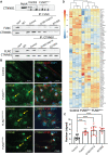
Fig. 3.. β-Catenin is associated with FLNC and the loss of the FLNC-activated β-catenin signaling pathway.
(A) β-Catenin was not detected in the coimmunoprecipitation pull-down lysate from FLNC KO iPSC-CMs. Each lane represents one differentiation batch. There is a reduction in the amount of β-catenin in the pull-down protein lysate from patients’ iPSC-CMs using antibody against FLNC. Each lane represents one iPSC-CM immunoprecipitation (IP) sample. IP: FLNC, IP with FLNC antibody; IP: CTNNB1, IP with coimmunoprecipitation and proteomic analysis identified β-catenin antibody. (B) Immunofluorescence staining against active β-catenin (green) showed increased nuclear localization of β-catenin in FLNC homozygous KO and patients’ iPSC-CMs. PCM1 (red) is a marker of cardiac myocytes. Nuclei (blue) was stained with DAPI. Yellow arrows indicate nuclear localization of β-catenin. All experiments were independently repeated three times. (C) Quantification of nuclear vs cytoplasmic CTNNB1 from the IF images in Fig. 3C. (D) Heatmap plot of DEGs involved in the β-catenin signaling pathway indicating β-catenin signaling activation in FLNC KO lines. PCM, pericentriolar material 1.
Fig. 4.
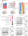
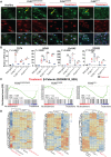
Fig. 4.. Dysregulation of cell-cell adhesion pathways resulted in PDGFRA activation in the pathogenesis of FLNC-related cardiomyopathy.
(A) Number of DEGs in FLNC heterozygous and homozygous KO iPSC-CMs compared to isogenic control iPSC-CMs. Percentage representation of the DEGs for each line is listed as up-regulated (red) and down-regulated (blue) in the bar graphs. (B) Venn diagram of shared DEGs between FLNC heterozygous and homozygous iPSC-CMs. (C) Enrichment analysis of Kyoto Encyclopedia of Genes And Genomes (KEGG) datasets identified dysregulation of pathways involved in cell-cell adhesion systems and membrane signaling. ECM, extracellular matrix. ER, Endoplasmic Reticulum. (D) Heatmap of DEGs in dysregulated cell-cell adhesion pathway (hsa04510). (E) GO analysis of DEGs revealed activation of the PDGF signaling pathway. Color codes indicate log10 transformed P values of the top 10 dysregulated pathways in GO molecular function. The number of genes involved in each pathway is represented by the size of the bubble. (F to H) PDGFRA and phosphorylated extracellular signal–regulated kinase (p-ERK) expression levels were significantly up-regulated in FLNC KO and patient’s iPSC-CMs as detected by immunoblot. Quantitative representation of each blot was presented as dot plots. All experiments were independently repeated three times.
Fig. 5.
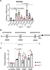
Fig. 5.. β-Catenin is a possible transcription activator of PDGFRA.
(A) Dosage-dependent decrease of PDGFRA mRNA levels upon inhibition of β-catenin using JW67 and JW76. All experiments were independently repeated three times. (B) In silico analysis of transcription factors binding sites on PDGFRA regulatory region. (C) Chromatin immunoprecipitation polymerase chain reaction (qPCR) showed enrichment of β-catenin–SOX2 binding to the promoter region of PDGFRA. NFκB, nuclear factor κB.
Fig. 6.
Fig. 6.. The loss of FLNC causes dysregulation to the cell-cell adhesion systems and inhibition of PDGFR signaling improved contractility of FLNC mutant iPSC-CMs.
(A) Immunofluorescence staining evidenced the loss of FLNC that resulted in segregation of GJA1 (red) away from the membrane compared to control iPSC-CMs. GJA1 segregated at the membrane upon treatment with crenolanib. α-Actinin was stained in green as a marker for Z-disc and cardiomyocytes. Nuclei (blue) were stained with DAPI. Yellow arrows indicate membrane localization of GJA1. (B) FLNCE2189X iPSC-CMs had the largest DEGs changes to the cell adhesion pathway compared to other FLNC-mutant lines upon crenolanib treatment. (C) Gene Set Enrichment Analysis (GSEA) indicated partial normalization of the cell adhesion pathway upon treatment with crenolanib in patients’ and FLNC KO iPSC-CMs. (D) Immunoblots indicated no significant changes to expression levels of GJA1, active β-catenin, and total β-catenin before and after PDGFRA inhibition. All experiments were independently repeated three times. (E) PDGFRA inhibition improved contractility of patients’ iPSC-CMs. (F) Crenolanib treatment over a period of 7 days showed improved contractility of patients’ iPSC-CMs (n = 20 repeated measured replicates). *P < 0.05, **P < 0.01, and ****P < 0.0001.
Fig. 7.
Fig. 7.. PDGFRA inhibition attenuated β-catenin signaling and its nuclear localization in FLNC mutant iPSC-CMs.
(A) Immunofluorescence staining against active β-catenin (green) showed increased nuclear localization of β-catenin in FLNC homozygous KO and patients’ iPSC-CMs. PCM1 (red) is a marker of cardiac myocytes. Nuclei (blue) were stained with DAPI. Treatment with crenolanib prevented nuclear localization of β-catenin in FLNC homozygous and patients’ iPSC-CMs. Yellow arrows indicate nuclear localization of β-catenin. (B) Reduced β-catenin targets transcripts by qPCR in FLNC mutant lines. (C) GSEA analysis showed partial normalization of the β-catenin signaling pathway in FLNC mutant lines. (D) Heatmaps showing partial normalization of expression profiles in genes involved in β-catenin signaling in FLNC mutant iPSC-CMs. ns, not significant. *P < 0.01, **P < 0.001, and ***P < 0.0001. All experiments were independently repeated three times.
Fig. 8.
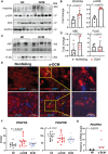
Fig. 8.. Up-regulation of PDGFRA contributes to GJA1 and β-catenin delocalization in the hearts of arrhythmogenic dilated cardiomyopathy.
(A) Up-regulation of PDGFRA and its effector p-ERK expression levels in the hearts of patients with a-DCM compared to nonfailing (NF) hearts (n = 5). (B) Quantitative representation of the immunoblots in (A). (C) Active β-catenin expression levels were not significantly different compared to NF hearts (n = 5). (D) Quantitative representation of the immunoblots in (C). (E) RNA-seq data showing up-regulation of PDGFRA transcripts but not PDGFRB in the hearts of a-DCM compared to DCM and NF. (F) Validation of increased PDGFRA transcripts observed in (E) by qPCR. (G) Immunofluorescence staining against CTNNB1 (red) and GJA1 showed CTNNB1 and GJA1 mobilized away from the ID of cardiac myocytes in the hearts of a-DCM compared to NF. Nuclei (blue) were stained with DAPI. Yellow arrows indicate GJA1 and β-catenin segregated away from the cell membrane. All experiments were independently repeated three times. ABC, Active beta-catenin.
Fig. 9.
Fig. 9.. Disease model of FLNC-related cardiomyopathy.
Our data indicate that β-catenin (β-cat) localized to the cell membrane with FLNC in the NF hearts (left). However, the loss of FLNC perturbed the cytoplasmic β-catenin and increased nuclear localization of β-catenin and activates transcription of PDGFRA, which leads to sustained up-regulation of PDGFRA signaling at the cell-cell membrane and, subsequently, activation of ERK signaling resulting in internalization of GJA1 and consequently arrhythmias in the heart of patients with a-DCM (right).

References

    1. Hershberger R. E., Siegfried J. D., Update 2011: Clinical and genetic issues in familial dilated cardiomyopathy. J. Am. Coll. Cardiol. 57, 1641–1649 (2011). - PMC - PubMed
    1. Nandi D., Rossano J. W., Epidemiology and cost of heart failure in children. Cardiol. Young 25, 1460–1468 (2015). - PMC - PubMed
    1. Towbin J. A., Lorts A., Arrhythmias and dilated cardiomyopathy. J. Am. Coll. Cardiol. 57, 2169–2171 (2011). - PubMed
    1. McKenna W. J., Maron B. J., Thiene G., Classification, epidemiology, and global burden of cardiomyopathies. Circ. Res. 121, 722–730 (2017). - PubMed
    1. Hershberger R. E., Hedges D. J., Morales A., Dilated cardiomyopathy: The complexity of a diverse genetic architecture. Nat. Rev. Cardiol. 10, 531–547 (2013). - PubMed