Review
. 2022 May 1;322(5):C875-C886.
doi: 10.1152/ajpcell.00448.2021. Epub 2022 Feb 23.

Elastin, arterial mechanics, and stenosis

Affiliations
Review

Elastin, arterial mechanics, and stenosis

Chien-Jung Lin et al. Am J Physiol Cell Physiol. .

Abstract

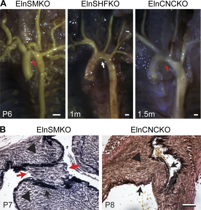
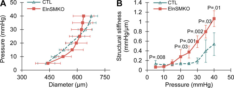
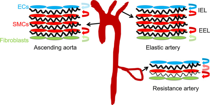
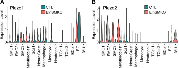
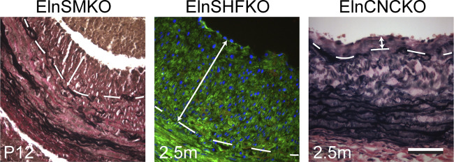
Elastin is a long-lived extracellular matrix protein that is organized into elastic fibers that provide elasticity to the arterial wall, allowing stretch and recoil with each cardiac cycle. By forming lamellar units with smooth muscle cells, elastic fibers transduce tissue-level mechanics to cell-level changes through mechanobiological signaling. Altered amounts or assembly of elastic fibers leads to changes in arterial structure and mechanical behavior that compromise cardiovascular function. In particular, genetic mutations in the elastin gene (ELN) that reduce elastin protein levels are associated with focal arterial stenosis, or narrowing of the arterial lumen, such as that seen in supravalvular aortic stenosis and Williams-Beuren syndrome. Global reduction of Eln levels in mice allows investigation of the tissue- and cell-level arterial mechanical changes and associated alterations in smooth muscle cell phenotype that may contribute to stenosis formation. A loxP-floxed Eln allele in mice highlights cell type- and developmental origin-specific mechanobiological effects of reduced elastin amounts. Eln production is required in distinct cell types for elastic layer formation in different parts of the mouse vasculature. Eln deletion in smooth muscle cells from different developmental origins in the ascending aorta leads to characteristic patterns of vascular stenosis and neointima. Dissecting the mechanobiological signaling associated with local Eln depletion and subsequent smooth muscle cell response may help develop new therapeutic interventions for elastin-related diseases.

Keywords: extracellular matrix; mechanobiology; neointima; smooth muscle cell; vascular stenosis.


Conflict of interest statement

No conflicts of interest, financial or otherwise, are declared by the authors.

Figures

Figure 1.
Figure 1.
Stenosis of the aorta in elastin conditional knockout mouse models. A: latex angiography in mouse mutants with Eln specifically deleted in smooth muscle cell (SMC) (ElnSMKO), secondary heart field (SHF) (ElnSHFKO), and cardiac neural crest (CNC) (ElnCNCKO). Note the presence (red arrows) and absence (white arrow) of stenosis in the different mouse models. Scale = 0.5 mm. B: histology of aortic stenosis. Verhoeff-Van Gieson staining shows neointima (red arrows) in ElnSMKO aortic stenosis, likely due to the fragmented internal elastic lamina (IEL). Tunica media hypertrophy (black arrowheads) is observed in both ElnSMKO and ElnCNCKO aorta. Black arrows point to the IEL. Scale = 50 µm. Adapted from Refs. and .
Figure 2.
Figure 2.
Aortic mechanical behavior in elastin conditional knockout mouse model. Diameter-pressure (A) and pressure-structural stiffness (B) behavior of postnatal day 1 (P1) control (Elnf/f) and ElnSMKO ascending aorta. Structural stiffness is the local slope of the diameter-pressure curve and is increased in ElnSMKO aorta between 20 and 40 mmHg. n = 6 (control) and 4 (ElnSMKO). Means ± SD. P values from two-tailed Student’s t test with unequal variance.
Figure 3.
Figure 3.
Cellular contributions to the internal (IEL) and external (EEL) elastic laminae differs throughout the vascular tree. In the ascending aorta, the IEL is primarily made by smooth muscle cells (SMCs). In other elastic arteries it is made by both endothelial cells (ECs) and SMCs, while in the resistance arteries it is mainly made by ECs. Cellular contributions are shown by colored arrows, with minor contributions shown in lighter colors. In all arteries, SMCs are the main cell type contributing to the EEL, however there is a minor contribution from fibroblasts in the ascending aorta and elastic arteries. Elastic laminae are shown in black.
Figure 4.
Figure 4.
Network diagram of significant cell-cell interactions via ligand-receptor pairs expressed in cell clusters (79) determined from previously published single-cell RNA-sequencing data of ElnSMKO ascending aorta (22). Arrows and color indicate direction (ligand to receptor) and thickness indicates the sum of the weighted paths between cell clusters.
Figure 5.
Figure 5.
Violin plots of Piezo1 (A) and -2 (B) expression in different cell clusters for postnatal day 8 (P8) control (Elnf/f or Elnf/+) and ElnSMKO ascending aorta determined from previously published single-cell RNA-sequencing data (22).
Figure 6.
Figure 6.
Neointima in the ascending aorta of elastin conditional knockout mouse models as demonstrated by VVG staining (ElnSMKO and ElnCNCKO) and lineage tracing with the ROSA26mT/mG allele (ElnSHFKO) (22). Internal elastic lamina (IEL) demarcated by dashed lines. Neointimal burden indicated by double arrows. Note the younger age of ElnSMKO mice. Scale = 50 µm.

References

    1. Sage H, Gray WR. Studies on the evolution of elastin. I. Phylogenetic distribution. Comp Biochem Physiol B 64: 313–327, 1979. doi: 10.1016/0305-0491(79)90277-3. - DOI - PubMed
    1. Kelleher CM, McLean SE, Mecham RP. Vascular extracellular matrix and aortic development. Curr Top Dev Biol 62: 153–188, 2004. doi: 10.1016/S0070-2153(04)62006-0. - DOI - PubMed
    1. Shapiro SD, Endicott SK, Province MA, Pierce JA, Campbell EJ. Marked longevity of human lung parenchymal elastic fibers deduced from prevalence of d-aspartate and nuclear weapons-related radiocarbon. J Clin Invest 87: 1828–1834, 1991. doi: 10.1172/JCI115204. - DOI - PMC - PubMed
    1. Sandberg LB, Soskel NT, Leslie JG. Elastin structure, biosynthesis, and relation to disease states. N Engl J Med 304: 566–579, 1981. doi: 10.1056/NEJM198103053041004. - DOI - PubMed
    1. Tamburro AM, Bochicchio B, Pepe A. The dissection of human tropoelastin: from the molecular structure to the self-assembly to the elasticity mechanism. Pathol Biol (Paris) 53: 383–389, 2005. doi: 10.1016/j.patbio.2004.12.014. - DOI - PubMed

Publication types

LinkOut - more resources