. 2022 Mar:77:103888.
doi: 10.1016/j.ebiom.2022.103888. Epub 2022 Feb 20.

SARS-CoV-2 vaccination elicits unconventional IgM specific responses in naïve and previously COVID-19-infected individuals

Affiliations

SARS-CoV-2 vaccination elicits unconventional IgM specific responses in naïve and previously COVID-19-infected individuals

Alessandra Ruggiero et al. EBioMedicine. 2022 Mar.

Abstract

Background: Currently, evaluation of the IgG antibodies specific for the SARS-CoV-2 Spike protein following vaccination is used worldwide to estimate vaccine response. Limited data are available on vaccine-elicited IgM antibodies and their potential implication in immunity to SARS-CoV-2.

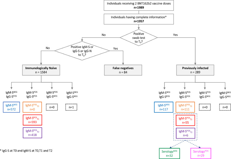
Methods: We performed a longitudinal study to quantify anti-S SARS-CoV-2 IgG and IgM (IgG-S and IgM-S) in health care worker (HCW) recipients of the BNT162b2 vaccine. Samples were collected before administration (T0), at the second dose (T1) and three weeks after T1 (T2). The cohort included 1584 immunologically naïve to SARS-CoV-2 (IN) and 289 with history of previous infection (PI).

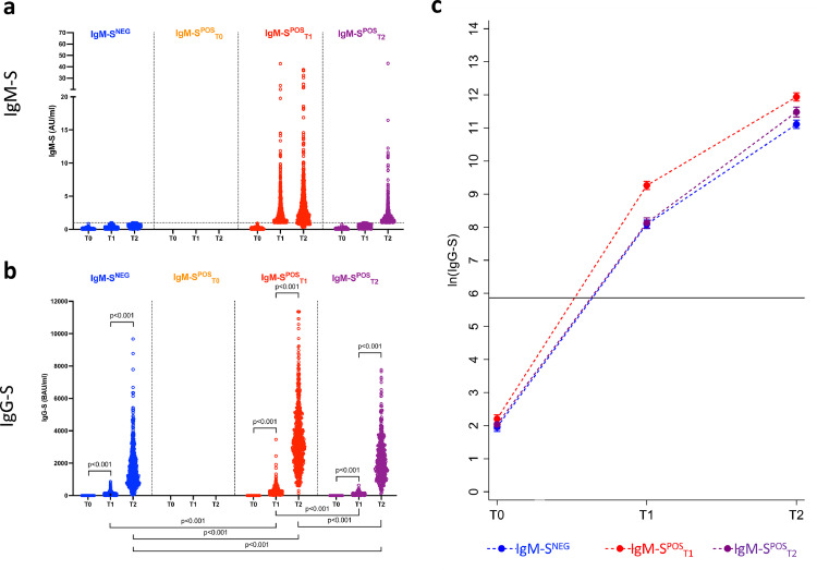
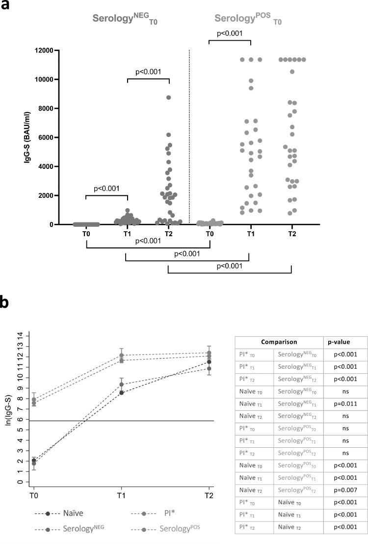
Findings: IN showed three patterns of responses: (a) IgG positive/IgM negative (36.1%), (b) coordinated IgM-S/IgG-S responses appearing at T1 (37.4%) and (c) IgM appearing after IgG (26.3%). Coordinated IgM-S/IgG-S responses were associated with higher IgG titres. In IgM-S positive PI, 64.5% were IgM-S positive before vaccination, whereas 32% and 3.5% developed IgM-S after the first and second vaccine dose, respectively. IgM-S positive sera had higher pseudovirus neutralization titres compared to the IgM-S negative.

Interpretation: Coordinated expression of IgG-S and IgM-S after vaccination was associated with a significantly more efficient response in both antibody levels and virus-neutralizing activity. The unconventional IgG-S positive/IgM-S negative responses may suggest a recruitment of cross coronaviruses immunity by vaccination, warranting further investigation.

Funding: Italian Ministry of Health under "Fondi Ricerca Corrente"- L1P5 and "Progetto Ricerca Finalizzata COVID-2020-12371675"; FUR 2020 Department of Excellence 2018-2022, MIUR, Italy; The Brain Research Foundation Verona.

Keywords: BTN162b2 vaccine; COVID-19; IgG; IgM; SARS-CoV-2; Spike.


Conflict of interest statement

Declaration of interests The authors declare no competing interests.

Figures

Fig 1
Figure 1
Neutralization assays in naïve and previously infected vaccinees. Pseudovirus neutralization assay expressed as infectious dose (ID50) in naïve (panel a) and previously infected (panel b) vaccinees according to time of examination (T0, T1 and T2) and IgM-S development after two doses of the BNT162b2 vaccine (IgM-SPOS, red dots and lines; IgM-SNEG subjects, blue dots and lines). Predicted means of ln(ID50) levels (with the 95% confidence interval) according to time of examination and IgM-S group in naïve (panel c) and previously infected (panel d) vaccinees were obtained by a two-level linear regression model. Statistically significant p-values of the difference in the predicted means between consecutive times of examination in the same IgM-S group and between IgM-S groups at the same time of examination are reported in panels a and b.
Fig 2
Figure 2
Study population. Classification and distribution of the different types of IgM-S and IgG-S responses in naïve and previously infected subjects who received the BNT162b2 vaccine. NEG: negative; POS: positive.
Fig 3
Figure 3
Development of IgM-S and IgG-S following vaccination. Scatterplots of IgM-S (y axis) and IgG-S (x axis) measures in naïve vaccines according to time of examination (T0, T1 and T2) and time of IgM-S positivity (IgM-SNEG, blue dots; IgM-SPOST1, red dots; IgM-SPOST2, purple dots).
Fig 4
Figure 4
IgG-S response in naïve vaccinees. IgM-S (panel a) and IgG-S (panel b) measures in naïve vaccinees according to time of examination (T0, T1 and T2) and time of IgM-S positivity (IgM-SNEG, n = 572, blue dots; IgM-SPOST1, n = 593, red dots; IgM-SPOST2, n = 418, purple dots). Being all naïve subjects, no individuals had detectable IgM-S at T0. Predicted means of ln(IgG-S) measures (with the 95% confidence interval) according to time of examination and time of IgM-S positivity (panel c) were obtained by a two-level linear regression model. Statistically significant p-values of the difference in the predicted means between consecutive times of examination at the same time of IgM-S positivity and between different times of IgM-S positivity at the same time of examination are reported in panel b. The horizontal lines indicate the cut-off value to discriminate positive and negative samples for each assay.
Fig 5
Figure 5
IgG-S response in previously infected vaccinees. IgM-S (panel a) and IgG-S (panel b) measures in previously infected vaccinees according to time of examination (T0, T1 and T2) and time of IgM-S positivity (IgM-SNEG, n = 117, blue dots; IgM-SPOST0, n = 111, orange dots; IgM-SPOST1, n = 55, red dots; IgM-SPOST2, n = 6, purple dots). Predicted means of ln(IgG-S) measures (with the 95% confidence interval) according to time of examination and time of IgM-S positivity (panel c) were obtained by a two-level linear regression model. Statistically significant p-values of the difference in the predicted means between consecutive times of examination at the same time of IgM-S positivity and between different times of IgM-S positivity at the same time of examination are reported in panel b. The horizontal lines indicate the cut-off value to discriminate positive and negative samples for each assay.
Fig 6
Figure 6
IgG-S response in previously infected vaccinees producing IgM-S. IgG-S measures (panel a) in previously infected vaccinees who produced IgM-S at T1 or at T2 following BNT162b2 vaccination according to time of examination (T0, T1 and T2) and negative or positive serology at T0 (SerologyNEG, n = 32, green dots; SerologyPOS, n = 29 magenta dots). Predicted means of ln(IgG-S) measures (with the 95% confidence interval) according to time of examination in (i) previously infected subjects who did not elicit IgM-S or had IgM-S at T0 (PI*, red line), (ii) subjects who did not have detectable IgM-S at T0 but produced them at T1 or at T2 following vaccination, and who had detectable IgG-S and/or IgG-N at T0 (SerologyPOS, magenta line), (iii) subjects as the previous ones, but with negative serology at T0 (SerologyNEG, green line), and (iv) naïve vaccinees (blue line) (panel B) were obtained by a two-level linear regression model. For SerologyNEG and SerologyPOS subjects, statistically significant p-values of the difference in the predicted means between consecutive times of examination in the same subject group and between different subject groups at the same time of examination are reported in panel b. For all four group of subjects, statistically significant p-values of the difference in the predicted means between different groups of subjects at the same time of examination are reported in panel b table. The horizontal lines indicate the cut-off value to discriminate positive and negative samples for each assay.

Comment in

References

    1. Krammer F., Srivastava K., Alshammary H., et al. Antibody responses in seropositive persons after a single dose of SARS-CoV-2 mRNA vaccine. N Engl J Med. 2021;384(14):1372–1374. - PMC - PubMed
    1. Li K., Huang B., Wu M., et al. Dynamic changes in anti-SARS-CoV-2 antibodies during SARS-CoV-2 infection and recovery from COVID-19. Nat Commun. 2020;11(1):6044. - PMC - PubMed
    1. Gong S., Ruprecht R.M. Immunoglobulin M: an ancient antiviral weapon – rediscovered. Front Immunol. 2020;11(1943) - PMC - PubMed
    1. Jin Y., Wang M., Zuo Z., et al. Diagnostic value and dynamic variance of serum antibody in coronavirus disease 2019. Int J Infect Dis. 2020;94:49–52. - PMC - PubMed
    1. Long Q.X., Liu B.Z., Deng H.J., et al. Antibody responses to SARS-CoV-2 in patients with COVID-19. Nat Med. 2020;26(6):845–848. - PubMed