

Targeted Antimicrobial Photodynamic Therapy of Biofilm-Embedded and Intracellular Staphylococci with a Phage Endolysin's Cell Binding Domain
- PMID: 35196798
- PMCID: PMC8865409
- DOI: 10.1128/spectrum.01466-21
Targeted Antimicrobial Photodynamic Therapy of Biofilm-Embedded and Intracellular Staphylococci with a Phage Endolysin's Cell Binding Domain
Abstract
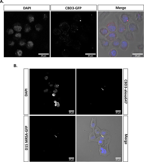
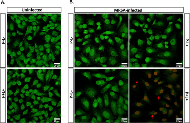
Bacterial pathogens are progressively adapting to current antimicrobial therapies with severe consequences for patients and global health care systems. This is critically underscored by the rise of methicillin resistant Staphylococcus aureus (MRSA) and other biofilm-forming staphylococci. Accordingly, alternative strategies have been explored to fight such highly multidrug resistant microorganisms, including antimicrobial photodynamic therapy (aPDT) and phage therapy. aPDT has the great advantage that it does not elicit resistance, while phage therapy allows targeting of specific pathogens. In the present study, we aimed to merge these benefits by conjugating the cell-binding domain (CBD3) of a Staphylococcus aureus phage endolysin to a photoactivatable silicon phthalocyanine (IRDye 700DX) for the development of a Staphylococcus-targeted aPDT approach. We show that, upon red-light activation, the resulting CBD3-700DX conjugate generates reactive oxygen species that effectively kill high loads of planktonic and biofilm-resident staphylococci, including MRSA. Furthermore, CBD3-700DX is readily internalized by mammalian cells, where it allows the targeted killing of intracellular MRSA upon photoactivation. Intriguingly, aPDT with CBD3-700DX also affects mammalian cells with internalized MRSA, but it has no detectable side effects on uninfected cells. Altogether, we conclude that CBD3 represents an attractive targeting agent for Staphylococcus-specific aPDT, irrespective of planktonic, biofilm-embedded, or intracellular states of the bacterium. IMPORTANCE Antimicrobial resistance is among the biggest threats to mankind today. There are two alternative antimicrobial therapies that may help to control multidrug-resistant bacteria. In phage therapy, natural antagonists of bacteria, lytic phages, are harnessed to fight pathogens. In antimicrobial photodynamic therapy (aPDT), a photosensitizer, molecular oxygen, and light are used to produce reactive oxygen species (ROS) that inflict lethal damage on pathogens. Since aPDT destroys multiple essential components in targeted pathogens, aPDT resistance is unlikely. However, the challenge in aPDT is to maximize target specificity and minimize collateral oxidative damage to host cells. We now present an antimicrobial approach that combines the best features of both alternative therapies, namely, the high target specificity of phages and the efficacy of aPDT. This is achieved by conjugating the specific cell-binding domain from a phage protein to a near-infrared photosensitizer. aPDT with the resulting conjugate shows high target specificity toward MRSA with minimal side effects.
Keywords: Staphylococcus aureus; Staphylococcus epidermidis; antimicrobial photodynamic therapy; biofilms; cell-binding domain; endolysin; intracellular Staphylococcus aureus; intracellular bacteria.
Conflict of interest statement
The authors declare no conflict of interest.
Figures
Similar articles
-
Empowering antimicrobial photodynamic therapy of Staphylococcus aureus infections with potassium iodide.J Photochem Photobiol B. 2021 Dec;225:112334. doi: 10.1016/j.jphotobiol.2021.112334. Epub 2021 Oct 13. J Photochem Photobiol B. 2021. PMID: 34678616
-
Ultrasonic irradiation enhanced the efficacy of antimicrobial photodynamic therapy against methicillin-resistant Staphylococcus aureus biofilm.Ultrason Sonochem. 2023 Jul;97:106423. doi: 10.1016/j.ultsonch.2023.106423. Epub 2023 Apr 29. Ultrason Sonochem. 2023. PMID: 37235946 Free PMC article.
-
Natural photosensitizers from Tripterygium wilfordii and their antimicrobial photodynamic therapeutic effects in a Caenorhabditis elegans model.J Photochem Photobiol B. 2021 May;218:112184. doi: 10.1016/j.jphotobiol.2021.112184. Epub 2021 Mar 29. J Photochem Photobiol B. 2021. PMID: 33848804
-
Photodynamic therapy and combinatory treatments for the control of biofilm-associated infections.Lett Appl Microbiol. 2022 Sep;75(3):548-564. doi: 10.1111/lam.13762. Epub 2022 Jun 27. Lett Appl Microbiol. 2022. PMID: 35689422 Review.
-
Antimicrobial photodynamic therapy (aPDT) for biofilm treatments. Possible synergy between aPDT and pulsed electric fields.Virulence. 2021 Dec;12(1):2247-2272. doi: 10.1080/21505594.2021.1960105. Virulence. 2021. PMID: 34496717 Free PMC article. Review.
Cited by
-
A TAT Peptide-Functionalized Liposome Delivery Phage System (TAT-Lip@PHM) for an Enhanced Eradication of Intracellular MRSA.Pharmaceutics. 2025 Jun 5;17(6):743. doi: 10.3390/pharmaceutics17060743. Pharmaceutics. 2025. PMID: 40574055 Free PMC article.
-
Harnessing light-activated gallium porphyrins to combat intracellular Staphylococcus aureus using an in vitro keratinocyte infection model.Sci Rep. 2025 Jan 8;15(1):1295. doi: 10.1038/s41598-024-84312-4. Sci Rep. 2025. PMID: 39779728 Free PMC article.
-
Photocatalysis and Photodynamic Therapy in Diabetic Foot Ulcers (DFUs) Care: A Novel Approach to Infection Control and Tissue Regeneration.Molecules. 2025 May 26;30(11):2323. doi: 10.3390/molecules30112323. Molecules. 2025. PMID: 40509211 Free PMC article. Review.
-
Photoimmuno-antimicrobial therapy for Staphylococcus aureus implant infection.PLoS One. 2024 Mar 8;19(3):e0300069. doi: 10.1371/journal.pone.0300069. eCollection 2024. PLoS One. 2024. PMID: 38457402 Free PMC article.
References
-
- World Health Organization (WHO). 2020. Antibiotic resistance. https://www.who.int/news-room/fact-sheets/detail/antibiotic-resistance.
-
- Kubica M, Guzik K, Koziel J, Zarebski M, Richter W, Gajkowska B, Golda A, Maciag-Gudowska A, Brix K, Shaw L, Foster T, Potempa J. 2008. A potential new pathway for Staphylococcus aureus dissemination: the silent survival of S. aureus phagocytosed by human monocyte-derived macrophages. PLoS One 3:e1409. doi:10.1371/journal.pone.0001409. - DOI - PMC - PubMed
Publication types
MeSH terms
Substances
LinkOut - more resources
Full Text Sources
Medical
Molecular Biology Databases
