. 2022 Apr;42(4):428-443.
doi: 10.1161/ATVBAHA.121.317365. Epub 2022 Feb 24.

YAP and TAZ in Vascular Smooth Muscle Confer Protection Against Hypertensive Vasculopathy

Affiliations

YAP and TAZ in Vascular Smooth Muscle Confer Protection Against Hypertensive Vasculopathy

Fatima Daoud et al. Arterioscler Thromb Vasc Biol. 2022 Apr.

Abstract

Background: Hypertension remains a major risk factor for cardiovascular diseases, but the underlying mechanisms are not well understood. We hypothesize that appropriate mechanotransduction and contractile function in vascular smooth muscle cells are crucial to maintain vascular wall integrity. The Hippo pathway effectors YAP (yes-associated protein 1) and TAZ (WW domain containing transcription regulator 1) have been identified as mechanosensitive transcriptional coactivators. However, their role in vascular smooth muscle cell mechanotransduction has not been investigated in vivo.

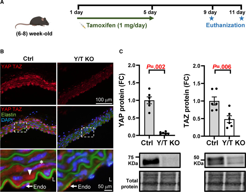
Methods: We performed physiological and molecular analyses utilizing an inducible smooth muscle-specific YAP/TAZ knockout mouse model.

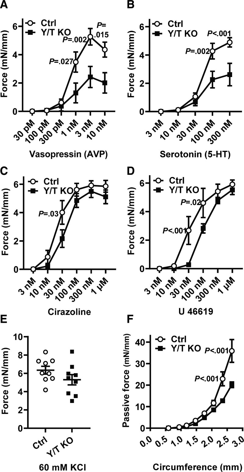
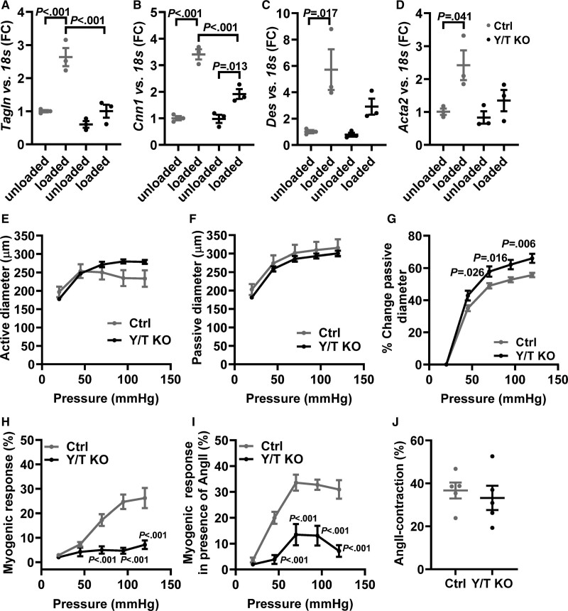
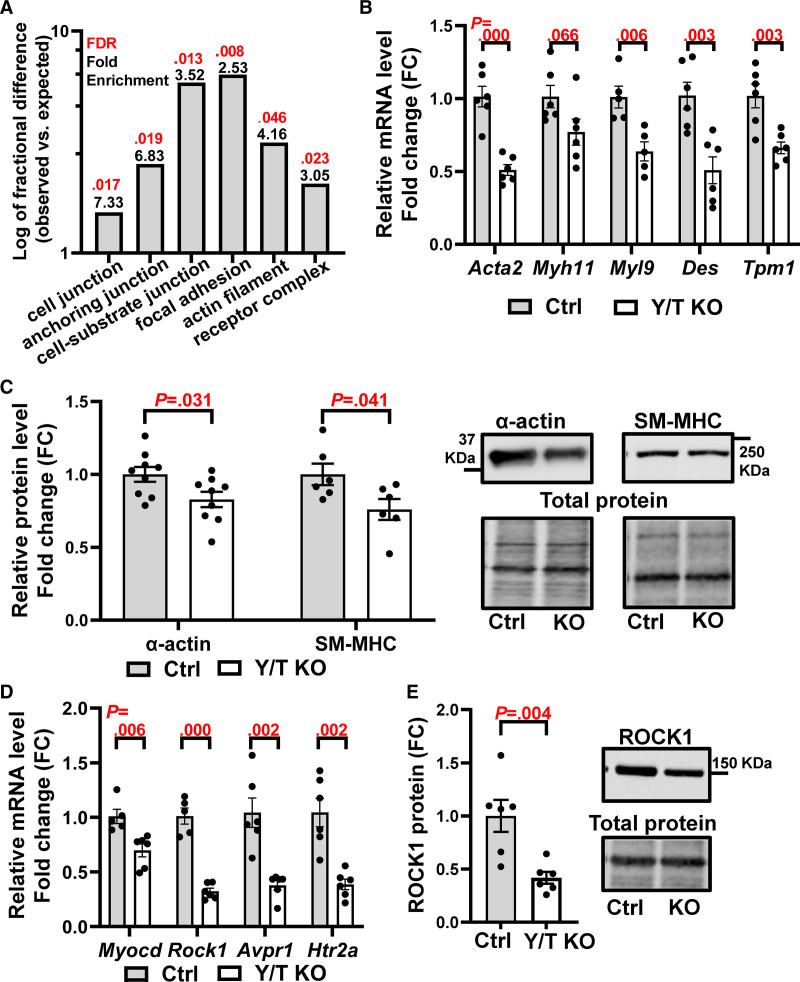
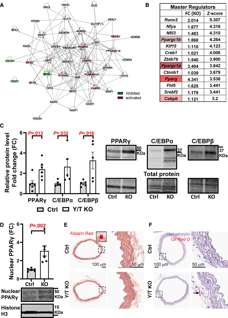
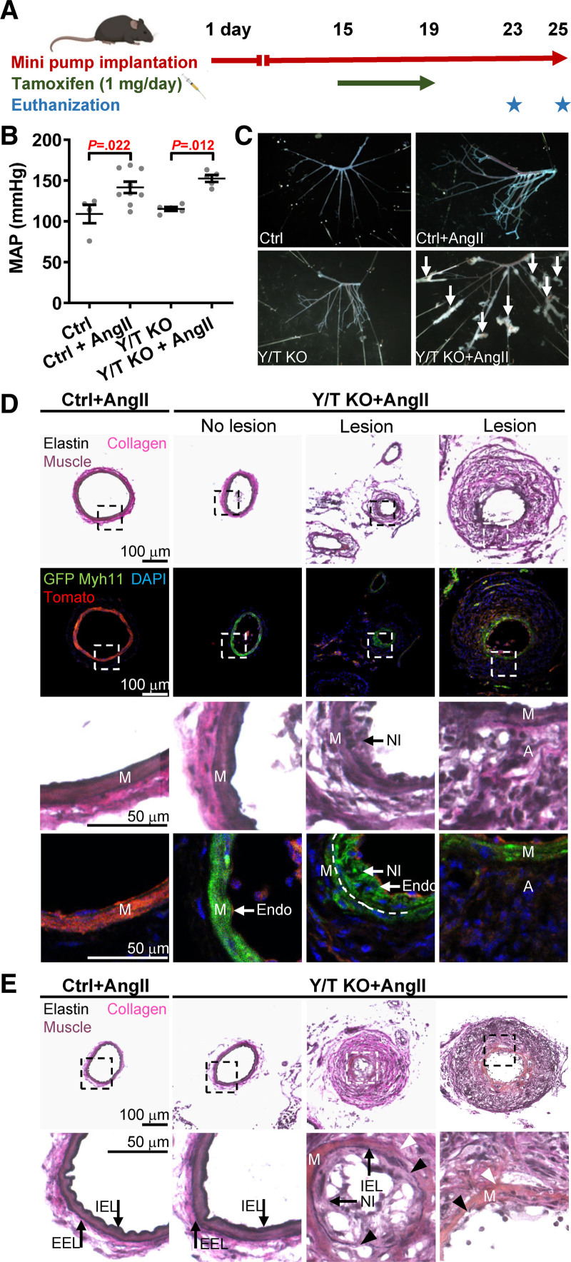
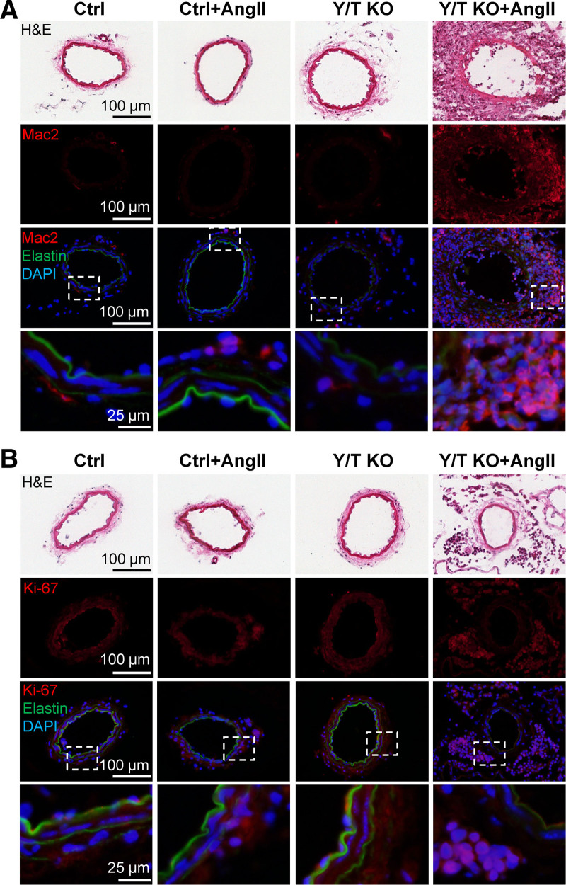
Results: Arteries lacking YAP/TAZ have reduced agonist-mediated contraction, decreased myogenic response, and attenuated stretch-induced transcriptional regulation of smooth muscle markers. Moreover, in established hypertension, YAP/TAZ knockout results in severe vascular lesions in small mesenteric arteries characterized by neointimal hyperplasia, elastin degradation, and adventitial thickening.

Conclusions: This study demonstrates a protective role of YAP/TAZ against hypertensive vasculopathy.

Keywords: adventitia; compliance; hyperplasia; muscle; neointima; smooth; vascular.


Figures

Figure 1.
Figure 1.
Reduced expression of YAP (yes-associated protein 1) and TAZ (WW domain containing transcription regulator 1) in vascular smooth muscle. A, Generation of Y/T KO (YAP and TAZ knockout) mice. Myh11-CreERT2 Yap1fl/fl Wwtr1fl/fl mice were injected with 1 mg tamoxifen for 5 consecutive days and euthanized on either day 9 or 11. B, Representative images of aortic cryosections, immunostained for YAP and TAZ. Asterisk represents nuclear localization, and arrowhead represents cytosolic localization. C, Western blot analysis, YAP and TAZ were normalized to total protein in abdominal aortic lysates (Ctrl [control], n=6; Y/T KO, n=6). The representative blots of YAP and TAZ are taken from the same lanes, hence the loading controls are identical. All data are presented as mean±SEM. DAPI indicates 4′,6-diamidino-2-phenylindole; Endo, endothelium; FC, fold change; and L, lumen.
Figure 2.
Figure 2.
Reduced contractile responses to various agonists and increased compliance of Y/T KO arteries. Tail arteries were mounted in a wire myograph and dose-response curves were generated by integrating force over 7 min for each dose of (A) AVP (vasopressin; Ctrl [control], n=7; Y/T KO, n=8), (B) 5-HT (serotonin; Ctrl, n=9; Y/T KO, n=9), (C) cirazoline, an α1-adrenergic agonist (Ctrl, n=9; Y/T KO, n=9), and (D) U 46619, a thromboxane A2 analog (Ctrl, n=6; Y/T KO, n=6). E, Force development over 7-min stimulation with 60 mmol/L KCl (Ctrl, n=9; Y/T KO, n=9). F, The length-tension relationship of the abdominal aorta was determined in Ca2+-free HEPES-buffered solution (Ctrl, n=10; Y/T KO, n=8; Ctrl:vehicle-treated mice). All force measurements were normalized to the length of each preparation. All data are presented as mean±SEM. 5-HT indicates 5-hydroxytryptamine/ serotonin.
Figure 3.
Figure 3.
Reduced expression of stretch-induced contractile markers and decreased myogenic response in Y/T KO (YAP/TAZ [yes-associated protein 1/WW domain containing transcription regulator 1] knockout) mice. The portal veins (n=3 in all groups and targets) were kept in organ culture for 24 h either stretched with gold weight (loaded) or not stretched (unloaded). Reverse transcription-quantitative polymerase chain reaction analysis of selected smooth muscle markers in portal veins from Y/T KO and Ctrl (control) mice: (A) Tagln, (B) Cnn1, (C) Des, and (D) Acta2. E–J, Mesenteric arteries were mounted in a pressure myograph. E, Intraluminal pressure was increased systematically, and active vessel diameter was recorded in Ca2+ containing HEPES-buffered Krebs solution. F, Passive vessel diameter was measured in Ca2+-free solutions. G, The relative change in passive diameter was used as an indicator of vascular distensibility. H, Myogenic response was calculated as the relative difference between active and passive diameter. I, A single dose of 100 nmol/L Ang II (angiotensin II) was added to the preparations followed by gradual increase in intraluminal pressure. Active diameter was monitored, and myogenic response was calculated (E–I; Ctrl, n=6; Y/T KO, n=6). J, Peak transient contraction relative to baseline after 100 nmol/L Ang II stimulation (Ctrl, n=5; Y/T KO, n=5). All data are presented as mean±SEM. FC indicates fold change.
Figure 4.
Figure 4.
Downregulation of genes involved in vascular smooth muscle differentiation and contraction in Y/T KO (YAP/TAZ [yes-associated protein 1/WW domain containing transcription regulator 1] knockout) mice. RNA sequencing was performed on aortae from Ctrl (control) and Y/T KO mice. A, Protein Analysis Through Evolutionary Relationships (PANTHER) overrepresentation test and PANTHER GO-slim cellular component of significantly downregulated genes with fold enrichment in black and false discovery rate in red. B, Confirmation of selected smooth muscle markers by RT-qPCR (Ctrl, n=5–6; Y/T KO, n=5–6). C, Quantification of Western blot analysis of SM-MHC (smooth muscle α-actin and myosin heavy chain) and their representative blots to the right. Targets were normalized to the total protein (Ctrl, n=6–9; Y/T KO, n=6–9). D, Reverse transcription-quantitative polymerase chain reaction analysis of Myocd, Rock1, Avpr1, and Htr2a mRNA expression (Ctrl, n=5–6; Y/T KO, n=6). E, Western blot analysis of ROCK1 (Rho kinase 1; Ctrl, n=6; Y/T KO, n=6). All data are presented as mean±SEM. FC indicates fold change; FDR, false discovery rate; and KO, knockout.
Figure 5.
Figure 5.
Upregulation of transcription factors that are involved in adipogenic differentiation: PPARγ (peroxisome proliferator activated receptor gamma), C/EBPα (CCAAT enhancer binding protein alpha), and C/EBPβ (CCAAT enhancer binding protein beta). A, Network of the predicted master regulators that are involved in transcription regulation. The interactions between the nodes are based on Qiagen ingenuity knowledge base, where the solid and dashed lines represent direct and indirect interaction, respectively. The intensity of coloring is based on the magnitude of Z score values ranging from strong inhibition (dark green) to strong activation (dark red). B, The top 13 master regulators and their fold change and Z score, which reflects their activation status. C, Quantification of Western blot analysis of PPARγ, C/EPBα, and C/EPBβ and representative blots to the right (Ctrl [control], n=5–6; Y/T KO [YAP/TAZ (yes-associated protein 1/WW domain containing transcription regulator 1) knockout], n=5–6). D, Quantification and representative blot showing PPARγ in the nuclear fraction and histone H3 as nuclear marker (Ctrl, n=6; Y/T KO, n=6). E, Alizarin Red staining for calcium deposits detection in aortae of Ctrl and Y/T KO mice. Inset represents positive Ctrl (bone). F, Assessment of lipid accumulation in the aorta by Oil Red O staining, positive staining in perivascular fat (arrow). Detailed legend description for IPA network is available online: https://qiagen.secure.force.com/KnowledgeBase/articles/Knowledge/Legend. All data are presented as mean±SEM. Ctrl indicates control; DAPI, 4′,6-diamidino-2-phenylindole; FC, fold change; and KO, knockout.
Figure 6.
Figure 6.
Vascular lesion development in hypertensive Y/T KO (YAP/TAZ [yes-associated protein 1/WW domain containing transcription regulator 1] knockout) mice. A, Timeline representation of osmotic pump implantation and knockout induction. B, Blood pressure measurement just before euthanization of Ctrl (control) and Y/T KO mice using the tail cuff method. C, Representative images of mesenteric arteries from Ctrl and Y/T KO mice after removal of surrounding fat and tissue. Lesions are highlighted with arrows. D, Bright-field and fluorescence images of cryosections of mesenteric arteries isolated from Ctrl (YAPfl/fl/TAZfl/fl Cre-negative ROSAmT/mG) and Y/T KO Cre reporter mice (YAPfl/fl/TAZfl/fl Cre/ERT2 ROSAmT/mG) at 9 or 11 d after the first tamoxifen injection. Top and third row: Verhoeff Van Gieson staining to visualize vascular lesions and the different layers of the vessel wall. Second and fourth row: fluorescence images demonstrating Cre recombination event by using ROSA mT/mG Cre reporter mice. Upon tamoxifen-induced activation of Myh11-Cre, the Tomato transgene (red) is excised and replaced by the expression of GFP (green fluorescence protein) in smooth muscle. Note that many of the cells of the neointima are positive for GFP, whereas the cells of the thickened adventitia and endothelial cells are negative. DAPI (4′,6-diamidino-2-phenylindole) was used as a nuclear stain. Dashed line represents the internal elastic lamina. E, Verhoeff Van Gieson staining of mesenteric arteries of Y/T KO mice demonstrates sites of degradation of both the external (white arrowheads) and internal (black arrowheads) elastic lamina (D and E; Ctrl+Ang II [angiotensin II], n=3; Y/T KO+Ang II, n=3). All data are presented as mean±SEM. A indicates adventitia; EEL, external elastic lamina; Endo, endothelium; IEL, internal elastic lamina; M, media; MAP, mean arterial pressure; Myh11‚ myosin heavy chain 11; and NI, neointimal.
Figure 7.
Figure 7.
Marked macrophage infiltration and increased rate of proliferation in the adventitia of the vascular lesions. Cryosections of mesenteric arteries from Ctrl (control) or Y/T KO (YAP/TAZ [yes-associated protein 1/WW domain containing transcription regulator 1] knockout) mice treated with Ang II (angiotensin II) or vehicle. A, Immunostaining for Mac2 (galectin 3) demonstrates pronounced infiltration of monocytes/macrophages in the adventitia of hypertensive Y/T KO vascular lesions. B, The thickened adventitia of Y/T KO vascular lesions is associated with intense cell proliferation (Ki-67), whereas the media displays no obvious signs of cell proliferation. DAPI (4′,6-diamidino-2-phenylindole) was used as a nuclear stain. Elastin autofluorescence is shown in green. H&E indicates hematoxylin and eosin.

References

    1. Bayliss WM. On the local reactions of the arterial wall to changes of internal pressure. J Physiol. 1902; 28:220–231. doi: 10.1113/jphysiol.1902.sp000911 - PMC - PubMed
    1. Mulvany MJ, Baumbach GL, Aalkjaer C, Heagerty AM, Korsgaard N, Schiffrin EL, Heistad DD. Vascular remodeling. Hypertension. 1996; 28:505–506 - PubMed
    1. Whelton PK, Carey RM, Aronow WS, Casey DE, Jr, Collins KJ, Dennison Himmelfarb C, DePalma SM, Gidding S, Jamerson KA, Jones DW, et al. . 2017 ACC/AHA/AAPA/ABC/ACPM/AGS/APhA/ASH/ASPC/NMA/PCNA guideline for the prevention, detection, evaluation, and management of high blood pressure in adults: executive summary: a report of the American College of Cardiology/American Heart Association Task Force on Clinical Practice Guidelines. Circulation. 2018; 138:e426–e483. doi: 10.1161/CIR.0000000000000597 - PubMed
    1. Lopez AD, Mathers CD, Ezzati M, Jamison DT, Murray CJ. Global and regional burden of disease and risk factors, 2001: systematic analysis of population health data. Lancet. 2006; 367:1747–1757. doi: 10.1016/S0140-6736(06)68770-9 - PubMed
    1. Calhoun DA, Jones D, Textor S, Goff DC, Murphy TP, Toto RD, White A, Cushman WC, White W, Sica D, et al. ; American Heart Association Professional Education Committee. Resistant hypertension: diagnosis, evaluation, and treatment: a scientific statement from the American Heart Association Professional Education Committee of the Council for High Blood Pressure Research. Circulation. 2008; 117:e510–e526. doi: 10.1161/CIRCULATIONAHA.108.189141 - PubMed

MeSH terms