. 2022 Feb 23;21(1):60.
doi: 10.1186/s12943-021-01447-y.

METTL3/IGF2BP3 axis inhibits tumor immune surveillance by upregulating N6-methyladenosine modification of PD-L1 mRNA in breast cancer

Affiliations

METTL3/IGF2BP3 axis inhibits tumor immune surveillance by upregulating N6-methyladenosine modification of PD-L1 mRNA in breast cancer

Weijun Wan et al. Mol Cancer. .

Abstract

Background: Continual expression of PD-L1 in tumor cells is critical for tumor immune escape and host T cell exhaustion, however, knowledge on its clinical benefits through inhibition is limited in breast cancer. N6-methyladenosine (m6A) plays a crucial role in multiple biological activities. Our study aimed to investigate the regulatory role of the m6A modification in PD-L1 expression and immune surveillance in breast cancer.

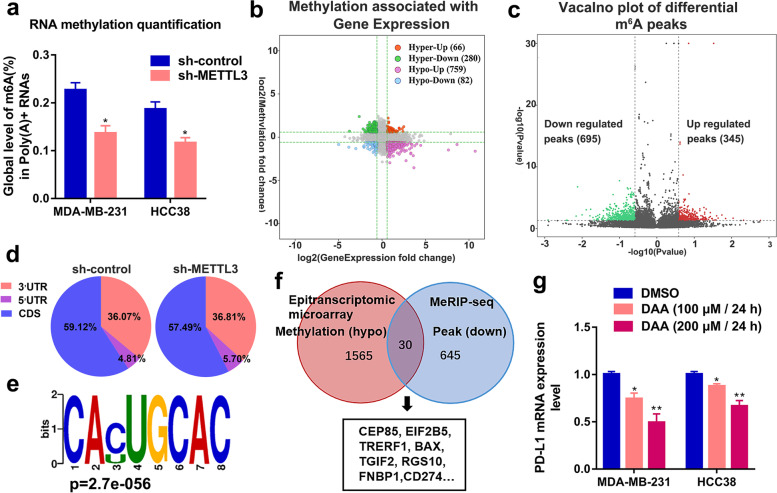
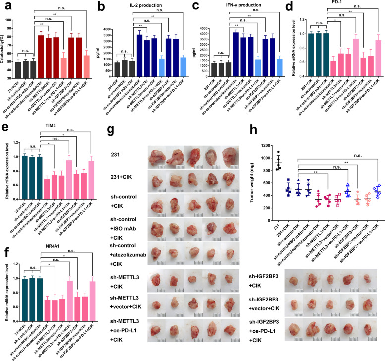
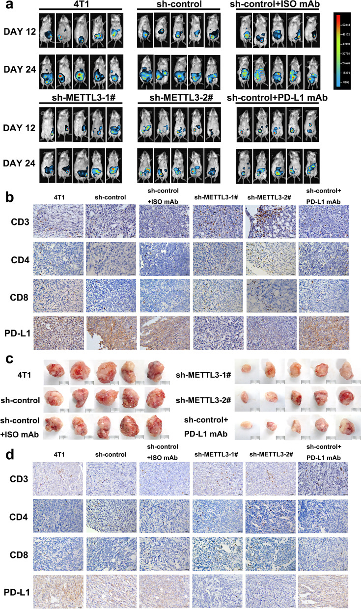
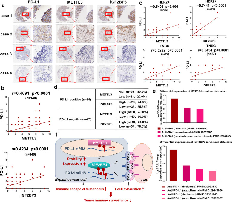
Methods: MeRIP-seq and epitranscriptomic microarray identified that PD-L1 is the downstream target of METTL3. MeRIP-qPCR, absolute quantification of m6A modification assay, and RIP-qPCR were used to examine the molecular mechanism underlying METTL3/m6A/IGF2BP3 signaling axis in PD-L1 expression. B-NDG and BALB/c mice were used to construct xenograft tumor models to verify the phenotypes upon METTL3 and IGF2BP3 silencing. In addition, breast cancer tissue microarray was used to analyze the correlation between PD-L1 and METTL3 or IGF2BP3 expression.

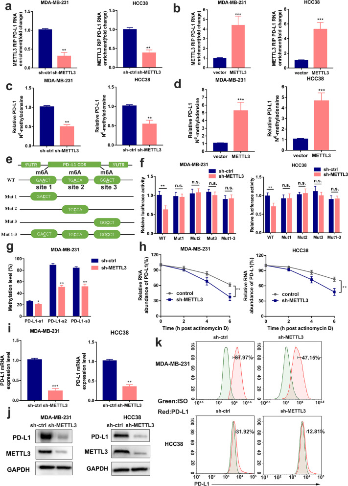
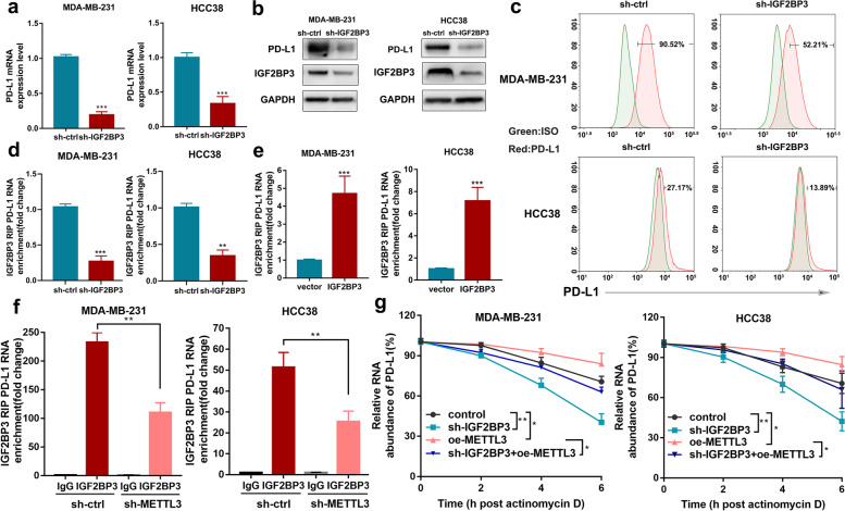
Results: We identified that PD-L1 was a downstream target of METTL3-mediated m6A modification in breast cancer cells. METTL3 knockdown significantly abolished m6A modification and reduced stabilization of PD-L1 mRNA. Additionally, METTL3-mediated PD-L1 mRNA activation was m6A-IGF2BP3-dependent. Moreover, inhibition of METTL3 or IGF2BP3 enhanced anti-tumor immunity through PD-L1-mediated T cell activation, exhaustion, and infiltration both in vitro and in vivo. PD-L1 expression was also positively correlated with METTL3 and IGF2BP3 expression in breast cancer tissues.

Conclusion: Our study suggested that METTL3 could post-transcriptionally upregulate PD-L1 expression in an m6A-IGF2BP3-dependent manner to further promote stabilization of PD-L1 mRNA, which may have important implications for new and efficient therapeutic strategies in the tumor immunotherapy.

Keywords: Breast cancer; Immune surveillance; METTL3; PD-L1; m6A.


Conflict of interest statement

The authors declare no competing interests.

Figures

Fig. 1
Fig. 1
PD-L1 is a downstream target of METTL3. a The global content of m6A was examined by RNA methylation quantification assay. b The starplot presented the distribution of genes with both differential (hyper or hypo) methylation level (Y axis; |fold change| ≥ 1.5) and differential (up or down) gene expression level (X axis; |fold change| ≥ 1.5) in sh-METTL3 with control groups. c Volcano plot of changed m6A peaks was identified by MeRIP-seq in control and METTL3-knockdown MDA-MB-231cells. d Distribution of total m6A peaks in sh-control and sh-METTL3 groups were shown. e Top sequence motif was identified from MeRIP-seq. f Venn diagram showed the down-modified genes following METTL3 knockdown. g The mRNA expression levels of MDA-MB-231 and HCC38 cells were tested by qRT-PCR after 3-deazaadenosine (DAA) treatment in the indicated concentration. *p < 0.05; **p < 0.01
Fig. 2
Fig. 2
METTL3 increases m6A modification and expression of PD-L1 mRNA. a-b The interaction between METTL3 and PD-L1 mRNA was analyzed by RIP-qPCR assay in MDA-MB-231 and HCC38 cells with METTL3 knockdown or overexpression. c-d The relative levels of m6A in PD-L1 were tested by MeRIP-qPCR from MDA-MB-231 and HCC38 cells with overexpression or knockdown of METTL3. e Putative m6A modification sites in the CDS sequence of PD-L1 and synonymous mutations in the PD-L1 CDS. f Relative activity of the WT or Mut luciferase reporters in METTL3-silenced MDA-MB-231 and HCC38 cells was determined (normalized to negative control groups). g The m6A levels of three specific sites of PD-L1 (correspond to the figure e) were determined by absolute quantification of m6A modification. h PD-L1 mRNA levels were determined by qRT-PCR in MDA-MB-231 and HCC38 cells (control and METTL3 disruption) after actinomycin D treatment (normalized to 0 h). i-k PD-L1 mRNA, protein and cell surface expression levels were detected by qRT-PCR, western blot and flow cytometry in sh-Ctrl or sh-METTL3 MDA-MB-231 and HCC38 cells. Values are the mean ± SD of three independent experiments. *p < 0.05; **p < 0.01; ***p < 0.001; n.s., no significance
Fig. 3
Fig. 3
IGF2BP3 mediates PD-L1 mRNA expression in an m6A-dependent manner. a-c PD-L1 mRNA, protein and cell surface expression levels were tested by qRT-PCR, western blot and flow cytometry in sh-ctrl or sh-IGF2BP3 MDA-MB-231 and HCC38 cells. d-e The interaction between IGF2BP3 and PD-L1 mRNA was analyzed by RIP-qPCR assay with overexpression or knockdown of IGF2BP3. f The binding of IGF2BP3 was tested by RIP-qPCR in sh-METTL3 and control cells. g PD-L1 mRNA levels were analyzed by qRT-PCR assay in MDA-MB-231 and HCC38 cells after actinomycin D treatment. Results were presented as mean ± SD of three independent experiments. *p < 0.05; **p < 0.01; ***p < 0.001
Fig. 4
Fig. 4
METTL3/IGF2BP3-downregulated antitumor immunity in breast cancer cells. a The cytotoxicity was measured by lactate dehydrogenase (LDH) release assay after incubation for 48 h. b-c The IFN-γ and IL-2 protein levels in co-culture medium were measured by ELISA after 48 h co-incubation. d-f The PD-1, TIM3 and NR4A1 mRNA expression levels were tested by qRT-PCR. g Images at the end points of subcutaneous xenograft tumors formed by MDA-MB-231 cells in B-NDG mice (n = 5 for each group; scale bar, 1 cm). h Tumors weight were measured in the xenograft mice. Results were presented as mean ± SD of three independent experiments. *p < 0.05; **p < 0.01; n.s., no significance
Fig. 5
Fig. 5
METTL3 knockdown enhances antitumor immunity and immune infiltration. a Bioluminescent images of intraperitoneal xenografted tumors from the indicated groups for Day 12 and Day 24. b The CD3+, CD4+ and CD8+ densities and PD-L1 expression were determined by Immunohistochemical analysis in the intraperitoneal xenografted tumors. Scale bar, 20 μm. c Images of subcutaneous tumors from the indicated groups (scale bar: 1 cm). d The CD3+, CD4+ and CD8+ densities and PD-L1 expression were determined by Immunohistochemical assay in the subcutaneous xenograft models. Scale bar, 20 μm
Fig. 6
Fig. 6
PD-L1 expression positively correlates with METTL3 and IGF2BP3 expression in breast cancer. a The expressions of PD-L1, METTL3 and IGF2BP3 were analyzed by IHC in a tissue microarray containing of 140 breast cancer tissues. Four Cases as representative IHC staining with positive- and negative-PD-L1 were shown. Scale bars, 100 μm. b The correlation of PD-L1 with METTL3 and IGF2BP3 in all breast cancer tissues (n = 140) were analyzed by IHC scores. Proportion scores were recorded as 0, 1, 2, 3, 4 corresponding to < 5%, 5–25%, 25–50%, 50–75%, and ≥ 75%. Intensity scores were recorded as 0, 1, 2, 3 corresponding to negative, weak, moderate, and strong staining. Finally, IHC scores was calculated as “proportion score × intensity score”. c The correlation between PD-L1 and METTL3 or IGF2BP3 were analyzed in HER2+ (n = 26) and TNBC (n = 27) subtypes. Spearman’s rank correlation test was used to analyze the P value. d Number of cases of METTL3 and IGF2BP3 were presented in two categories (PD-L1 positive and PD-L1 negative) in 140 tissues. e The differential expression of METTL3 or IGF2BP3 between responders and non-responders in cilnial data sets. The Y-axis represents the log2 Fold change values (responders vs. non-responders). f A schematic model illustrating the mechanism of METTL3/IGF2BP3-mediated N6-methyladenosine modification of PD-L1 mRNA in breast cancer

References

    1. Sung H, Ferlay J, Siegel R, Laversanne M, Soerjomataram I, Jemal A, et al. Global cancer statistics 2020: GLOBOCAN estimates of incidence and mortality worldwide for 36 cancers in 185 countries. 2021. - PubMed
    1. Li ZL, Zhang HL, Huang Y, Huang JH, Sun P, Zhou NN, et al. Autophagy deficiency promotes triple-negative breast cancer resistance to T cell-mediated cytotoxicity by blocking tenascin-C degradation. Nat Commun. 2020;1:3806. - PMC - PubMed
    1. Esteva F, Hubbard-Lucey V, Tang J, Pusztai L. Immunotherapy and targeted therapy combinations in metastatic breast cancer. Lancet Oncol. 2019;20:e175–e186. - PubMed
    1. Postow M, Callahan M, Wolchok JD. Immune checkpoint blockade in Cancer therapy. J Clin Oncol. 2015;33:1974–1982. - PMC - PubMed
    1. Topalian S, Drake C, Pardoll DM. Targeting the PD-1/B7-H1(PD-L1) pathway to activate anti-tumor immunity. Curr Opin Immunol. 2012;24:207–212. - PMC - PubMed

Publication types