. 2022 Mar 1;119(9):e2118695119.
doi: 10.1073/pnas.2118695119.

Abolishing the prelamin A ZMPSTE24 cleavage site leads to progeroid phenotypes with near-normal longevity in mice

Affiliations

Abolishing the prelamin A ZMPSTE24 cleavage site leads to progeroid phenotypes with near-normal longevity in mice

Yuexia Wang et al. Proc Natl Acad Sci U S A. .

Abstract

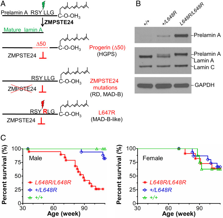
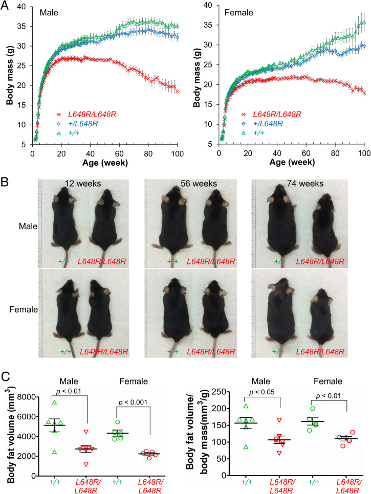
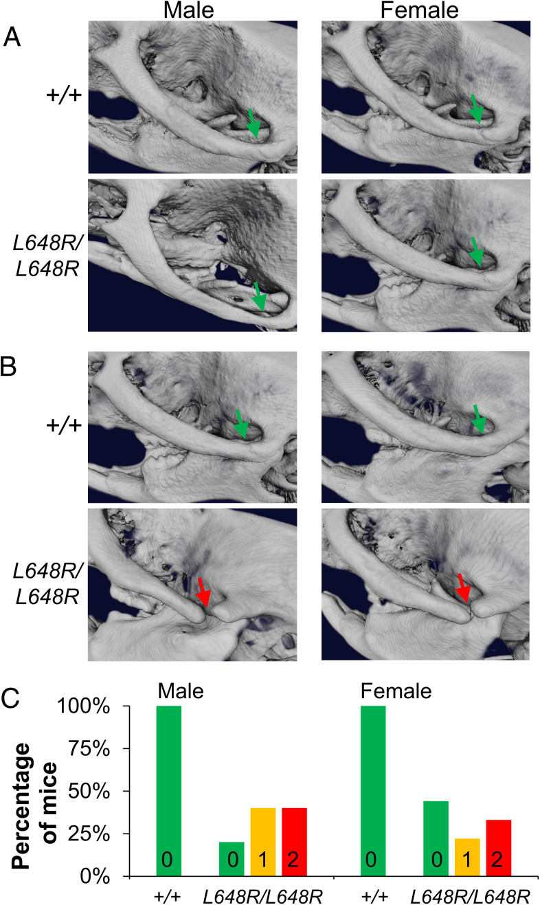
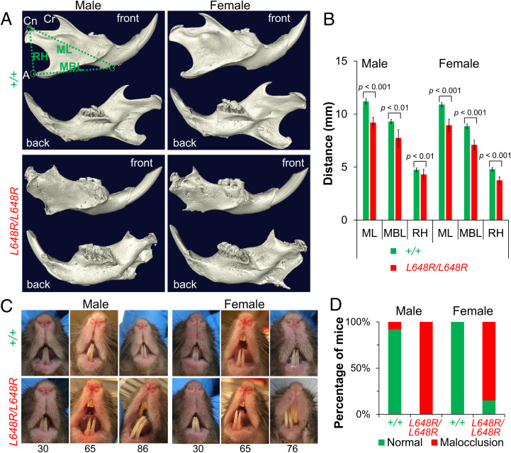
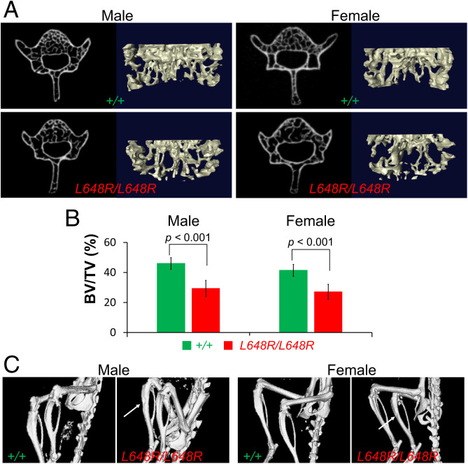
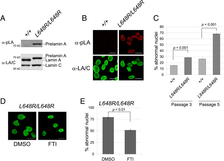
Prelamin A is a farnesylated precursor of lamin A, a nuclear lamina protein. Accumulation of the farnesylated prelamin A variant progerin, with an internal deletion including its processing site, causes Hutchinson-Gilford progeria syndrome. Loss-of-function mutations in ZMPSTE24, which encodes the prelamin A processing enzyme, lead to accumulation of full-length farnesylated prelamin A and cause related progeroid disorders. Some data suggest that prelamin A also accumulates with physiological aging. Zmpste24-/- mice die young, at ∼20 wk. Because ZMPSTE24 has functions in addition to prelamin A processing, we generated a mouse model to examine effects solely due to the presence of permanently farnesylated prelamin A. These mice have an L648R amino acid substitution in prelamin A that blocks ZMPSTE24-catalyzed processing to lamin A. The LmnaL648R/L648R mice express only prelamin and no mature protein. Notably, nearly all survive to 65 to 70 wk, with ∼40% of male and 75% of female LmnaL648R/L648R mice having near-normal lifespans of 90 wk (almost 2 y). Starting at ∼10 wk of age, LmnaL648R/L648R mice of both sexes have lower body masses than controls. By ∼20 to 30 wk of age, they exhibit detectable cranial, mandibular, and dental defects similar to those observed in Zmpste24-/- mice and have decreased vertebral bone density compared to age- and sex-matched controls. Cultured embryonic fibroblasts from LmnaL648R/L648R mice have aberrant nuclear morphology that is reversible by treatment with a protein farnesyltransferase inhibitor. These novel mice provide a model to study the effects of farnesylated prelamin A during physiological aging.

Keywords: aging; bone; lamin; nuclear envelope; progeria.


Conflict of interest statement

Competing interest statement: H.J.W. has received consulting income from Eiger BioPharmaceuticals.

Figures

Fig. 1.
Fig. 1.
Survival of mice with a Lmna L648R mutation corresponding to the human mutation LMNA L647R that encodes an uncleavable variant of prelamin A. (A) Prelamin A is normally processed to lamin A after proteolytic cleavage catalyzed by the zinc metalloprotease ZMPSTE24 between tyrosine (Y) and leucine (L) 647 (648 in mouse), removing a peptide containing the carboxyl-terminal farnesylated cysteine (C). HGPS-causing LMNA mutations generate a prelamin A variant with an internal deletion of 50 amino acids (Δ50) called progerin, which lacks the ZMPSTE24 cleavage site and retains a farnesylated carboxyl-terminal cysteine. ZMPSTE24 loss-of-function mutations cause RD or MAD-B, in which unprocessed farnesylated prelamin A accumulates. LMNA mutation causing a MAD-B-like disorder generates a leucine (L) to arginine (R) substitution at residue 647 of prelamin A blocks ZMPSTE24 processing, leading to expression of a farnesylated variant with only a single amino acid difference. (B) Immunoblots of protein extracts from livers of Lmna+/+ (+/+), Lmna+/L648R (+/L648R), and LmnaL648R/L648R (L648R/L648R) mice. Blots were probed with an antibody specific for prelamin A (Top), an anti-lamin A/C antibody that recognized prelamin A, lamin A, and lamin C (Middle), or anti-GAPDH antibody as loading control (Bottom). (C) Survival curves for male L648R/L648R (n = 19), +/L648R (n = 17), and +/+ (n = 10) mice and female L648R/L648R (n = 11), +/L648R (n = 15), and +/+ (n = 8) mice.
Fig. 2.
Fig. 2.
Growth of LmnaL648R/L648R mice. (A) Body mass versus age of male LmnaL648R/L648R (L648R/L648R) (n = 20), Lmna+/L648R (+/L648R) (n = 17), and Lmna+/+ (+/+) (n = 13) mice and female L648R/L648R (n = 13), +/L648R (n = 15), and +/+ (n = 11) mice. Values are means and error bars indicate SEM. (B) Photographs comparing the sizes of male and female +/+ and L648R/L648R mice at the indicated ages. (C) Body fat volume (Left) and body fat volume normalized to body mass (Right) of 52-wk-old male and female +/+ and L648R/L648R mice. Each triangle or circle represents value for an individual animal; long horizontal bars represent mean and errors bars indicate SEM.
Fig. 3.
Fig. 3.
Degenerative deformities in zygomatic arches of LmnaL648R/L648R mice. (A) Representative 3D renderings of the micro-CT images of male and female Lmna+/+ (+/+) and LmnaL648R/L648R (L648R/L648R) mice showing the formation of the zygomatic arches (green arrows) at 4 wk. (B) Degenerative deformity (red arrows) in the zygomaticotemporal suture of male and female L648R/L648R mice compared to the proper maintenance (green arrows) in the +/+ mice at 30 wk. (C) Percentages of male +/+ (n = 7), female +/+ (n = 7), male L648R/L648R (n = 10), and female L648R/L648R (n = 9) mice with degenerative deformity in 0, 1, or 2 zygomatic arches at 30 wk of age.
Fig. 4.
Fig. 4.
Mandibular defects in LmnaL648R/L648R mice. (A) Representative 3D renderings of the segmented micro-CT–scanned images showing mandibles of male and female Lmna+/+ (+/+) and LmnaL648R/L648R (L648R/L648R) mice. A: angular process; Cn: condylar process; Cr: coronoid process; MBL: mandibular body length; ML: mandibular length; RH: ramus height. (B) Comparisons of ML, MBL, and RH between male +/+ (n = 4) and L648R/L648R (n = 4) mice and between female +/+ (n = 4) and L648R/L648R (n = 4) mice. Values are means and error bars indicate SEM. (C) Representative photographs of teeth of male and female +/+ and L648R/L648R mice at the ages in weeks indicated. Dental malocclusion is minimal to mild at 30 wk of age and more severe at older ages. (D) Percentages of male +/+ (n = 13), female +/+ (n = 10), male L648R/L648R (n = 18) and female L648R/L648R (n = 13) mice with malocclusion at ≥65 wk of age.
Fig. 5.
Fig. 5.
Decreased vertebral bone density and tibial defects in LmnaL648R/L648R mice. (A) Representative micro-CT–scanned transverse sections and 3D reconstructions of the L5 vertebrae from male and female Lmna+/+ (+/+) and LmnaL648R/L648R (L648R/L648R) mice. (B) Comparison of vertebra L5 bone density (bone volume/total volume; BV/TV %) between male +/+ (n = 5) and L648R/L648R (n = 5) mice and between female +/+ (n = 7) and L648R/L648R (n = 6) mice. Values are means and error bars indicate SEM. (C) Micro-CT–generated representative images of hind legs of living male and female +/+ and L648R/L648R mice. Arrows indicate thinner and more irregular surfaces of tibias of L648R/L648R mice.
Fig. 6.
Fig. 6.
Farnesylation-dependent abnormal nuclear morphology in LmnaL648R/L648R MEFs. (A) Immunoblots of protein extracts from MEFs generated from Lmna+/+ (+/+) and LmnaL648R/L648R (L648R/L648R) mouse embryos. Blots were probed with an antibody specific for prelamin A (α-pLA, Upper) and anti-lamin A/C antibody that recognizes prelamin A, lamin A, and lamin C (α-LA/C, Lower). (B) Immunofluorescence photomicrographs showing the indicated MEFs at passage 5 stained with antibodies that specifically recognize prelamin A (Upper) or prelamin A, lamin A, and lamin C (Lower). (Scale bar, 20 µm.) (C) Quantification of aberrant nuclear morphology in MEFs at passage 3 and passage 5. Three independently grown MEF cultures were fixed, stained, and ∼100 nuclei counted from each. Values are means (n = 3) and error bars indicate SEM. Representative images of nuclei that are counted as abnormal—including those that are blebbed, misshapen, or highly crenylated—are shown in SI Appendix, Fig. S6. (D) Immunofluorescence photomicrographs showing LmnaL648R/L648R MEFs at passage 6 treated with DMSO vehicle or FTI as described in Materials and Methods. (Scale Bar, 20 µm.) (E) Percentages of nuclei with aberrant nuclear morphology in LmnaL648R/L648R MEFs treated with DMSO or FTI. For quantification, three independently grown MEF cultures were fixed, stained, and ∼100 nuclei counted from each. Values are means (n = 3) and error bars indicate SEM.

References

    1. Davies B. S., Fong L. G., Yang S. H., Coffinier C., Young S. G., The posttranslational processing of prelamin A and disease. Annu. Rev. Genomics Hum. Genet. 10, 153–174 (2009). - PMC - PubMed
    1. Worman H. J., Michaelis S., Permanently farnesylated prelamin A, progeria, and atherosclerosis. Circulation 138, 283–286 (2018). - PMC - PubMed
    1. Young S. G., Meta M., Yang S. H., Fong L. G., Prelamin A farnesylation and progeroid syndromes. J. Biol. Chem. 281, 39741–39745 (2006). - PubMed
    1. De Sandre-Giovannoli A., et al. , Lamin a truncation in Hutchinson-Gilford progeria. Science 300, 2055 (2003). - PubMed
    1. Eriksson M., et al. , Recurrent de novo point mutations in lamin A cause Hutchinson-Gilford progeria syndrome. Nature 423, 293–298 (2003). - PMC - PubMed

LinkOut - more resources